Brachytherapy and GYN malignancy Brachytherapy Brachytherapy brachy from

Brachytherapy and GYN malignancy
 consists of placing sealed")
Brachytherapy • Brachytherapy (brachy, from the Greek for “short distance”) consists of placing sealed radioactive sources close to or contact with the target tissue. • Interstitial, intracavity, or transluminal approach. • Temporary, or permanent implant. • Low or high dose rate.
 • High radiation dose can")
Introduction • Discovery in 1898 • Short distance (cm) • High radiation dose can be delivered locally to the tumor with rapid dose fall-off in the surrounding normal tissue

Radioactive sources
")
Radioactive sources Radium-226 • Average energy 0. 83 Mev (0. 5 mm of platinum) • A filtration of at least 0. 5 mm platinum is sufficient to absorb all the α particles and most of the β particles emitted by the radium and its daughter products. • Half life ~1600 years • It was loaded into cells about 1 cm long and 1 mm in diameter. • Radium sources are manufactured as needles or tubes in a variety of lengths and activities

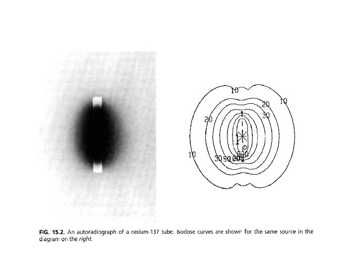
Radioactive sources Cesium-137 • Substitute for radium in both interstitial and intracavitary brachytherapy • Energy 0. 662 Mev nearly the same penetrating power as radium • Half life 30 years (clinically used 7 years without replacement) It was doubly encapsulated in stainless-steel needles and tubes.

Radioactive sources Cobalt-60 • High specific activity • Small sources required for some special applicators 137 • More expensive than Cs and short half life (5. 26 years) • The sources can be used to replace 226 Ra in intracavitary application

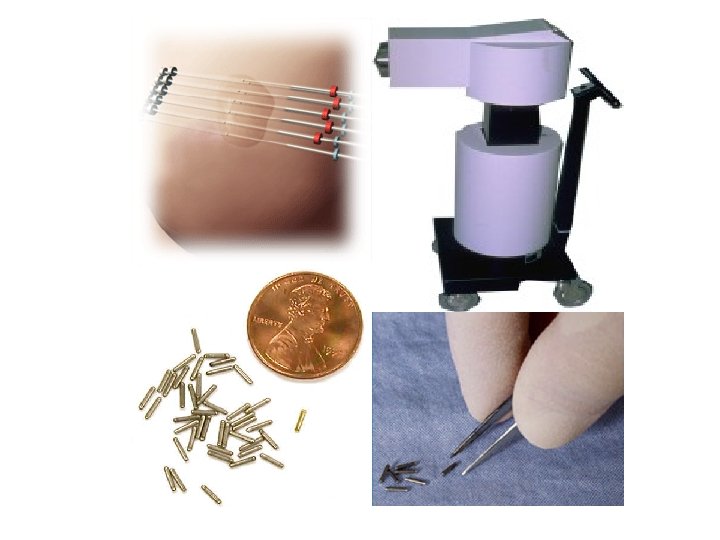
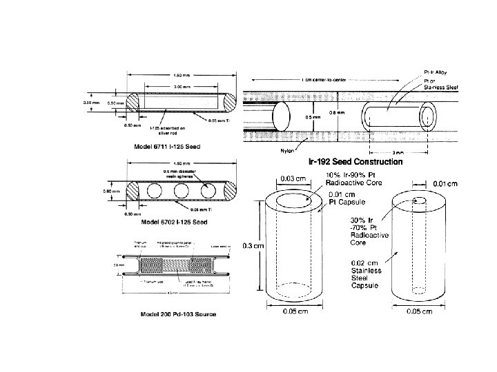
Radioactive sources Iridium-192 • It has a complicated γ ray spectrum with an average energy of 0. 38 Me. V. → It required less shielding for personnel protection. • It has the disadvantage of a short half-life (73. 8 days) • It is fabricated in the form of thin flexible wires which can be cut to desired lengths

Radioactive sources Iodine-125 • Widely used for permanent implants. • Longer half-life: 59. 4 days (convenient for storage) • Low photon energy (0. 028 Me. V) → less shielding. • Disadvantages: dosimetry of 125 I is much more complex.

Brachytherapy Permanently Implanted Energ y T 1/2 Dose Rate Rn 1. 2 Me V 3. 83 0. 75 G/h Au 412 ke V 2. 70 1. 07 G/h I 28 ke. V 59. 6 0. 07 G/h Pd 22 ke. V 17 0. 19 G/h Source 222 198 125 13
 HDR")
Radioactive sources • ICRU 38 LDR sources: 0. 4 -2 Gy/hr (137 Cs) HDR sources: ≥ 12 Gy/hr (60 Co, 192 Ir) • 226 Ra leakage Radon gas. • 137 Cs better than 226 Ra less shielding and microsphere form with leakage gas. • 137 Cs better than 60 Co less shielding and cheap. • 192 Ir better than 137 Cs lower energy require less shielding for personnal protection and higher specific activity. • 103 Pd better than 198 Au and 125 I less shielding and biologic advantage.
 Patient • Long history of use. • Ability")
Radioactive sources Low Dose Rate (LDR) Patient • Long history of use. • Ability to predict rate of late complications High Dose Rate (HDR) • No long term confinement to bed. • No indwelling bladder catheters. • Not labeled “radiation risk zone” to relative, visitors, and staff. • Avoid several anesthesias. Clinical • Improves chances of atching tumors in • Maintain position of the sources sensitive phase of cell cycle. Physica • Longer treatment times allow for leisurely review of and potential l modifications to the treatment. • Plan prior to the delivery of a significant portion of treatment. • Favorable dose-rate effect on repair of normal tissues. • Infrequent replacement and calibration of sources because of long isotope half -life. during the brief treatment. • Patient preparation. • No specialized nursing. • Ability to treat great patient loads. • Short treatment times and minimal radiation protection problems. • Possibility of optimizing dose distribution by altering the dwell times of the source at different

Brachytherapy and GYN Malignancy

Reference point from which lymph node position were measured on lymphoangiograms and the range of location Int. J Radiat Oncol Biol Phys 34: 167 -172, 1996

Distribution of pelvic node metastases in patients with Ib-IIa cervical cancer Gynecol Oncol 62: 19 -24, 1996 Tumor size <=4 cm Local advanced tumor

External beam radiotherapy for GYN Malignancy

Pelvic irradiation portal in cervical cancer 4 -field box technique

Pelvic irradiation portal in cervical cancer 4 -field box technique

")
Combination of external beam pelvic irradiation and intracavitary brachytherapy (ICRT)
")
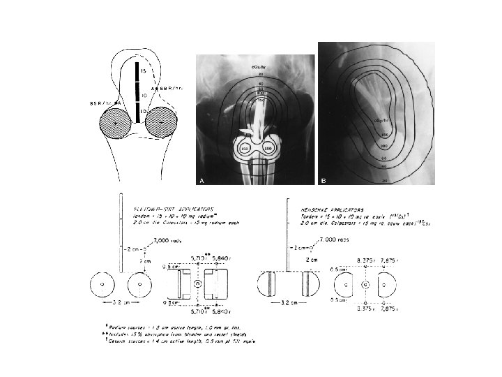
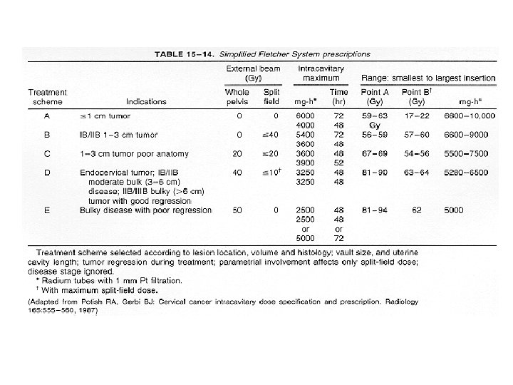
Brachytherapy in definitive radiotherapy of cervical cancer (Intracavity radiotherapy, ICRT)
")
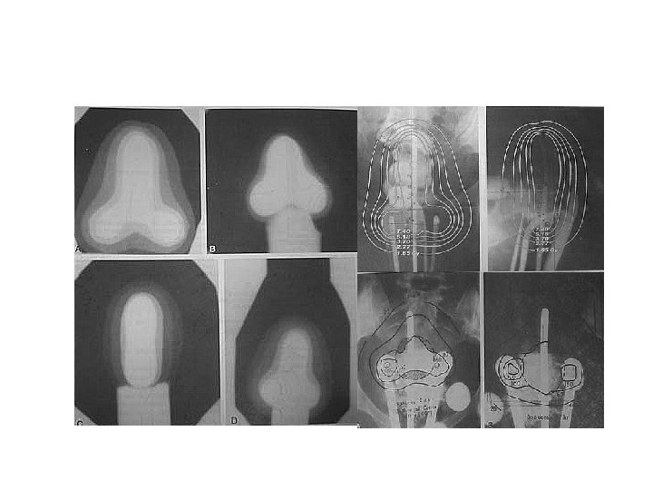
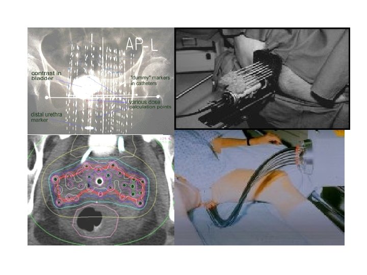
Intracavitary Radiotherapy (ICRT)
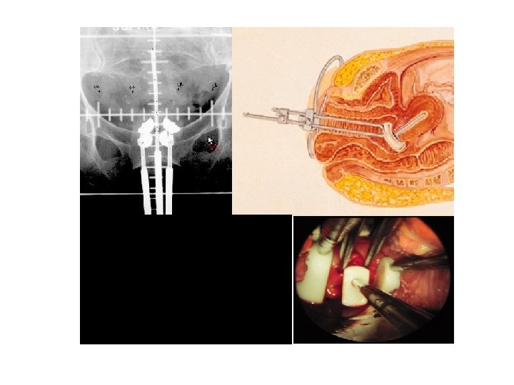
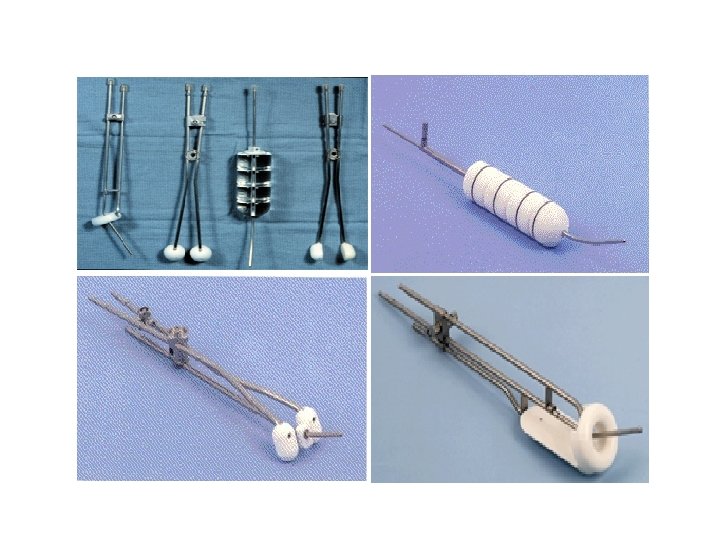
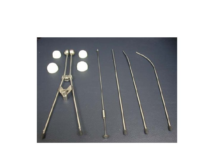

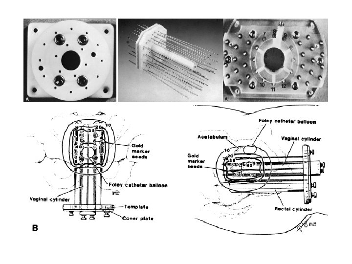
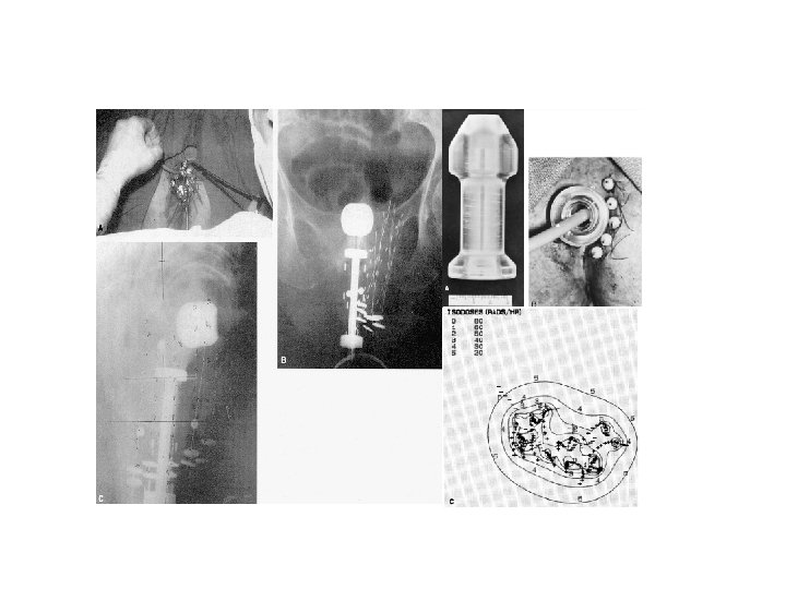
Applicator of ICRT
")
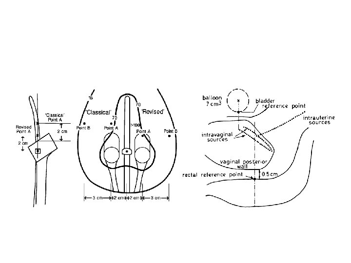
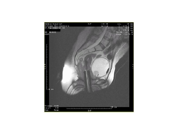
Intracavitary insertion (ICRT)

")
Postoperative brachytherapy (Intravaginal radiotherapy)
")
Intravaginal radiotherapy (IVRT)


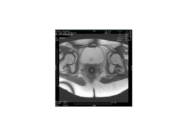
Female urethral cancer

Endometrial cancer
- Slides: 46