
Brachial plexus injury is classified According to the

Brachial plexus injury is classified According to the Etiology into: a) Obstetric Brachial plexus injury, it is considered birth related trauma. Injury to the brachial plexus occurs at the time of delivery and may be associated with other injuries i. e. fr. clavicle or humerus. b) Traumatic Brachial plexus injury, sometimes it is also called adult type, but this name is somewhat incorrect as Traumatic Brachial plexus injury can happen in pre-adolescent children as well as in adults.
Introduction • In 1764 Smellie was the first to describe Birth related brachial plexus palsy. • But in 1872 it was Erb who localized the most common lesion to the fifth and sixth cervical roots i. e. upper trunk lesion and gave his name to this condition.
Incidence Reported incidence of OBPI ranging from 0. 4 to 4 per 1000 of live births, more frequent in males (55. 3%) than in females (44. 7%), more often unilateral (93. 5%) than bilateral (6. 5%). The right side was involved in (62. 8%); the left in (30. 7%).
Risk Factors and Mechanism of injury • Large birth weight. • Prolonged labor. • Difficult delivery. • Shoulder dystocia. • Breech presentation. • Forceps delivery. • Maternal diabetes. • Previous births with brachial plexopathy.

Risk Factors and Mechanism of injury In general traction on the brachial plexus causes its injury (3), (31); which may be due to aggressive head tilt or shoulder depression or traction on upper limb to deliver the aftercoming head (31), (5). i. e. Shoulder dystocia is the mechanical factor that results in an upper trunk lesion (Figure 8) (32). A breech delivery often results in a stretch of the lower plexus from traction applied to the trunk with the arm abducted. Also, a difficult arm extraction in a breech delivery can result in an avulsion injury to the upper trunk. The severity of the palsy depends on which roots of the brachial plexus have been injured and the extent of injury.
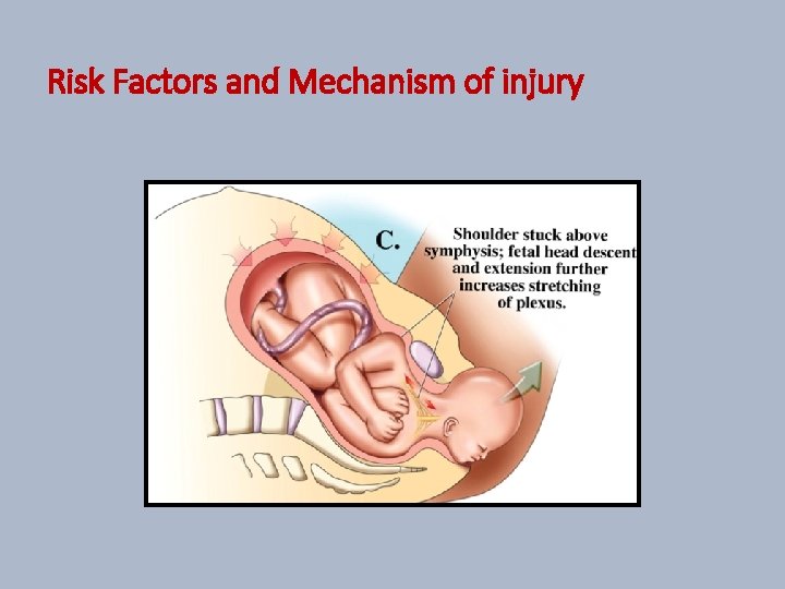
Risk Factors and Mechanism of injury

Surgical Anatomy

Classification and Clinical Picture According to the anatomical lesion into: 1. 2. 3. 4. Upper trunk (C 5, 6) injury. Upper and Middle trunks (C 5, 6, 7) injury. Lower trunk (C 8, T 1) injury. Total Brachial plexus palsy (C 5, 6, 7, 8, T 1) injury. with or without Horner's syndrome.
Classification and Clinical Picture According to the time of presentation and surgical intervention into: A. Early cases: Where surgical intervention took place before the age of twelve months i. e. 1 year. B. Late or delayed presenting cases: Where surgical intervention took place at or after the age of twelve months i. e. 1 year.
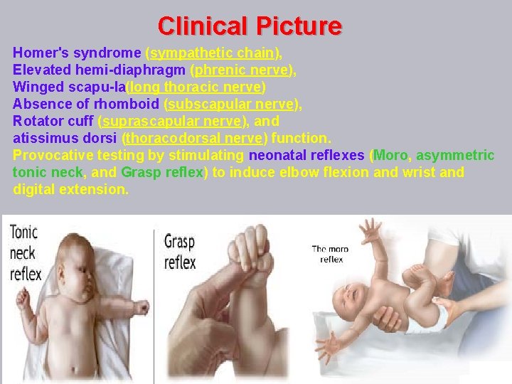
Clinical Picture Homer's syndrome (sympathetic chain), Elevated hemi diaphragm (phrenic nerve), Winged scapu la(long thoracic nerve) Absence of rhomboid (subscapular nerve), Rotator cuff (suprascapular nerve), and atissimus dorsi (thoracodorsal nerve) function. Provocative testing by stimulating neonatal reflexes (Moro, asymmetric tonic neck, and Grasp reflex) to induce elbow flexion and wrist and digital extension.
DIFFERENTIAL DIAGNOSIS v. OBPP should be differentiated from other neurological disorders e. g. cerebral palsy, post-traumatic monoplegia in these conditions usually it is not difficult to differentiate cases with OBPP from other neurological disorders by history and clinical picture. v. OBPP should be differentiated from the cases with pseudo-paralysis due to clavicle or humerus fracture or from cases with septic shoulder x-rays can be used to help in the diagnosis and ultra-sound may be superior to the x-rays in the diagnosis of proximal humerus physeal fracture and in cases with septic arthritis of shoulder (31).

Assessment §Diagnosis and clinical evaluation of OBPP cases is a mandatory step, pre-operative classification of the cases according to Narakas’s classification is very important in pre-operative planning and in predicting the outcome. §Using valid and internationally accepted scores for assessment of various joints and their movements provide more accurate and quantitative method to estimate the amount of recovery and for comparing the outcome with the results of other authors. §Assessment of shoulder joint, elbow joint, wrist joint, forearm movements and hand functions are needed.

Assessment § Examination of different muscles of the upper limbs gives idea about which roots are injured i. e. examination of shoulder abduction (deltoid muscle function) to evaluate C 5 root, examination of elbow flexion (biceps muscle function) to evaluate C 6 root, examination of elbow and wrist extension (triceps muscle and forearm extensor function) to evaluate C 7 root, examination of finger flexion (forearm flexor muscles function) to evaluate C 8 root (5). § The most commonly used score for assessment of various joint movements is the Active Movement Scale (AMS) Toronto proposed by Clark & Curtis (1995) from Hospital for Sick Children (Table 1). § This score is widely used by many authors for assessment of shoulder, elbow, wrist, fingers and thumb movement.

Assessment Table 1: Active Movement Scale (AMS) Toronto proposed by Clark & Curtis (1995) from Hospital for Sick Children Observation Muscle grade Gravity eliminated No contraction 0 Contraction, no motion 1 Motion ≤ 1/2 range 2 Motion > 1/2 range 3 Full motion 4 Against gravity Motion ≤ 1/2 range 5 Motion > 1/2 range 6 Full motion 7

Assessment of shoulder function Estimation of the range of movements (ROM) of various shoulder movements is important especially abduction, adduction, internal and external rotation. Scores used for assessment of shoulder function: 1. Active Movement Scale (AMS) Toronto (Table 1). 2. The Modified Mallet score (Figure 12). 3. Gilbert scale table (Table 2).
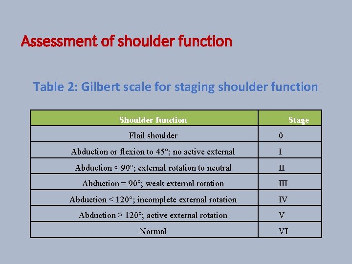
Assessment of shoulder function Table 2: Gilbert scale for staging shoulder function Stage Flail shoulder 0 Abduction or flexion to 45°; no active external I Abduction < 90°; external rotation to neutral II Abduction = 90°; weak external rotation III Abduction < 120°; incomplete external rotation IV Abduction > 120°; active external rotation V Normal VI

Assessment of shoulder function Figure 12: The Modified Mallet score
Assessment of elbow function 1. Active Movement Scale (AMS) Toronto (Table 1). 2. Gilbert and Raimondi scale for evaluating elbow recovery (Table 3).
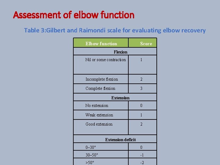
Assessment of elbow function Table 3: Gilbert and Raimondi scale for evaluating elbow recovery Elbow function Score Flexion Nil or some contraction 1 Incomplete flexion 2 Complete flexion 3 Extension No extension 0 Weak extension 1 Good extension 2 Extension deficit 0– 30° 0 30– 50° -1 >50° -2
Assessment of forearm pronation/supination movements and the presence of fixed pronation or supination deformities. Assessment of wrist function by: Active Movement Scale (AMS) Toronto (Table 1). For assessment of wrist flexion and extension.
Assessment of hand function 1. Active Movement Scale (AMS) Toronto (Table 1). 2. Raimondi scale for evaluating hand function (Table 4).
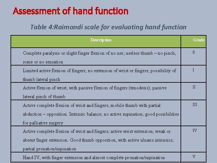
Assessment of hand function Table 4: Raimondi scale for evaluating hand function Description Complete paralysis or slight finger flexion of no use; useless thumb – no pinch; Grade 0 some or no sensation Limited active flexion of fingers; no extension of wrist or fingers; possibility of I thumb lateral pinch Active flexion of wrist, with passive flexion of fingers (tenodesis); passive II lateral pinch of thumb Active complete flexion of wrist and fingers; mobile thumb with partial III abduction – opposition. Intrinsic balance; no active supination; good possibilities for palliative surgery Active complete flexion of wrist and fingers; active wrist extension; weak or IV absent finger extension. Good thumb opposition, with active ulnaris intrinsics; partial pronation/supination Hand IV, with finger extension and almost complete pronation/supination V
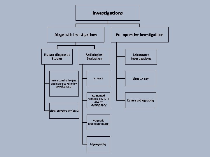
Investigations Diagnostic investigations Electro-diagnostic Studies Nerve conduction(NC) and nerve conduction velocity(NCV) Radiological Evaluation Pre-operative investigations Laboratory investigations X-RAYS chest x-ray Computed tomography (CT) and CT Myelography Echo-cardiography Electromyography(EMG Magnetic resonance image Myelography
Natural History and Prognosis • Many studies have discussed the natural history of OBPP and they stressed on the high probability of spontaneous recovery reaching up to 90%. • The rate of recovery is proportional to the extent of roots involved and the nature of injury. According to the extent of roots involved • In general, the less roots affected the more favorable outcome. • Upper trunk (C 5 and C 6) palsy is associated with the best results of spontaneous recovery and the least later functional impairments, Upper and middle trunks (C 5, C 6 and C 7) palsy is associated with intermediate rates of spontaneous recover, Total OBPP (C 5, C 6, C 7, C 8 and T 1) especially with Horner syn. carries the worst prognosis due to the high possibility of root avulsions. The involvement of C 7 root injury has special impact on the prognosis of global shoulder function. According to the nature of injury • According to the Seddon classification of nerve injuries the nature of injury may be one of these three types (Figure 13): • Neuropraxia, Axonotmesis, Neurotmesis.

Conservative Treatment • Spontaneous recovery of OBPP is expected in most cases of OBPP, recovery reaches up to 90% and is directly related to the nature of injury i. e. full recovery in weeks is expected with neuropraxia, intermediate recovery with axonotmesis which may not reach full recovery, poor or no recovery with neurotmesis. also, the it is related to the extent of injury Spontaneous recovery of OBPP is related to the extent of injury i. e. up to 95% of spontaneous recovery with C 5, 6 injury, worst results with total palsy especially if associated with phrenic n. palsy and/or Horner’s syndrome which usually indicate root avulsions. • Once OBPP is diagnosed physiotherapy should be initiated to encourage recovery, prevent joint stiffness and preserve the muscle power until recovery and re-enervation.
Timing of surgical intervention • Although many authors described in their researches that the majority of OBPP cases have spontaneous recovery reaching up to 90% ; still cases with some partial spontaneous recovery may have great functional impairment in their affected upper limbs especially in hand function which may greatly affect the child future and makes a great burden to his family and the community even in advanced countries. • Because of the high axonal regeneration potential in children It is well known that early Microsurgical exploration and reconstruction in these cases yields more favorable outcome in patient`s upper limb functions in comparison to untreated cases. • Most of the surgeons believe that the golden time for Microsurgical intervention is between 6 and 9 months or at the age of 6 months if there is no recovery of biceps function by that time (12), (6).
Timing of surgical intervention • some authors prefer to do surgical intervention at the age of 3 months in cases of total OBPP with flail hand upper limbs especially if associated with phrenic n. palsy and/or Horner’s syndrome which usually indicate root avulsions. • Many researches did not demonstrate significant difference in the functional outcome between surgical interventions at the age of 6 months versus 3 months, indeed more morbidity is encountered if surgical intervention took place before the age of 6 months, meanwhile researches shows spontaneous recovery between the ages of 3 and 6 months. • Satisfactory results can be achieved up to 12 months of age. • Some authors claimed that there is no role of primary surgery in late(delayed) presenting cases, while on the other hand some reports still showing improved function after delayed primary surgical reconstruction.
Timing of surgical intervention • This may be explained by the fact that axons crossing the neuroma in cases with OBPP and reaching distal targets even if they did not have proper arrangement of fibers or giving good function (6), they still offering important innervation necessary to spare the motor end plates and preventing the occurrence of denervation fibrosis and wasting with loss of all motor units. This preserve the muscles and gives success to the process of re-innervation even if it was delayed. • In general, the best results are with early surgical intervention in cases with only upper trunk injury especially without root avulsions. • So OBPP cases can be classified according to the time of presentation and surgical intervention into: a) Early cases: Where surgical intervention took place before the age of twelve months i. e. 1 year. b) Late or delayed presenting cases: Where surgical intervention took place at or after the age of twelve months i. e. 1 year.
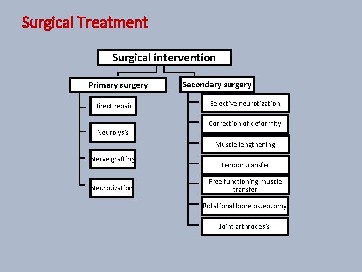
Surgical Treatment Surgical intervention Primary surgery Direct repair Neurolysis Secondary surgery Selective neurotization Correction of deformity Muscle lengthening Nerve grafting Neurotization Tendon transfer Free functioning muscle transfer Rotational bone osteotomy Joint arthrodesis
Complication 1 - Intra- operative and/or early post-operative: General surgical complications e. g. bleeding. complications of prolonged anesthesia e. g. hypothermia, post-operative chest infection and respiratory complications, pleural injury, pneumo-and/or heamo-thorax, great vessel injury e. g. subclavian a. & v. , axillary a. & v. , innominate a. & v. , carotid a. & v. , and jugular v. 2 -Late post-operative complications: Wound infection, dehiscence, scarring and keloid formation. Early loss of some pre-operative motor function (in cases with partial recovery with neuroma), as cutting of the neuroma and its reconstruction leading to the loss of the previous haphazard spontaneous healing.
Post-operative care and rehabilitation • The arm was adducted and the hand was kept close to the opposite shoulder using crepe bandage without embarrassing respiratory movements. The neck was supported by Philadelphia collar to prevent side bending of the neck that might endanger the nerve sutures performed. All patients were admitted to the microsurgical IC Unit. They were observed for vital signs and were kept sedated for the first 24 hours. Parenteral antibiotics were given for 3 days and then continued orally for one week. The average ICU stay was 2. 1 day (1 -6). Oral feeding was allowed as soon as the infant was aware and fully conscious. • Rehabilitation program was tailored and individually designed by the physiotherapist. It included local massage, local heating, passive and active range of motion, according to state of recovery. Also, it included electric stimulation of paralyzed muscles to prevent fibrosis and contracture. A home program was designed and written in Arabic and supported by illustrated drawings for the needed movement and methods of playing and even posture during sleep. A copy of this program was given and taught to all mothers.
Thank you

- Slides: 34