BRACHIAL PLEXUS INJURIES REVIEW OF BRACHIAL PLEXUS Contents

BRACHIAL PLEXUS INJURIES

REVIEW OF BRACHIAL PLEXUS

Contents of Discussion n n n n Spinal Nerves Nerve Plexus BP – Origin & Relations Formation Parts of BP Distribution - Nerve Supply – areas Anatomical Variations Applied Anatomy

Spinal Nerves n Spinal nerves attach to the spinal cord via roots n Dorsal root n n Has only sensory neurons Attached to cord via rootlets Dorsal root ganglion Ventral root n n Has only motor neurons No ganglion - all cell bodies of motor neurons found in gray matter of spinal cord

Spinal Nerves n 31 pair n n each contains thousands of nerve fibers All are mixed nerves have both sensory and motor neurons n Connect to the spinal cord n Exit from SC – Supplying the muscles & structures of the body

Spinal Nerves 8 pairs of cervical nerves from C 1 to C 8 12 pairs of thoracic nerves from T 1 -T 12 5 pairs of lumbar nerves from L 1 to L 5 5 pairs of sacral nerves from S 1 to S 5 1 pair of coccygeal nerves located at C zero (Co)

Formation of Rami n Rami are lateral branches of a spinal nerve Rami contain both sensory and motor neurons n Two major groups n Dorsal ramus n Neurons innervate the dorsal regions of the body n Ventral ramus n Larger n Neurons innervate the ventral regions of the body n Braid together to form plexuses (plexi) 12 -7 n

Nerve Plexuses n Nerve plexus n n A nerve plexus is nothing more than a system or network of connected nerve fibers that link spinal nerves with specific areas of the body. A network of ventral rami. Ventral rami (except T 2 -T 12) n n Branch and join with one another Form nerve plexuses In cervical, brachial, lumbar, and sacral regions n No plexus formed in thoracic region of s. c. n 12 -8

Branches of Spinal Nerves n Dorsal Ramus n n Neurons within muscles of trunk and back Ventral Ramus (VR) n Braid together to form plexuses n n n Communicating Rami: communicate with sympathetic chain of ganglia n 12 -9 Cervical plexus - VR of C 1 -C 4 Brachial plexus - VR of C 5 -T 1 Lumbar plexus - VR of L 1 -L 4 Sacral plexus - VR of L 4 -S 4 Coccygeal plexus -VR of S 4&S 5 Covered in ANS unit

Brachial Plexus - Origin n n Formed by ventral rami of spinal nerves C 5 -T 1 Five ventral rami form n n 12 -10 Roots / Trunks that separate into Divisions that then form Cords that give rise to Branches Major nerves n Axillary n Radial n Musculocutaneous n Ulnar n Median

Brachial Plexus n 15 cms long , spinal column to axilla. n Brachial plexus is responsible for cutaneous (sensory) and muscular (motor) innervation of the entire upper limb & pectoral girdle. n It proceeds through the neck, the axilla and into the arm.
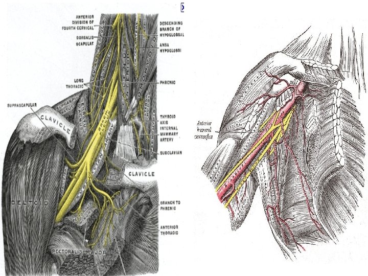

Relations - BP n In the neck, the brachial plexus lies in the posterior triangle, being covered by the skin, Platysma, and deep fascia; where it is crossed by the supraclavicular nerves, the inferior belly of the Omohyoid, the external jugular vein, and the transverse cervical artery.

Relations - BP n When it emerges between the Scalene anterior and medius -* its upper part lies above third part of the subclavian artery, * while the trunk formed by the union of C 8 & T 1 is placed behind the artery.

Relations - BP n The plexus next passes behind the clavicle, the Subclavius, and the transverse scapular vessels, and lies upon the first digitation of the Serratus anterior, and the Subscapularis.

Relations n In the axilla it is placed lateral to the first portion of the axillary artery; it surrounds the second part of the artery, one cord lying medial to it, one lateral to it, and one behind it; at the lower part of the axilla it gives off its terminal branches to the upper limb.

FORMATION OF THE BRACHIAL PLEXUS

PUT IT ALL TOGETHER…. . .

ROOTS n n Originates from C 5 -C 8 and most of T 1 Receives gray rami communicates from the sympathetic trunk. Carry postganglionic sympathetic fibers onto root for distribution of periphery. Root and trunk enter posterior triangle of neck by passing between anterior scalene and middle scalene muscles and lies between superior and posterior to subclavian artery.

TRUNKS n n n C 5, C 6 roots pass down wards between Scalenus medius and Scalenus anterior muscles and unite to form SUPERIOR TRUNK C 7 root pass between Scalenus muscles and at laeral border of scalenus anterior emerges as MIDDLE TRUNK C 8, T 1 roots unite behind a fascial sheet (sibson”s fascia) and beneath the subclavian artery form LOWER TRUNK

DIVISION n n n Lateral to the 1 st rib , where three trunks are located behind the axillary artery , they separate into 3 anterior and 3 posterior divisions The 3 anterior division form parts of brachial plexus that ultimately give rise to peripheral nerves associated with the anterior compartment of arm or forearm. The 3 posterior division combine to form parts of the brachial plexus that give rise to nerves associated with the posterior compartments.

CORDS n n 3 posterior divisions unite to form posterior cord Anterior divisions of upper and , middle trunks (C 5 -C 7) unite to form lateral cord Anterior division of lower trunk forms medial cord(C 8 -T 1) Cords – named after their relation with AA & passes through the thoracic outlet and give off major branches

BRANCHES - Roots n From the Roots n Dorsal Scapular nerve Derived from C 5 root Motor nerve to the Rhomboideus major and minor muscles

Roots n Long Thoracic nerve Derived from C 5, 6, 7 Innervates the serratus anterior muscle

BRANCHES OF UPPER TRUNK NERVE TO SUBCLAVIUS Root value – C 5, C 6 SUPRASCAPULAR NERVE Root value – C 5, C 6

Branches – LC & MC LATERAL PECTORAL NERVE MEDIAL PECTORAL NERVE Root value- C 5, C 6, C 7 Root value- C 8, T 1

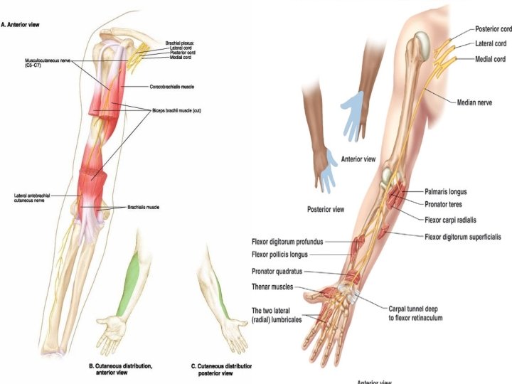
Lateral Cord n Direct branches: Lateral pectoral nerve: C 5 -7 To clavicular head of pectoralis major muscle n Terminal nerves: Musculocutaneous: C 5 -7 Lateral root of median nerve: C 5 -7

Medial Cord: Direct Branches n Medial pectoral nerve: C 8 -T 1 To sternal head of pectoralis major muscle and pectoralis minor muscle. n Medial cutaneous nerve to arm Medial cutaneous nerve to forearm n

n n MEDIAL CUTANEOUS NERVE OF ARM Root value- C 8, T 1 MEDIAL CUTANEOUS NERVE OF FOREARM Root value – C 8, T 1

Medial Cord: Terminal Nerves n n Ulnar C 8 -T 1 Medial root of median nerve C 8 -T 1

ULNAR NERVE

Posterior Cord Direct Branches n Upper subscapular nerve C 5 -6 To subscapularis muscle n Thoracodorsal nerve: C 6 -7 To latissimus dorsi muscle n Lower subscapular nerve: C 5 -6 To subscapularis and teres major muscles

n n n UPPER SUBSCAPULAR Root value-C 5, C 6 LOWER SUBSCAPULAR Root value- C 5, C 6 NERVE TO LATISSIMUS DORSI Root value-C 6, C 7, C 8

Posterior Cord Terminal Nerves n Axillary nerve: C 5 -6 Motor: To deltoid and teres minor muscles. Sensory: Skin on arm over deltoid muscle:

AXILLARY NERVE

Posterior Cord Terminal Nerves n Radial nerve: C 5 -T 1 Motor: Posterior compartments of arm and forearm. Brachioradialis muscle Sensory: Back of arm, forearm, hand

RADIAL NERVE


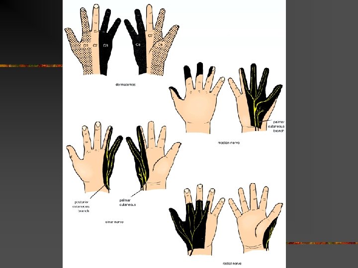
Cutaneous distribution

Anatomic Variations n n The plexus may include anterior rami from C 4 or T 2 and these are designated as Pre fixed- C 4 added Post fixed- T 2 added. The connective tissue sheath that invests the plexus especially in the axillary region has a convoluted and septated structure that can lead to non uniform distribution of local anaesthetics.

n The musculocutaneous nerve may fuse to or have communications with the median nerve , which can result in its absence from within the coracobrachialis muscle. n Communication between median and ulnar nerves is common in the forearm with the median nerve replacing the innervations to various muscles normally supplied by the ulnar nerve. n Variations with respect to vessels within the arm may be present like double axillary veins , high origin of radial artery and double brachial arteries.

n n The interscalene groove may have variations in the relationship between the plexus roots and trunks and the muscles. For eg. - the C 5 or C 6 roots may traverse through or anterior to the anterior scalene muscles. In many specimens no inferior trunk exists , a single cord or a pair of cords may develop. In some cases no discrete posterior cord forms , with the posterior divisions diverging to form terminal branches.

APPLIED ANATOMY

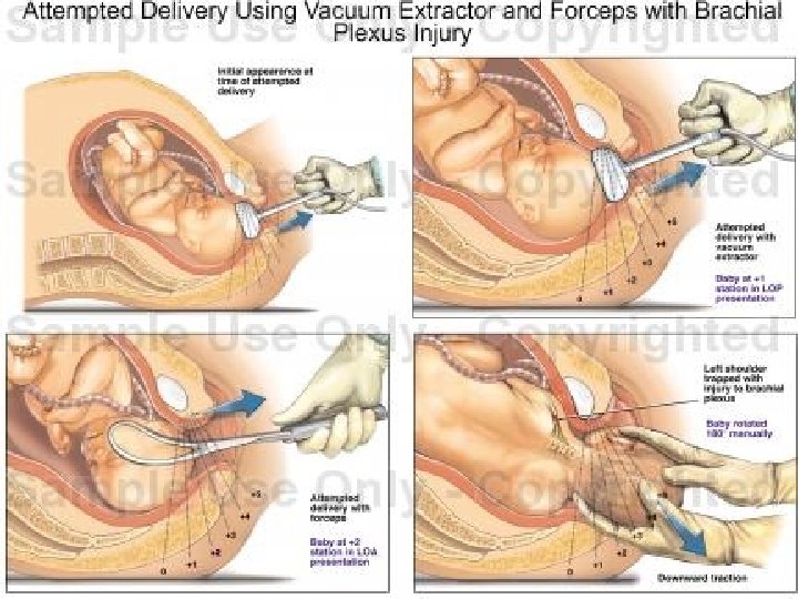
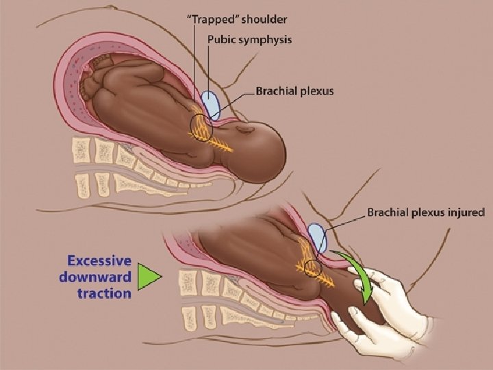
Brachial Plexus Injury n Obstetric palsy - Injury to all or portion of a child brachial plexus occurring at that time of the delivery. n Excessive lateral traction on the head so that the head is pulled away from the shoulder. n Divide into : n Erb’s Duchenne Palsy n Klumpkee’s Palsy

Erb’sparalysis • C 5 -C 6 • Birth injury / Trauma • Arm hangs by the side + Rotate medially • Forearm pronated + extended • Flexed wrist + fingers • Deltoid/supraspinatus/ infraspinatus/biceps/ brachialis. LOS – in arm

n Erb’s Palsy – Nerves Affected
: resulting from")
Brachial Plexus Injuries � Upper Lesions of the Brachial Plexus (Erb’s Palsy): resulting from excessive displacement of the head to opposite side and depression of shoulder on the same side.

� This causes excessive traction or even tearing of C 5 and 6 roots of the plexus. It occurs in infants during a difficult delivery or in adults after a blow to or fall on shoulder.

Effects: Motor: paralysis of � Ø Ø Ø Ø the supraspinatus, infraspinatus, subclavius, biceps brachii, part of brachialis, coracobrachialis; deltoid teres minor. Sensroy: sensory loss on the lateral side of the arm.

� Deformity: waiter tip postion a. limb will hang by the side, b. medially rotated by sternocostal part of the pectoralis major; c. pronated forearm (biceps paralysis) Ø
")
Erb-Duchenne palsy (waiter's tip)
 Ø Ø traction injuries by excessive")
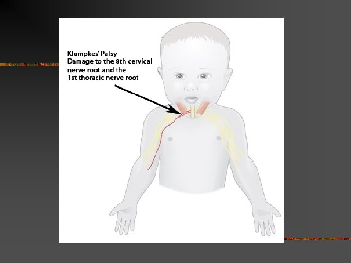
Lower Lesions of the Brachial Plexus (Klumpke Palsy) Ø Ø traction injuries by excessive abduction of the arm i. e. occurs if person falling from a height clutching at an object to save himself or herself. Can be caused by cervical rib. T 1 is usually torn (ulnar and median nerves)

Motor Effects: paralysis of all the small muscles of the hand. Sensory effects: loss of sensation along the medial side of the arm. deformity: claw hand caused by hyperextension of the metacarpophalangeal joints and flexion of the interphalangeal joints.

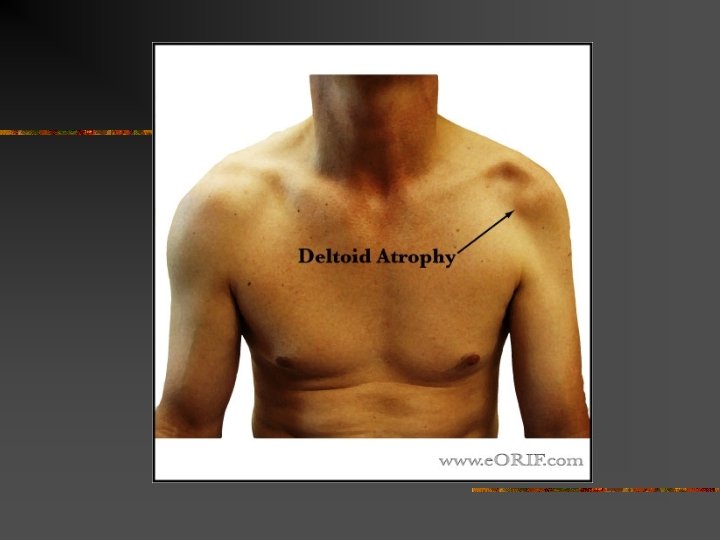
Axillary Nerve injury Causes: Ø crutch pressing upward into the armpit, Ø Downward shoulder dislocations Ø fractures of the surgical neck of the humerus.

n. Motor effects: Ø Deltoid paralysis Ø teres minor paralysis. n Sensory effects: Ø loss of sensation at lower ½ of deltoid n Deformity: Ø Wasting of deltoid

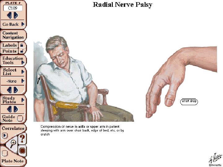
Radial Nerve injury Injury in axilla : � crutch pressing up into armpit � drunkard falling asleep with one arm over the back of a chair. � fractures of proximal humerus.

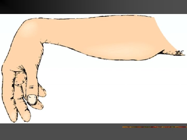
n Ø Ø Ø n Motor effects: paralysis of triceps Anconeus extensors of the wrist Extensors of fingers. Brachioradialis supinator muscle Deformity: Wrist and finger drop

n. Sensory Ø small effects : area of sansation loss at arm and forearm Ø sensory loss over lateral part of the dorsum of the hand (lat. 3. 5 fingers without distal phalynges)

Injuries at Spiral Groove Ø Caused by fracture shaft of humerus. n Motor effects: paralysis of Ø extensors of the wrist Ø Extensors of fingers

Deformity: Ø Wrist and finger drop n Sensory effects: Ø anesthesia is present over the dorsal surface of the hand (lat. 3. 5 fingers) n

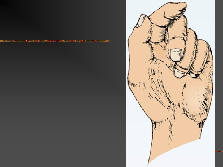
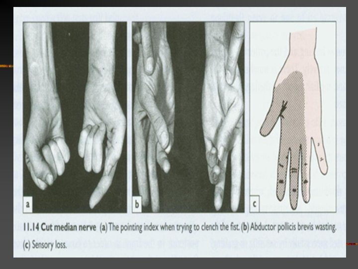
Median Nerve injury � Motor effects: paralysis of Ø pronator muscles Ø long flexor muscles of the wrist and fingers, Ø Exception: a. b. flexor carpi ulnaris medial half flexor digitorum profundus.

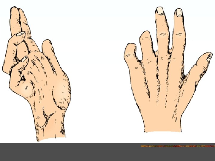
�Deformity: �apelike 1. 2. 3. 4. hand thenar muscles wasted thumb is laterally rotated and adducted. index and to a lesser extent the middle fingers tend to remain straight on making Weakening of lat. 2 fingers

�Sensory: � Sensory loss on the lat. 3. 5 fingers on palmar side � Sensory loss over distal phalanges of lat. 4 fingers on dorsal surface

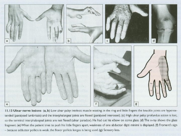
Ulnar nerve injury Motor effects: paralysis of Ø flexor carpi ulnaris Ø medial half of the flexor digitorum profundus Ø All interossei Ø 3 -4 lumbricals n loss of abduction and adduction of fingers n Wasting of hypothenar n

n Deformity: Ø partial claw hand n Sensory effects : Ø Sensory loss over 1. 5 fingers on both surfaces

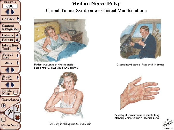
CARPAL TUNNEL FORMED BETWEEN THE CONCAVITY OF THE CARPAL BONES AND A LIGAMENT THAT COVERS THIS( FLEXOR RETINACULAM) n TENDONS OF THE FLEXORS PASS THROUGH n MEDIAN NERVE ALSO PASSES THROUGH n CROWDED TUNNEL CARPAL TUNNEL SYNDROME CAUSED DUE TO COMPRESSION OF THE NERVE IN THE TUNNEL CAUSES 1. SWELLING OF THE TEDONS( OVERUSE) 2. PREGNANCY( EDEMA) 3. ARTHRITIS SYMPTOMS- TINGLING OR NUMBNESS-LATERAL PART OF HAND, WEAKNESS IN THUMB MOVEMENT TREATMENT- REST, SPLINTING, ANTI-INFLAMMATORY DRUGS, SURGERY n

SUMMARY

Begin with a letter “Y”, an “I” and a “Y”.

Add a “strike” and a “spare”

Draw “arches”.

Draw horizontal lines to separate the parts. Roots Trunks Divisions Cords Branches

Begin labeling. C 5 Roots Trunks C 6 C 7 Upper C 8 T 1 Middle Lower Medial Posterior Cords Lateral Divisions Branches Musculocutaneous SLOW Axillary Median Radial Ulnar

Add details. . . Branches off the posterior cord spell “ULTRA” C 5 C 6 C 7 C 8 Roots Trunks Upper T 1 Middle Lower Upper subscapular Lower subscapular Thoracodorsal Medial Posterior Cords Lateral Divisions Branches Musculocutaneous SLOW Axillary Median Radial Ulnar

“ 3 M” comes off the medial cord. C 5 C 6 C 7 C 8 T 1 Roots Trunks Middle Upper Lower Upper subscapular Lower subscapular Thoracodorsal Medial Posterior Cords Lateral Divisions Medial pectoral n. Medial cutan. n. of arm Medial cutan. n. forearm Branches Musculocutaneous SLOW Axillary Median Radial Ulnar

The lateral pectoral n. comes off the lateral cord. C 5 C 6 C 7 C 8 T 1 Roots Trunks Upper Middle Lower Upper subscapular Lower subscapular Thoracodorsal Medial Lateral pectoral n. Posterior Cords Lateral Divisions Medial pectoral n. Medial cutan. n. of arm Medial cutan. n. forearm Branches Musculocutaneous Axillary Median Radial Ulnar

There are 4 supraclavicular branches. C 5 C 6 C 7 C 8 T 1 Roots Long Thoracic n. Dorsal Scapular n. Trunks Upper Middle N. to subclavius Suprascapular n. Lower Upper subscapular Lower subscapular Thoracodorsal Medial Lateral pectoral n. Posterior Cords Lateral Divisions Medial pectoral n. Medial cutan. n. of arm Medial cutan. n. forearm Branches Musculocutaneous SLOW Axillary Median Radial Ulnar

That’s it! The Brachial Plexus C 5 C 6 C 7 C 8 T 1 Roots Long Thoracic n. Dorsal Scapular n. Trunks Upper Middle N. to subclavius Suprascapular n. Lower Upper subscapular Lower subscapular Thoracodorsal Medial Lateral pectoral n. Posterior Cords Lateral Divisions Medial pectoral n. Medial cutan. n. of arm Medial cutan. n. forearm Branches Musculocutaneous Axillary Median Radial Ulnar

THANKS
- Slides: 91