Bowel Anatomy Atresia Volvulus Meckels diverticulum Dr Amit

Bowel Anatomy Atresia, Volvulus, Meckel’s diverticulum Dr Amit Gupta Associate Professor Dept Of Surgery

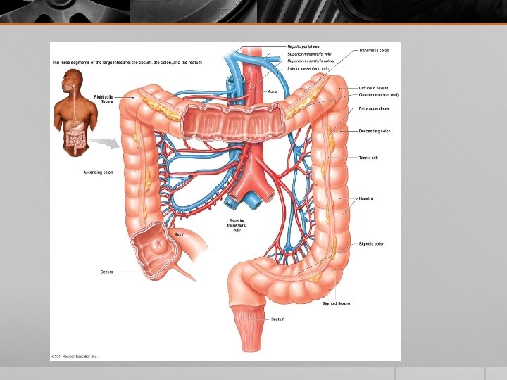
Anatomy § Small intestine: 6 meters § Large intestine: 1. 5 meters. § The first 25 cm of the small intestine (duodenum) is retroperitoneal § Jejunum marks the entry of the small intestine into the peritoneal cavity, terminates where the ileum enters the colon at the ileocecal valve § Large intestine is subdivided into cecum, ascending, transverse, & descending colon. § Sigmoid colon begins at the pelvic brim and loops within the peritoneal cavity § Rectum begins at about the level of the third sacral vertebra. § Reflection of the peritoneum from the rectum over the pelvic floor creates a cul de sac known as the pouch of douglas
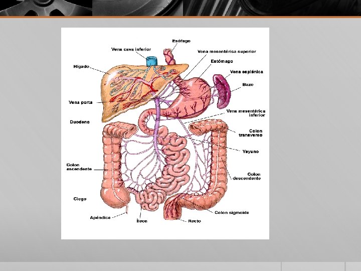

Vasculature § Superior mesenteric artery: proximal jejunum to hepatic flexure of colon § Inferior mesenteric artery: remainder of colon to the level of rectum § Superior hemorrhoidal branch of the inferior mesenteric artery: upper rectum § Hemorrhoidal branches of the internal iliac or internal pudendal artery: lower rectum

§ venous drainage follows essentially the same distribution § connected by an anastomotic capillary bed between the superior and inferior hemorrhoidal veins, providing a connection between the portal and systemic venous systems. § Since the colon is a retroperitoneal organ in the ascending and descending portions, it derives considerable accessory arterial blood supply and lymphatic drainage from a wide area of the posterior abdominal wall.

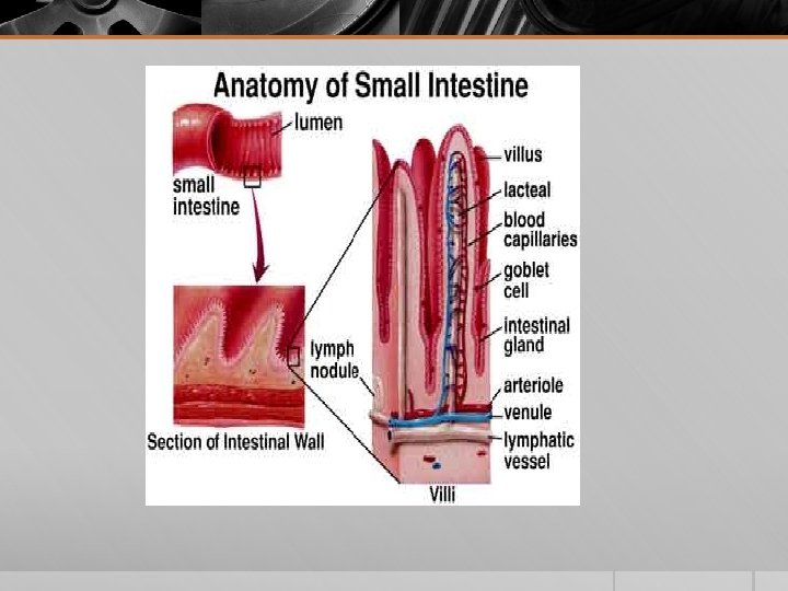
Small Intestinal Mucosa § The most distinctive feature of the small intestine is its mucosal lining, which is studded with innumerable villi § Villi: site for terminal digestion and absorption of foodstuffs § Between the bases of the villi are the pit like crypts of Lieberkühn § Crypts of Lieberkühn: contain stem cells that replenish and regenerate the epithelium § In normal individuals, the villus-to-crypt height ratio is about 4 to 5: 1. § Within the duodenum are abundant submucosal mucous glands, termed Brunner glands. § Brunner glands: secrete bicarbonate ions, glycoproteins, and pepsinogen II and are virtually indistinguishable from the pyloric mucous glands.

Colonic Mucosa § The function of the colon is to reclaim luminal water and electrolytes. § Colonic mucosa has no villi and is flat. § Mucosa is punctuated by numerous straight tubular crypts § Crypts contain abundant goblet cells, endocrine cells, and stem cells. § Paneth cells are occasionally present at base of crypts in the cecum& ascending colon § The regenerative capacity of the intestinal epithelium is remarkable. § Cellular proliferation is confined to the crypts § Turnover of the colonic surface epithelium takes 3 to 8 days

Atresia

Incidence of Atresia § Duodenum : 35% § Jejunum : 15% § Ileum : 25% § Colon : 10% § Multiple sites : 15%

Duodenal Atresia § Failure of vacuolization of duodenum from it’s solid cord stage at 8 -10 th week gestation § Types : § Duodenal stenosis § Mucosal web § Gap separated by fibrous cord § Complete gap
 § Malrotation § Annular pancreas § Biliary atresia")
Associated anomalies § Down Syndrome (30%) § Malrotation § Annular pancreas § Biliary atresia § Congenital heart disease § Anorectal malformations

Clinical features § Presents in first 24 hrs of life. § 85% distal to ampulla of vater § Characterized by bilious emesis § Abdominal distension is absent § Visible gastric peristalsis

Imaging § Check for patent anus/anorectal anomalies § Abdominal x-ray: Double Bubble sign: § Air in the stomach, and 1 st and 2 nd portions of duodenum. § If there is no distal air, the diagnosis is secure. § If there is distal air, and urgent UGI contrast study is needed to rule out midgut volvulus.

“Double Bubble” Sign

Management § Nasogastric decompression § Duodenoduodenostomy § Duodenojejunostomy

Types of jejunoileal atresia

Jejuno-ileal atresia § Type I: § Mesenteric development and intestinal length are normal § Mucosa and submucosa forms a web or membrane within the intestinal lumen § Windsock Effect Increased pressure proximal to the obstruction causes the web to prolapse, pushing the web through the bowel distally

Jejuno-ileal atresia § Type II: § Mesenteric development and overall intestinal length is normal § Small bowel lumen is not continuous § Proximal small bowel ends in bulbous blind pouch § Distal small bowel is flattened § Proximal and distal small bowel connected by fibrous cord.

Jejuno-ileal atresia § Type IIIa: § Similar to Type II § Small V-shaped mesenteric defect is present, bowel length is shortened § Proximal blind end is markedly dilated & aperistaltic § No fibrous cord connects the proximal & distal small bowel

Jejuno-ileal atresia § Type IIIb: § Similar to Type IIIa § Significant mesenteric defect is present Superior mesenteric artery is largely absent Small bowel supplied by a single ileocolic or right colic artery § Known as Christmas tree or apple peel deformity Bowel wraps around a single perfusing artery

Jejuno-ileal atresia § Type IV: § Multiple JIAs of any combination Types I – III Appears as a string of sausages due to multiple lesions § Likely result of multiple ischemic insults or inflammatory process

Pathophysiology JIA is usually a vascular insult versus failure to recanalization of Duodenal Atresia Intrauterine vascular accident Necrosis of bowel segment in a sterile environment Resorption and disappearnce of tissue Malrotation, Volvulus (abnormal twisting of SMA), Gastroschisis, Omphalocele All result in blood supply / gut segment separation

symptoms, physical exam findings. § Within the first day of life § Depends on level of obstruction Proximal Distal § Vomiting green bile § Bowel sounds absent in distal small intestine § No meconium § Abdominal distention

Proximal gut dilatation
 § First")
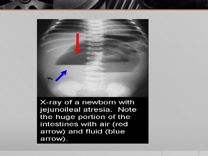
Imaging § Prenatally § Ultrasonography will likely show excess amniotic fluid (polyhydramnios) § First day of life § Abdominal X-ray: Air Fluid level, Dilated proximal gut § Water soluble contrast enema: exclude multiple strictures, Flattened distal gut, Micro colon

Triple Bubble Presentation

Initial treatment § Immediately a tube is placed orally into the stomach to evacuate excess fluid and gasses § Prevents vomiting and aspiration § Relieves GI discomfort § IV Fluids and Nutrients are provided until surgery is available § Surgical intervention is necessary to repair the bowel obstruction and blood supply

Surgical management § Aim: Preserve as much bowel length as possible § Dilated proximal part § Resection and anastomosis § Tapering enteroplasty if remaining bowel is short § Multiple strictures: § Multiple anastomosis over an endoluminal tube

Prognosis § Types I, II, and IIIa have good prognosis § Fairly normal small bowel length results in almost normal bowel function § Types IIIb, IV is associated with complications § Shortened small bowel is associated with short gut syndrome and malabsorption

Volvulus

§ Volvulus occurs when an air-filled segment of the colon twists about its mesentery. § Sigmoid colon is involved in up to 90% of cases, but volvulus can involve the cecum (<20%) or transverse colon. § Volvulus may reduce spontaneously, but more commonly produces bowel obstruction § Chronic constipation may produce a large, redundant colon (chronic megacolon) that predisposes to volvulus § Symptoms: abdominal distention, nausea, and vomiting. § Symptoms rapidly progress to generalized abdominal pain and tenderness. § Fever and leucocytosis are heralds of gangrene and/or perforation.

Sigmoid volvulus § Can often be differentiated from cecal or transverse colon volvulus by the appearance of plain x-rays of the abdomen. § Sigmoid volvulus produces a characteristic bent inner tube or coffee bean appearance § Gastrografin enema shows a narrowing at the site of the volvulus and a pathognomonic bird's beak

Plain X ray showing coffee bean appearance
")
Gastrografin enema showing "bird-beak" sign (arrow)

Management § Initial management: resuscitation followed by endoscopic detorsion § Detorsion: by rigid proctoscope, but a flexible sigmoidoscope or colonoscope might also be effective. § Elective sigmoid colectomy should be performed after the patient has been stabilized § Surgical exploration: evidence of gangrene or perforation, presence of necrotic mucosa, ulceration, or dark blood noted on endoscopy examination § If dead bowel is present at laparotomy, a sigmoid colectomy with end colostomy (Hartmann procedure) may be the safest operation to perform.

Cecal Volvulus § Nonfixation of the right colon. § Rotation occurs around the ileocolic blood vessels and vascular impairment occurs early. § Plain x-rays of the abdomen show a characteristic kidney-shaped, air-filled structure in the left upper quadrant (opposite the site of obstruction), and a gastrografin enema confirms obstruction at the level of the volvulus. § Cecal volvulus can almost never be detorsed endoscopically. § Surgical exploration is necessary when the diagnosis is made. § Right hemicolectomy with a primary ileocolic anastomosis can usually be performed safely and prevents recurrence

Cecal Volvulus

Meckel’s Diverticulum

§ Failure of involution of the vitelline duct, which connects the lumen of the developing gut to the yolk sac, produces a Meckel diverticulum. § True diverticulum: contains all three layers of the normal bowel wall: mucosa, submucosa, and muscularis propria. § It may be a small pouch or a blind segment having a lumen greater in diameter than that of the ileum and a length of up to 6 cm.

Rule of 2’s § 2% of the population have one § 1/2 of symptomatic lesions usually present before the age of 2 years old, others most commonly in the first 2 decades of life § Diveriticuli in adult patients only become symptomatic in about 2% § 2 times more common in males than females § Usually found within 2 feet of the ileocecal valve § Usually are about 2 inches in length § 1/2 contain heterotrophic mucosa (usually gastric, occasionally pancreatic)

Clinical Features: • Lower GI bleeding • Intestinal obstruction • Local inflammation with or without perforation • Rare presentations: Neoplasms Diagnosis: • Most accurate test, especially in children, is “Meckel’s scan”- sodium 99 -tc-pertechinetate, taken up by gastric mucosa • Abdominal CT scan • If CT is negative barium studies should be done • If bleeding with a negative scan, angiography may be helpful

Treatment § If symptomatic: prompt surgical intervention to resect the diverticulum or segment of ileum containing the diverticulum. § If not symptomatic: and found incidentally at surgery in children under 2 y/o, resection is recommended. § In asymptomatic adults, resection is controversial since only about 2% of these patient’s will become symptomatic and there is about a 2% incidence of short or long term complications
- Slides: 45