Bony framework of the skull Sphenoid ethmoid and

Bony framework of the skull Sphenoid, ethmoid and temporal bone Semmelweis Egyetem ÁOK Humánmorfológiai és Fejlődésbiológiai Intézet Dr. Csáki Ágnes 2019. 11. 19.

NEUROCRANIUM CALVARIA BASIS CRANII INTERNA FOSSA CRANII ANTERIOR FOSSA CRANII MEDIA FOSSA CRANII POSTERIOR BASIS CRANII EXTERNA VISCEROCRANIUM - FACIAL CRANIUM ORBIT CAVUM NASI PTERYGOPALATINE FOSSA TEMPORAL FOSSA INFRATEMPORAL FOSSA MANDIBLE, TEMPOROMANDIBULAR JOINT

Neurocranium CRANIUM/SKULL Os frontale Os sphenoidale Os temporale Calvaria Os parietale Os occipitale Viscerocranium cranial base /basis cranii Os ethmoidale Maxilla Os zygomaticum Os lacrimale Os nasale Concha nasalis inferior Vomer Mandibula Os hyoideum (Malleus, Incus, Stapes)
 sutura squamosa (temporoparietalis) sutura")
SYNDESMOSIS sutura serrata (sagittal, coronal, lambdoid s. , etc. ) sutura squamosa (temporoparietalis) sutura plana (in the facial skeleton: nasomaxillary suture, etc. ) SYNCHONDROSIS (sphenopetrosa, petrooccipitalis around the pyramid) SYNOSTOSIS (sphenooccipitalis - clivus) ARTICULATIO temporomandibularis Articulatio incudomallearis és incudostepedialis(in the tympanic cavity)
 some meeting points of")
CALVARIA top part of the skull (skull-cap or cranial vault) some meeting points of the sutures: bregma (frontal angle), lambda (occipital angle), Squamous suture plane sutures
 Diploe } compact bone outer and inner layer + diploe: spongy")
Lamina corticalis (compact) Diploe } compact bone outer and inner layer + diploe: spongy bone between them with bone marrow spaces The thickness varies with age, sex, race in different parts
 areas of fibrous connective tissue (soft spots), allows the skull to flex")
FONTANELLE (FONTICULUS) areas of fibrous connective tissue (soft spots), allows the skull to flex during birth Anterior (major, greater) fontanelle: frontal, sagittal, and coronal sutures (closures in the 2. year) Occipital (posterior, minor, lesser) fontanelle: lambdoid and sagittal sutures (in the 2. month) Sphenoidal fontanelles: squamous and coronal sutures Mastoid fontanelles: squamous and lambdoid sutures Fonticulus anterior Fonticulus posterior Fonticulus spheniodalis Fonticulus mastoideus

Fejlődési rendellenességek Scaphocephalia early closure of sagittal suture Brachycephalia early closure of coronal and lambdoid suture

NEUROCRANIUM 2 parietal bone 2 temporal b. 1 occipital b. 1 shenoid b. 1 frontal b. transverse upper arch sagittal lower arch

INTERNAL BASE

ANTERIOR CRANIAL FOSSA Anterior ethmoid canal frontal bones (pars orbitalis+lesser wing of the sphenoidal bone Ciribriform plate Crista galli

MIDDLE CRANIAL FOSSA from lesser wing of sphenoid bone to pars petrosa of the temporal bone (groove for the sup. petrosal sinus)
+occipital bone (squama)")
POSTERIOR CRANIAL FOSSA posterior surface of the pars petrosa (termporal bone)+occipital bone (squama)

BASIS CRANII INTERNA

Synchondrosis sphenooccipitalis fusion around the 18 th year Abbildung: G. Baksa

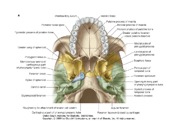
EXTERNAL BASE:

Temporal b. Sphenoid b. Ethmoid b. Palatine b. maxilla

OS TEMPORALE I CAVUM TYMPANI CANALIS FACIALIS CANALICULUS TYMPANICUS CANALIS MUSCULOTUBARIUS CANALIS CAROTICUS

Pars squamosa Pars petrosa Pars tympanica Styloid process
 Arcuate eminence")
OS TEMPORALE II. Sulci arteriosi Tegmen tympani (roof of the tympanic cavity) Arcuate eminence Subarcuate fossa vestibular aqueduct Grooves for petrosal nerves Groove for sup. petrosal sinus Inner surface Porus acusticus internus Processus styloideus

OS TEMPORALE III. Canalis musculotubarius Canalis caroticus Fossa mandibularis Fossula petrosa! Fissura petrotympanica Tympanic canaliculus Cochlear canaliculus Porus acusticus externus Fossa jugularis (Jugular notch, foramen) Processus mastoideus Inferior surface Foramen stylomastoideum

SAGGITAL SECTION: Pyramid part is excavated to form the cavity which is closed by the tympanic part, contains the middle ear. The roof of tympanic cavity, the tegmen tympani turns downward and the edge of it is seen on the external base beetwen the two fissures

and groove for the occipital artery

CAROTID CANAL Canalis caroticus
 Synchondrosis sphenopetrosa")
Path of the internal carotid artery (Sphenoid body) Synchondrosis sphenopetrosa
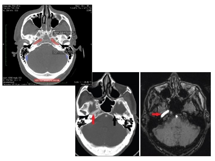

carotid agenesia , internal view normal

TYMPANIC CAVITY Tympanic cavity Fossa subarcuata

Tympanic cavity

Cavum tympani

Lateral wall

Medial wall

FACIAL CANAL

Scheme of the cavity anterior wall: canalis caroticus with canaliculi caroticotympanici and musculotubarius canal (pharyngotympanic tube) inferior wall: jugular fossa, canaliculus tympanicus posterior wall: entrance to the air cells (antrum mastoideum), canaliculus chordae tympani, canaliculus stapedius superior wall: canalis nervi petrosi minoris

OS SPHENOIDALE body with sinus lesser wings greater wings: orbital surface temporal surface cerebral surface infratemporal surface maxillary surface pterygoid processes

SCAPHOID FOSSA

DORSAL SURFACE – SELLA TURCICA

Sella turcica
")
Maxillary surface Round foramen Pterygoid canal Dorsal surface Sella turcica (tuberculum, fossa and dorsum) Carotid sulcus

Maxillary surface foramen rotundum pterygoid canal apertura sphenoidalis – nasal cavity pterygoid process : lateral and medial plate with the hook rostrum- vomer crista – perp. plate of ethmoid
 PALATINE BONE Horizontal and")
MAXILLA four sided-pyramid body (orbital, facial, temporal and nasal surfaces) PALATINE BONE Horizontal and perpendicular plate with orbital process

MAXILLA Maxilla corpus maxillae processus frontalis facies anterior for. infraorbitalis fossa canina crista lacrimalis anterior Os palatinum processus alveolaris processus palatinalis facies infratemporalis tuber maxillae (foramina alveolares) hiatus maxillaris
 can. nasolacrimalis INFERIOR NASAL")
MAXILLA, PALATINE BONE facies nasalis: hiatus maxillaris (sin. max. ) can. nasolacrimalis INFERIOR NASAL CONCHA Maxilla Os palatinum

OS ETHMOIDALE Frontal section Superior wiev lamina perpendicularis crista galli lamina cribrosa labyrinthus ethmoidalis lamina orbitalis conha nasalis superior, media bulla ethmoidalis , processus uncinatus hiatus semilunaris (sinus frontalis)

Os ethmoidale Os nasale Os ethmoidale Os sphenoidale Os frontale Os zygomaticum Os lacrimale Os palatinum Maxilla

Ethmoid bulla Uncinate process

LATERAL WALL

CAVUM NASI MEDIAL WALL: SEPTUM NASI SUPERIOR AND INFERIOR WALL

CHOANAE

LATERAL WALL

LATERAL WALL

SECTION OF THE NASAL CAVITY
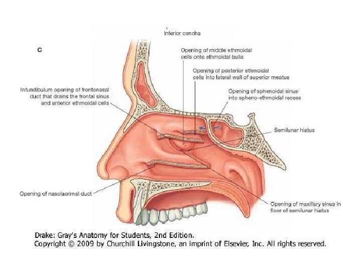

Connections of the nasal cavity

CAVUM NASI Meatus superior Middle, Inferior and common

Sinus paranasales

SINUS PARANASALES

Spheno-ethmoid recess

FOSSA TEMPORALIS AND INFRATEMPORALIS

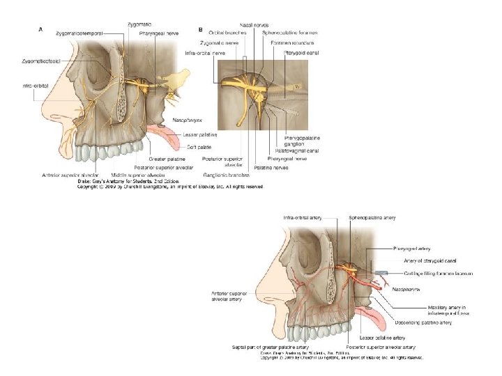
FOSSA PTERYGOPALATINA

PTERYGOPALATINE FOSSA

Sphenopalatine foramen

CONNECTIONS OF THE PTERYGOPALATINE FOSSA „Exit” sphenopalatine foramen nasal cavity inferior orbital fissure orbit greater and lesser palatine canal oral cavity (palatovaginal canal) pharynx) „Entrance” foramen rotundum middle cranial fossa pterygoid canal external base of the skull pterygomaxillary fissure infratemporal fossa

ORBIT Roof: frontal bone -orbital part sphenoid bone - lesser wing Floor: body of the maxilla palatine bone !! Medial wall: maxilla, frontal process lacrimal bone ethmoid bone - orbital plate sphenoid body Lateral wall: zygomatic bone sphenoid bone - greater wing) Entrance: aditus orbitae

Connections of the orbit Optic canal fossa cranii media (optic nerve + ophtalmic art. ) Sup. orb. fissure fossa cranii media (oculomtory n. , trochlear n. , abducens n. , ophtalmic n. , sup. ophtalmic v. ) Inf. orbital fissure fossa pterygopalatina and infraorbital fossa (infraorbital a. and n. ) Anterior ethmoidal foramen (ant. ethmoidal a. and n. ) fossa cranii ant. Post. ethm. foramen ethmoidal cellulae ethmoid. a. and n. ) nasolacrimal canal (nasolacrimal duct) (post. nasal cavity Infraorbital foramen fossa canina (infraorbital nerve) Supraorbital foramen and frontal notch (supraorbital and frontal nerve) front
 Supraorbital foramen and frontal notch (supraorbital and frontal")
Infraorbital foramen fossa canina (infraorbital nerve) Supraorbital foramen and frontal notch (supraorbital and frontal nerve) front
")
nasolacrimal canal nasal cavity, inferior meatus (nasolacrimal duct)

References: Kahle W, Leonhardt H, Platzer W: Color Atlas/Text of Human Anatomy, 1992, Thieme, Stuttgart Putz R, Pabst R (editors): Sobotta Atlas of Human Anatomy, 1993, Urban & Schwarzenberg, München Romanes GJ (editor): Cunningham’s Textbook of Anatomy, 1991, Oxford University Press, Oxford Standring S (editor-in-chief): Gray’s Anatomy, 2005, Elsevier, Edinburgh Szentágothai J, Réthelyi M: Funkcionális anatómia, 2002, Medicina, Budapest Vízkelety T: Az ortopédia tankönyve, 1995, Semmelweis Kiadó, Budapest

1 Crista galli 2 Lamina perpendicularis 3 Lamina cribrosa 4 Labyrinthus ethmoidalis 5 Cellulae ethmoidales 6 Concha nasalis superior 7 Concha nasalis media 8 Lamina orbitalis / papyracea
 is. Az")
Megszületéskor még hiányoznak melléküregek, nevezetesen a homloküreg és a sinus sphenoidalis (iköböl) is. Az iköböl kb. a 2 -3. életév körül fejlődik ki teljesen, míg a homloküreg még később alakul ki, a végső méretét (arányát) a 8 -9. életév körül éri el. Az arcüregek születéskor babnyi nagyságúak, aljuk az alsó orrkagyló szintjében van. Az első három évben a melléküregek oldalirányban és hátrafelé nőnek. A maradandó fogak kifejlődése után a szájpadok felé nő, s az arcüreg alja az orrnyílással lesz egy magasságban 10– 12 éves korra. Az arcüreg végleges nagysága 15– 18 éves korra alakul ki. A fentiek segítik azt megérteni, hogy egy csecsemőnek miért csak rostasejt- és arcüreggyulladása lehet, és miért nem lehet iköböl, vagy homloküreg.
- Slides: 72