Bones and Skeletal Tissues 2013 Pearson Education Inc

Bones and Skeletal Tissues

© 2013 Pearson Education, Inc. Location of Bones and Cartilages

© 2013 Pearson Education, Inc. Types of Skeletal Cartilages • Three types 1. Hyaline cartilage �Provides support, flexibility, and resilience �Collagen fibers only; most abundant type �Articular skeleton, costal (ribs), respiratory, nasal cartilage 2. Elastic cartilage �Similar to hyaline cartilage, but contains elastic fibers �External ear and epiglottis 3. Fibrocartilage �Thick collagen fibers—has great tensile strength �Menisci of knee; vertebral discs

© 2013 Pearson Education, Inc. Important Point! • cartilage is not "turned into" bone, it is replaced by bone in a complicated process **hyaline cartilage forms the model for the bones in a developing embryo. Growth and reshaping occurs in two ways: interstitial growth and appositional growth.

© 2013 Pearson Education, Inc. Growth of Cartilage • Appositional growth: takes place "at the edge" of the cartilage mass • Interstitial growth takes “in the middle" of the mass of cartilage. • Calcification of cartilage- Depositing calcium ▫ Occurs during normal bone growth �Youth and old age ▫ Hardens, but cacified cartilage is not bone!

© 2013 Pearson Education, Inc. Review Questions: pg 196 M. C 3, 4, 10, Short Answer: 15, 18,

Classification of Bones © 2013 Pearson Education, Inc. • 206 named bones in Adult skeleton • Divided into two groups: See fig. 7. 1 ▫ Axial skeleton and Appendicular skeleton

© 2013 Pearson Education, Inc. Classification of Bones by Shape • Long bones ▫ Longer than they are wide ▫ Limb, wrist, ankle bones • Short bones ▫ Cube-shaped bones (in wrist and ankle) ▫ Sesamoid bones (within tendons, e. g. , Patella) ▫ Vary in size and number in different individuals • Flat bones ▫ Thin, flat, slightly curved ▫ Sternum, scapulae, ribs, most skull bones • Irregular bones ▫ Complicated shapes ▫ Vertebrae, coxal bones

Figure 6. 2 Classification of bones on the basis of shape. © 2013 Pearson Education, Inc. Flat bone (sternum) Long bone (humerus) Irregular bone (vertebra), right lateral view Short bone (talus)

Functions of Bones 1. Support: Standing upright, sitting 2. Protection: soft organs 3. Movement: muscles attached 4. Storage: fat, calcium, phosphorus 5. Blood Cell Formation: hematopoiesis 6. Hormone production ▫ Osteocalcin �Regulates bone formation

© 2013 Pearson Education, Inc. Bones • Are organs ▫ Contain different types of tissues �Bone (osseous) tissue, nervous tissue, cartilage, fibrous connective tissue, muscle and epithelial cells in its blood vessels • Three levels of structure ▫ Gross anatomy ▫ Microscopic ▫ Chemical

© 2013 Pearson Education, Inc. Gross Anatomy: Bone Texture Compact and Spongy bone • Compact ▫ Dense outer layer; smooth and solid • Spongy: also called trabecular ▫ Less dense- open spaces

© 2013 Pearson Education, Inc. Structure of Short, Irregular, and Flat Bones • Thin plates of spongy bone covered by compact bone • Plates sandwiched between connective tissue membranes ▫ Periosteum (outer layer) and endosteum (inner layer) • No shaft or epiphyses • Bone marrow throughout spongy bone; no marrow cavity

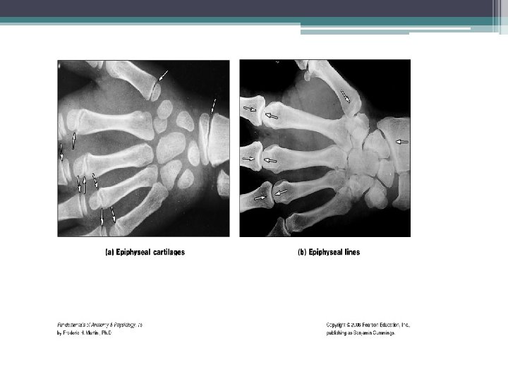
© 2013 Pearson Education, Inc. Long Bones • Epiphyses ▫ Bone ends ▫ External compact bone; internal spongy bone ▫ Articular cartilage covers articular surfaces ▫ Between is epiphyseal line • Remnant of childhood bone growth at epiphyseal plate Diaphysis ▫ Tubular shaft forms long axis ▫ Compact bone surrounding medullary cavity

© 2013 Pearson Education, Inc. Bone Membranes: • Endosteum • Periosteum

© 2013 Pearson Education, Inc. Review Questions: # 5, 21, 22

© 2013 Pearson Education, Inc. Hematopoietic Tissue in Bones • Red marrow ▫ Found within trabecular cavities of spongy bone and in flat bones (e. g. , sternum) ▫ In medullary cavities and spongy bone of newborns ▫ Adult long bones have little red marrow �Heads of femur and humerus only ▫ Yellow marrow can convert to red, if necessary
 © 2013 Pearson Education, Inc.")
Table 6. 1 Bone Markings (1 of 2) © 2013 Pearson Education, Inc.
 © 2013 Pearson Education, Inc.")
Table 6. 1 Bone Markings (2 of 2) © 2013 Pearson Education, Inc.

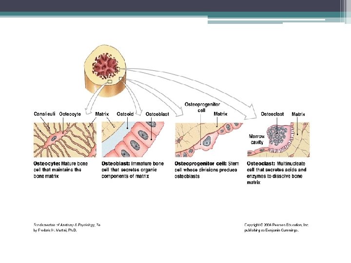
© 2013 Pearson Education, Inc. Cells of Bone Tissue

© 2013 Pearson Education, Inc. Anatomy of Compact Bone • Osteon or Haversian system ▫ Structural unit of compact bone ▫ Hollow tubes of bone matrix called lamellae �Collagen fibers in adjacent rings run in different directions �Withstands stress – resist twisting

Figure 6. 7 Microscopic anatomy of compact bone. Compact bone Spongy bone © 2013 Pearson Education, Inc. Central (Haversian) canal Perforating (Volkmann’s) canal Endosteum lining bony canals and covering trabeculae Osteon (Haversian system) Circumferential lamellae Lamellae Nerve Vein Artery Canaliculi Osteocyte in a lacuna Perforating (Sharpey’s) fibers Periosteal blood vessel Periosteum Lamellae Central canal Lacunae Interstitial Lacuna lamella (with osteocyte)

© 2013 Pearson Education, Inc. Chemical Composition of Bone: Inorganic Components • Hydroxyapatites (mineral salts) ▫ 65% of bone by mass ▫ Mainly of tiny calcium phosphate crystals in and around collagen fibers ▫ Responsible for hardness and resistance to compression

© 2013 Pearson Education, Inc. Bone Fun Facts • Half as strong as steel in resisting compression • As strong as steel in resisting tension • Last long after death because of mineral composition ▫ Reveal information about ancient people ▫ Can display growth arrest lines �Horizontal lines on bones �Proof of illness - when bones stop growing so nutrients can help fight disease
- Slides: 26