BONE TUMORS BY ALI IBRAHIM MD Bone tumors

BONE TUMORS BY ALI IBRAHIM, MD

Bone tumors n n n Patient presentation: *Pain. *Swelling. *Pathological fracture & deformity. *Disturbance of function. *Incidental discovery.

Bone tumors n Clinical features &evaluation: 1 -Careful history& physical examination beside routine X-ray &laboratory facilities. n 2 -Pre-biopsy evaluation : C T scan and/or M R I. n 3 -Actual biopsy. n 4 -Search for metastatic disease. n
-Clinical features and evaluation B)-Investigations: 1 -Imaging of musculoskeletal neoplasm's : a) Plain")
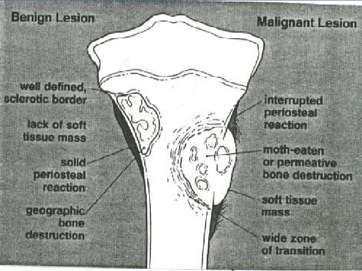
DIAGNOSIS A)-Clinical features and evaluation B)-Investigations: 1 -Imaging of musculoskeletal neoplasm's : a) Plain radiography b) C T scan c) M R I d) Isotope bone scan e) Arteriography 2 -Laboratory studies & tumor markers 3 -Biopsy


Simple bone cyst n n Occurs before 15 years of age. 50% develop in the proximal end of the humerus. Other common sites include: the proximal femur , the proximal and distal tibia , iliac wing and calcaneus. Common presentation : incidental discovery when radiographs are taken for other reasons or by pathological fracture after minor trauma.

Simple bone cyst n n Solitary or unicameral bone cyst: Expansion of bone & thinning of the bone cortex through endosteal erosion. Pathological fracture. Extensive bone destruction resemble a neoplasm

Simple bone cyst n n The cavity is filled by a serous fluid. The fluid will be stained by hemorrhage if pathological fracture occurred. Radiographically appear as symmetric well demarcated radiolucent expansile lesions at bone metaphyses, often extending up to the physeal plate and the cortex may be thinned. as the bone grows away from the cyst , the lesion may come to lie in the diaphysis. When pathological fracture occurs a fragment of thin cortex may separate and fall in the cavity {fallen leaf sign}. No periosteal reaction except after stress fracture

Simple bone cyst

Simple bone cyst

Simple bone cyst

Simple bone cyst n n n With skeletal growth and maturation, simple bone cysts tends to spontaneous healing. Most fractures of simple bone cysts heal rapidly with closed treatment as callus formation induces cystic healing as well as fracture consolidation. Methods of treatment: 1 - observation and restriction of activity until the cyst heals in asymptomatic lesions. 2 - curettage and bone grafting in active lesions.

Aneurysmal bone cyst n n ABCs arise primarily as a skeletal osteolytic lesion or may arise as a reaction to hemorrhage within a pre-existing tumoral lesion. The primary lesion is found mostly in the second decade of life. Common sites are metadiaphysis of long bones preferably in the lower limb bones. The lesion usually is eccentric subperiosteal in origin, so pathological fracture is unusual presentation, but it may arise centrally within the bone.

Aneurysmal bone cyst n n The diagnostic features are : Marked expansion of the involved bone, cystic bone destruction and periosteal new bone formation. The lesion rapidly destroys the original bone cortex and is contained only by a thin rim of periosteal new bone. During curettage there may be a considerable bleeding from the fleshy lining membrane [welling or pouring of blood ]

Aneurysmal bone cyst n Radiographic features: is a well defined radiolucent subperiosteal osteolytic lesion elevating and inflating the periosteum and progressively eroding the cortex, with scarce periosteal reaction observed as a thin shell of reactive bone at the metaphysis of long bones [egg shell]. The cyst initially appears as an eccentric osteolytic area.

Aneurysmal bone cyst

Aneurysmal bone cyst

Aneurysmal bone cyst n n Methods of treatment : The treatment of choice is curettage and autogenous grafting. [during curettage copious bleeding may be encountered ].

Aneurysmal bone cyst
![osteochondroma n n Osteocartilaginous exostoses: [cartilage capped exostosis] the most common tumors of bone.](http://slidetodoc.com/presentation_image_h2/7c9a0d13c07485d204a0011d147c4c4f/image-21.jpg "osteochondroma n n Osteocartilaginous exostoses: [cartilage capped exostosis] the most common tumors of bone.")
osteochondroma n n Osteocartilaginous exostoses: [cartilage capped exostosis] the most common tumors of bone. Are hamartomas and occur during skeletal growth as a small overgrowth of cartilage at the edge of the physeal plate and develops by enchondral ossification into a bony protuberance still covered by the cap of cartilage. They continue to grow and mature , undergoing enchondral ossification. Once skeletal maturity occurs, growth of exostosis ceases.

osteochondroma n n n Radiographic features are usually classic and other studies are rarely required to make the diagnosis. The pathogonomonic feature of an exostosis is that the medullary bone is contiguous in the stalk of the exostosis and the underlying bone. Lesions are usually pedunculated but can be sessile, the lesion initially is adjacent to the physeal mechanism and becomes more diaphyseal as the child ages. Pedunculated lesions extend away from the epiphysis of origin toward the diaphysis of the involved bone. It looks smaller than it feels because the cartilage cap is usually invisible

osteochondroma

osteochondroma

osteochondroma n n The exostosis is covered with a cartilaginous cap and undergoes enchondral ossification to from the underlying cancellous bone. The thickness of the cap is variable depending on the age of the person and diminishes following skeletal maturity. The cells of the cartilaginous cap are arranged in columns similar to an epiphyseal mechanism.

osteochondroma n n n Common problems of osteochondromas: 1 - Fracture of the stalk in pedunculated type. 2 - Entrapment of the adjacent neurovascular structures. 3 - Adventitious bursa formation over the cartilaginous cap which is liable for bursitis. 4 - Malignant transformation to chondro – sarcoma { more in lesions arising in the pelvis, scapula, ribs and spine }. 5 - Pressure atrophy on adjacent bone when arise from the leg or forearm bones.

osteochondroma

osteochondroma n n Malignant transformation : Malignant transformation of a single osteochondroma is a rare event {<1%} and is less common than in patients with MHE {6%}. Lesions arising in the pelvis, scapula, ribs and spine are at higher risk for malignant transformation than those in the appendicular skeleton. Transformation does not occur before skeletal maturity

osteochondroma n n n Methods of treatment : Surgical excision should be reserved for lesions that cause discomfort or deformity or are cosmetically unappealing. To avoid the local recurrence, the entire cartilaginous cap must be excised.

osteochondroma n n n Multiple Heritable Exostoses : MHE is a syndrome of being multiple , heritable and associated with skeletal deformity and short stature, and by having a significant frequency of transformation into secondary peripheral chondrosarcoma. The ratio of solitary exostoses to MHE is at least 10: 1. Exostoses usually are identified earlier in persons with MHE [usually by 10 years of age] than with solitary lesions. MHE are transmitted as an autosomal dominant trait.

osteochondroma n n n Multiple Heritable Exostoses: The lesions tend to be diffuse and relatively symmetric. The long bones are affected most severely, with the greatest involvement around the knee, shoulder, hip, wrist and ankle. The pathogenesis and histopathologic features are the same as for solitary exostoses.

osteochondroma n n Multiple Heritable Exostoses: Radiographically: the lesions are larger than the solitary one and the underlying bones are shorter than normal, with a widened metaphysis [ due to failure of bone remodeling as the periosteum is tethered in all direction at the metaphysis ].

osteochondroma

osteochondroma n n n Multiple Heritable Exostoses: Malignant transformation occurs in around 16% and central lesions [ pelvis, scapula, ribs, and spine] are at great risk of malignant transformation as with solitary osteochondromas. Excision of one or more exostoses often is necessary in persons with MHE because of discomfort or for mechanical reasons as fracture of a stalk in pedunculated lesion or due to bursitis and pressure on nearby vital structures.

Enchondromas are hamartomatous collections of mature hyaline cartilage within bone. n They usually present as solitary lesions. n Multiple lesions known as enchondromatosis or Ollier`s disease. n It arise from the lack of normal enchondral ossification below the growth plate. n n They are asymptomatic unless a pathological

Enchondroma n n n They commonly involve the small tubular bones in the hand foot, the proximal humerus, the femur and the ribs. A well defined radiolucentral lesion within the metaphysis or diaphysis of the involved bone, sometimes the bone is slightly expanded. Calcification may be present and usually is stippled or flecks, “smoke ring or popcorn” ossification.

Enchondroma

Enchondroma

Giant cell tumor n n n Although benign, GCTs show a tendency for significant bone destruction, local recurrence, and occasionally metastasis. * Frequency: GCTs represent approximately 5% of all primary bone tumors and 18 -21% of all benign bone tumors. The most common bone tumor in the young adults aged 25 to 40 years, GCTs occur most commonly in the third decade of life. giant cell tumors only occur after the epiphyseal plates have closed and a diagnosis of GCT in a patient with open growth plates should be questioned.

Giant cell tumor n Approximately 50% of GCTs are located about the knee at the distal femur and proximal tibia, with the proximal humerus and distal radius representing the third and fourth most common sites.

Giant cell tumor n Radiologic features: n * The lesions are expansile, osteolytic, radiolucent without sclerotic margins and usually without a periosteal reaction and eccentrically located within n the bone. There is a well-defined defect in the metaphysis and epiphysis, with destruction of the medullary cavity and adjacent cortex. The destruction may stop just short of the joint.

Giant cell tumor

Giant cell tumor

Giant cell tumor

Giant cell tumor

Osteosarcoma n n Osteosarcoma is the most common primary malignant tumor of bone, characterized by the direct formation of bone or osteoid by tumor cells. The incidence of osteosarcoma peaks in those aged 10 -20 years; (maximum period of skeletal growth).

Osteosarcoma n n osteosarcoma occurs most commonly in the metaphyses of long tubular bones, particularly around the knee joint (distal femur, proximal tibia). The proximal humeral metaphysis is another common site. The disease commonly extends from the metaphysis into the adjacent diaphysis or epiphysis.

Osteosarcoma n n n Radiographic appearances shows a mixture of lytic and sclerotic areas. Soft tissue extension of osteosarcoma is common and seen on radiographs as a soft-tissue mass. Cloudlike areas of sclerosis due to malignant osteoid production and calcification may be seen within the mass. Periosteal reactions are commonly seen once the tumor extends through the cortex ( Codman triangles and multilaminated, spiculated, and sunburst reactions).

Osteosarcoma

Osteosarcoma

Osteosarcoma

Chondrosarcoma n n Chondrosarcoma is a malignant tumor that produces cartilage matrix. Primary chondrosarcoma is very uncommon, arises centrally in the bone, and is found in young adult. Secondary chondrosarcoma arises from benign cartilage defects such as osteochondroma or enchondroma. Chondrosarcoma can also be classified as intramedullary, which generally arise from enchondromas, and surface which arise from osteochondromas

Chondrosarcoma n n It is most common in the femur, humerus, ribs and on the surface of the pelvis. chondrosarcoma is a fusiform, lucent defect with scalloping of the inner cortex and periosteal reaction. Extension into the soft tissue may be present as well as punctate or stippled calcification of the cartilage matrix. May shows a lobulated appearance like a cauliflower mass.

Chondrosarcoma

Chondrosarcoma

Chondrosarcoma

Ewing's sarcoma n n Ewing's sarcoma is a highly malignant tumor that is found in the lower extremity more than the upper extremity, but any long tubular bone may be affected. The most common sites are the metaphysis and diaphysis of the femur followed by the tibia and humerus. Ewing's sarcoma is most common in the first and second decade.

Ewing's sarcoma n The clinical presentation of Ewing's sarcoma includes pain and swelling of weeks or months duration. Erythema and warmth of the local area are sometimes seen. Osteomyelitis is often the initial diagnosis based on intermittent fevers, leukocytosis, anemia and an increased ESR.

n Ewing's sarcoma Radiologically, Ewing's sarcoma is often associated with a lamellated or "onion skin" periosteal reaction.

Ewing's sarcoma
 n n n Multiple myeloma is a malignant tumor of plasma")
Multiple Myeloma (Plasmacytoma) n n n Multiple myeloma is a malignant tumor of plasma cells that causes widespread osteolytic bone damage. Multiple myeloma is the most common primary tumor of bone and is found in the spine, skull, ribs, sternum and pelvis but may affect any bone with hematopoietic red marrow. The average patient age is over fifty years old. Monoclonal immunoglobulin is found on serum protein electrophoresis. Bence. Jones proteins are present in urine.

Multiple Myeloma n n The radiological appearance of multiple myeloma is characterized by irregular lytic defects of different sizes. These lytic areas are often described as "punched out" and have no periosteal reaction. Erosion begins intramedullarly and progresses through the cortex. It may causes generalized osteoporosis.

Multiple Myeloma

Multiple Myeloma

BONE SECONDARIES COMMONEST MALIGNANT BONE TUMOURS SITE OF PRIMARY: breast, prostate, thyroid, kidney, lung 2/3 come from cancer breast or prostate 16 % from thyroid, kidney, lung 17 % unknown primary SITE OF SECONDARIES: bones rich in cancellous bone trunk bones e. g spine, pelvis, skull, ribs root bones e. g upper humerus, upper femur ROUTES : 1 - Local invasion…e. g rectum invades pelvis 2 - Blood born… from tumour drain via vena cava to heart , to lung to systemic circulation to bone 3 - LYMPPHATICS…
")
BONE SECONDARIES TYPES : Osteolytic 90 %. . . Bone destruction ( breast ) osteosclerotic … Bone sclerosis ( prostate ) PRESENTATION: 1 - Pain 2 - Pathologic fracture ( fr. Spine ) 3 - Swelling 4 - Anaemia, cachexia TREATMENT : Hormonal Radio & Chemo. . Surgical. Internal fixation. Amputation. Resection

Thank you

- Slides: 68