Bone Tissue Prof Abdulameer AlNuaimi Email a alnuaimisheffield

Bone Tissue Prof. Abdulameer Al-Nuaimi E-mail: a. alnuaimi@sheffield. ac. uk E-mail: abdulameerh@yahoo. com

Bone Tissue • Bone is type of connective tissue which composed of calcified extracellular matrix (ECM) and cells. • Provides solid support for the body. • Protects many vital internal organs such as those in the cranial and thoracic cavities. • Has cavities containing bone marrow. • Reservoir of calcium, phosphate, and other ions to maintain constant concentrations in body fluids.

Bone Tissue

Bone Tissue Composed of: • Cells: 1. Osteocytes 2. Osteoblasts 3. Osteoclasts • Extracellular matrix (Bone matrix)

Bone Cells Bone is composed of three major cell types: • Osteocytes: found in lacunae between bone matrix layers lamellae, with cytoplasmic processes extending into small canaliculi between lamellae. • Osteoblasts: synthesize the organic components of the matrix. • Osteoclasts: are multinucleated, giant cells involved in the resorption and remodeling of bone tissue. • Bone resorption is the process by which osteoclasts break down bone and release the minerals, resulting in a transfer of calcium from bone to the blood. • Bone remodeling (bone metabolism) where mature bone tissue is removed from the skeleton (resorption) and new bone tissue is formed (ossification or new bone formation).

Bone Tissue

Bone Cells

Osteaoprogenitor Cells • Mesenchymal periosteum. stem cells, • Transform to osteoblasts. • Secrete bone matrix. • Vital role in growth and repair. found in the

Osteoblasts • Active: Cuboidal or polygonal shape, found in the periosteum and endosteum. • Synthesize and secrete the organic components of bone matrix (type I collagen fibers, proteoglycans, and several glycoproteins such as osteonectin). • Osteoblasts secret osteoid (newly made uncalcified matrix). This is followed by subsequent deposition of calcium salts into the newly formed matrix. Therefore osteoblasts are responsible for bone calcification. Osteoblast may change to osteocyte.

Bone Cells

Osteocytes • Located in Lacunae • Extend cytoplasmic processes through canaliculi for nutrition. • Maintain bone matrix. • Death of Osteocytes will lead to bone resorption • Communicate by gap junctions.

Osteocytes • It’s flat, almond-shaped with less RER, smaller Golgi complexes, and more condensed nuclear chromatin (comparing to osteoblasts). • Osteocyte products such as sclerostin and certain cytokines help regulate bone remodeling. • The extensive lacunar-canalicular network and their communication with all other bone cells suggest additional roles for osteocytes in calcium homeostasis. • As sensors for detection of mechanical stresses on bone, important in directing bone remodeling.

Bone Cells

Osteoclasts • Large, multinucleated, branched, motile cells. • Play a major role in matrix resorption during bone growth and remodeling • The surface of osteoclast facing matrix have projections ruffled border. • Acidophilic cytoplasm, many lysosomes, many mitochondria. • Found in resorption cavities

Osteoclasts • Osteoclast secretes collagenase and other enzymes and pumps protons to produce an acidic environment locally promoting the localized digestion of matrix proteins.

Bone Matrix • Inorganic material represents about 50% of the dry weight of bone matrix. • Calcium hydroxyapatite is most abundant, but bicarbonate, citrate, magnesium, potassium, and sodium ions are also found. • Significant quantities of amorphous (noncrystalline) calcium phosphate are also present.

Bone Matrix • The organic matter embedded in the calcified matrix includes type I collagen, proteoglycan aggregates, and bonespecific multiadhesive glycoproteins such as osteonectin. • Calcium-binding glycoproteins, osteocalcin, and the phosphatases released in matrix vesicles by osteoblasts promote calcification of the matrix.

Bone Matrix • The association of minerals with collagen fibers during calcification is responsible for the hardness and resistance of bone tissue. • After a bone is decalcified, its shape is preserved, but it becomes as flexible as a tendon.

Periosteum and Endosteum • All bones are lined on both internal and external surfaces by membranes made of layers of connective tissue containing osteogenic cells. • The se membranes are called 1 - Endosteum is a membrane on the internal surface surrounding the marrow cavity 2 - Periosteum is a membrane surround the external surface of the bone.

Periosteum • The principal functions of periosteum are to nourish the osseous tissue and provide a continuous supply of new osteoblasts for bone growth or repair. • The periosteum is organized much like the perichondrium. • The outer layer is dense connective tissue, with small blood vessels, collagen bundles, and fibroblasts.

Periosteum • Bundles of periosteal collagen fibers, called perforating fibers, penetrate the bone matrix, binding the periosteum to bone. • The inner region of periosteum is a more cellular layer containing osteoblasts, and mesenchymal stem cells (osteoprogenitor cells).

Endosteum • Internally the very thin endosteum covers small trabeculae of bony matrix that project into the marrow cavities. • Although considerably thinner than the periosteum, endosteum also contains osteoprogenitor cells, osteoblasts

Bone type • Types of bone tissue is classified according to the component of the bony tissue into 1 - Lamellar Bone A-Compact Bone B- Spongy Bone 2 - Non Lamellar (Woven Bone)
 are composed of")
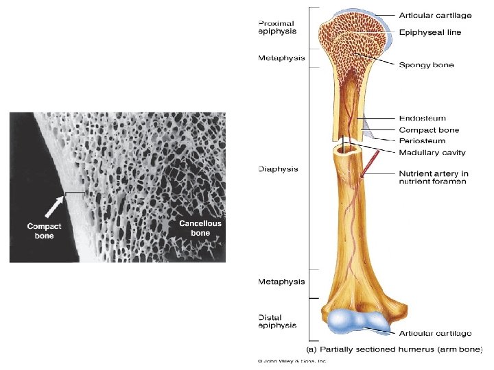
Type of Bone • In long bones, the bulbous ends, (epiphyses) are composed of spongy bone. • The cylindrical part (diaphysis) is almost totally composed of compact bone, with a thin region of spongy bone on the inner surface around the central medullary cavity.

• Short bones such as those of the wrist and ankle usually composed of spongy bone surrounded completely by compact bone. • The flat bones that form the calvaria (skullcap) have two layers of compact bone called plates, separated by a thicker layer of spongy bone called the diploë.

• Most bones in adults, compact and cancellous, are organized as lamellar bone, characterized by multiple layers or lamellae of calcified matrix. • The highly ordered organization of collagen fibers in lamellae adds greatly to the strength of lamellar bone. • When we examine a long bone, The dense area near the surface is called compact (cortical) bone, which represents 80% of the total bone mass. • Deeper areas with numerous interconnecting cavities, called cancellous (spongy) bone, constituting about 20% of total bone mass.

Compact Bone • Compact bone is formed of units called osteons or Haversian systems. • Osteons contains central canal called Haversian canal contains blood vessels, lymphatic vessels, nerves. • Surrounding this canal are concentric rings of osteocytes along with the calcified matrix. • Osteons are aligned in the same direction along lines of stress. These lines can slowly change as the stresses on the bone changes.
.")
An osteon (or Haversian system).

• Each osteon consists of a central canal surrounded by 4 -10 concentric lamellae. • The central canals communicate with the marrow cavity and the periosteum and with one another through transverse perforating canals (or Volkmann canals). External circumferential lamellae

• Scattered among the intact osteons are numerous irregularly shaped groups of parallel lamellae called interstitial lamellae. • The interstitial lamellae are lamellae remaining from osteons partially destroyed by osteoclasts during growth and remodeling of bone.
 the lamellae")
• In compact bone (e. g, the diaphysis of long bones) the lamellae exhibit a typical organization consisting of multiple external circumferential lamellae and often some inner circumferential lamellae. • External circumferential lamellae are located immediately beneath the periosteum. • Inner circumferential lamellae are located around the marrow cavity.
 bone does not contain osteons. • It consists of")
Spongy Bone • Spongy (cancellous) bone does not contain osteons. • It consists of trabeculae surrounding many red marrow filled spaces. • It forms most of the structure of short, flat, and irregular bones, and the epiphyses of long bones. • Spongy bone tissue is light and supports and protects the red bone marrow.

Woven Bone • Non-lamellar and characterized by random disposition of type I collagen fibers and is the first bone tissue to appear in embryonic development and in fracture repair. Composition of non lamellar bone • Irregular, interwoven array of collagen fibers. • Has a lower mineral content (it is more easily penetrated by x-rays). • Have higher proportion of Osteocytes than mature lamellar bone.

Woven Bone • Usually temporary and is replaced in adults by lamellar bone, except in a very few places in the body (e. g near the sutures of the calvaria and in the insertions of some tendons). • Woven bone forms more quickly but has less strength than lamellar bone.

Bone types and their organization

Bone remodeling • Bone remodeling is continuous throughout life and involves a process of bone resorption and bone formation. • In compact bone, remodeling resorbs parts of old osteons and produces new ones. • Resorption involves the actions of osteoclasts, often working in groups to remove old bone in tunnel-like cavities having the approximate diameter of new osteons.

Bone remodeling • Tunnels are quickly invaded by many osteoprogenitor cells from the endosteum or periosteum and sprouting loops of capillaries. • Osteoblasts develop, line the wall of the tunnels, and begin to secrete osteoid in a cyclic manner, forming the concentric lamellae of bone with trapped osteocytes. • In healthy adults • 5%-10% of the • bone turns over annually

Histology of Muscular System Prof. Abdulameer Al-Nuaimi E-mail: a. al-nuaimi@sheffield. ac. uk E. mail: abdulameerh@yahoo. com

Muscle Tissue • Muscle tissue is composed of cells that have property of contractility. • Actin and myosin microfilaments are associated proteins that generate the forces necessary for the muscle contraction • Origin of all muscle cells is the mesoderm.

Muscle Tissue • sarcoplasm is The cytoplasm of muscle cells. • The sarcoplasmic reticulum (SR) is a membrane bound structure found within muscle cells, that is similar to the endoplasmic reticulum in other cells. The main function of the SR is to store calcium (Ca 2+). Calcium levels are kept relatively constant, with the concentration of calcium within a cell being 100, 000 times smaller than the concentration of calcium outside the cell • sarcolemma is the muscle cell membrane sarcoplasmic reticulum

sarcoplasmic reticulum is a network of tubules that extend throughout muscle cells, wrapping around the myofibrils (contractile units of the cell). Cardiac and skeletal muscle cells, contain structures called transverse tubules (T-tubules), which are extensions of the cell membrane that travel into the centre of the cell. T-tubules are closely associated with a specific region of the SR, known as the terminal cisternae in cardiac muscle or junctional SR in skeletal muscle, with a distance of roughly 12 nanometers, separating them. This is the primary site of calcium release. The longitudinal SR are thinner projects, that run between the terminal cisternae/junctional SR, and are the location where ion channels necessary for calcium absorption are most abundant. These processes are fundamental for the process of excitation-contraction coupling in skeletal, cardiac and smooth muscle

Longitudinal Section of skeletal muscle, showing t-tubules running deep into the centre of the cell between two terminal cisternae/junctional SR. The thinner projections, running horizontally between two terminal cisternae are the longitudinal sections of the SR

Muscle Tissue • Characteristics – Contracts or shortens with force – Moves entire body and pumps blood • Types – Skeletal • Striated and voluntary – Cardiac • Striated and involuntary – Smooth • Non striated and involuntary 4 -44
 muscle consists of bundles of very long, cylindrical multinucleated")
Skeletal Muscle • Skeletal (striated) muscle consists of bundles of very long, cylindrical multinucleated cells with crossstriations. • Their contraction is quick, forceful, and usually under voluntary control. • Elongated nuclei are found peripherally just under the sarcolemma, a characteristic nuclear location unique to skeletal muscle fibers (cells).

Skeletal Muscle • During embryonic muscle development, mesenchymal myoblasts fuse, forming myotubes with many nuclei. • Myotubes then further differentiate to form striated muscle fibers. • A small population of reserve progenitor cells (Satellite ells) remains adjacent to most fibers of differentiated skeletal muscle.

Organization of a Skeletal Muscle Thin layers of connective tissue surround and organize the contractile fibers in skeletal muscle. • The epimysium • The perimysium • The endomysium

Organization of a Skeletal Muscle The epimysium, an external sheath of dense connective tissue, surrounds the entire muscle. Septa of this tissue extend inward, carrying the larger nerves, blood vessels, and lymphatics of the muscle.

Organization of a Skeletal Muscle The perimysium is a thin connective tissue layer that immediately surrounds each bundle of muscle fibers termed a fascicle. Each fascicle of muscle fibers makes up a functional unit in which the fibers work together. Nerves, blood vessels, and lymphatics penetrate the perimysium to supply each fascicle.

Organization of a Skeletal Muscle The endomysium, very thin, delicate layer of reticular fibers and scattered fibroblasts, surrounds the external lamina of individual muscle Fibers. Exist within fascicles. Capillaries form a rich network in the endomysium bringing O 2 to the muscle fibers. Muscle fibre (cell)

Organization of a Skeletal Muscle Collagen in the connective tissue layers of muscle transmit the mechanical Forces generated by the contracting muscle cells (fibers). Some skeletal muscles taper at their ends, where the epimysium is continuous with the dense regular connective tissue of a tendon at myotendinous junctions.

Organization Within Muscle Fibers • Internally each muscle fiber is filled with myofibrils, composed of thousands of thick myosin filaments and thin actin filaments, highly organized into contractile units called sarcomeres. • The thick and thin filaments interdigitate • Troponin is the protein controlling the sliding filaments

Myosin Actin

The sarcomeric subunits of one myofibril are in nearly perfect alignment with those of the myofibrils next to it. This alignment gives rise to certain optical properties which cause the cell to appear striped or striated. In smooth muscle cells, this alignment is absent, hence there are no apparent striations and the cells are called smooth Myofibril Myosin Actin Myocyte (muscle cell) Striation of skeletal muscle

Thank You
- Slides: 55