Bone histology DEFINITION Bone is a dynamic highly

Bone histology

DEFINITION Bone is a dynamic, highly vascular specialized connective tissue, which supports the body and provides leverage for movements.

Gross structure of a long bone

• Periosteum – outer connective tissue covering • Endosteum – inner connective tissue covering • Marrow cavity – bone marrow • Bone marrow – 2 types- yellow and red • Red marrow – ends of bone – blood cell formation • Yellow marrow - shaft – adipose tissue

COMPOSITION OF BONE Cells Osteoprogenitor Fiber s Matrix Organi c Inorgani c cells Osteoblasts Osteocytes Osteoclasts Lamella
 Inorganic material Calcium")
Lamella Organic material Matrix • Ground substance • Collagen (type 1) Inorganic material Calcium & phosphate Hydroxy – apatite crystals

OSTEOPROGENITOR CELLS / OSTEOGENIC CELLS • Stem cells of mesenchymal origin. • Present – endosteum, periosteum & haversian canal • Function - Proliferate and convert themselves into osteoblasts.

OSTEOBLASTS • Oval in shape. • Derived – osteogenic cells • Found – wherever there is bone formation • Function üSynthesis and secretion of matrix. üCalcification of bone matrix.

OSTEOCYTES • Derived from osteoblasts. • Mature bone cells • Star shaped • Present in lacunae. • Functions: – Play a role in maintenance of surrounding matrix

OSTEOCLASTS • Large & multinucleated cell • Cytoplasm – lysosomes & mitochondria • Contains acid phosphatase • Present – sites of bone resorption called pits of Howships • Function – helps in bone

Fibers • Type I collagen fibers • Gives tensile strength

Ground substance • Proteoglycans • GAG’s – hyaluronic acid, keratan sulphate, chondroitin sulfates. • organic component – ground substance and fibers • Inorganic component- mineral salts- calcium phosphates, calcium carbonates, calcium fluoride, citrate, magnesium and sodium.

MICROSCOPIC STRUCTURE Lamella – thin plate of bone, made up of collagen fibers, mineral salts and matrix Lacunae – small spaces between adjacent lamellae, osteocytes are located here. Canaliculi – minute canals which helps to connect adjacent lacunae.

Spongy bones

Structure of a Flat Bone

• Compact bone forms the outer shell of all bone and also the shafts in long bones. • Spongy bone is found at the expanded parts of long bones and fills most irregular bones. • Spongy or cancellous bone consists of a lattice of thin threads of bone called trabeculae, which are lamellae that are arranged as rods or plates and is less dense than compact bone. Red bone marrow is found between the trabuculae.

Bone is covered by………? ?

Periosteum The external surface of the bone is covered by a connective tissue membrane called periosteum except articular surface. The periosteum consist of outer fibrous layer & inner cellular layer. The periosteum is united to underlying bone by Sharpey’s fibers, but it is stronger where there is attachment of tendons & ligaments. Abundant periosteal arteries nourish outer part of the cortex.

Endosteum • Thin lining of the bone towards marrow cavity

Structure of Compact bone 1. Haversian system of lamellae 2. Interstitial lamellae 3. Circumferential lamellae

Haversian system of lamellae • Osteon • 4 -15 concentric lamellae around central canal Haversian Canal • Lacunae contains osteocytes
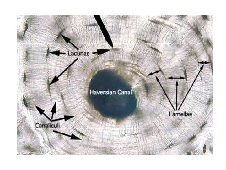

Haversian canal • Contains – – Small amount loose connective tissue – Capillaries, nerves and – Lymphatics 23

Volkmann’s canal …? ? ? vv

Interstitial lamellae • Older bone tissue is continuously replaced by new bone tissue • Fragments of older osteocytes lodges between the adjacent haversian systems.

Circumferential lamellae • Outer circumferential lamellae • Inner circumferential lamellae

Cement line…? ? • Surrounding each osteon, there is a thin layer of mineralised bone matrix which is devoid of collagen fibers

T. S. of compact bone A Haversian system Osteocyte in lacuna Haversian canal Concentric lamellae Volkmann’s canal Interstitial lamellae

d: UsersHemaDesktopIMG-20180907 WA 0001. jpg

L. S OF BONE

L. S. of compact bone Volkmann’s canal Osteocyte in lacuna Haversian canal Lamellae
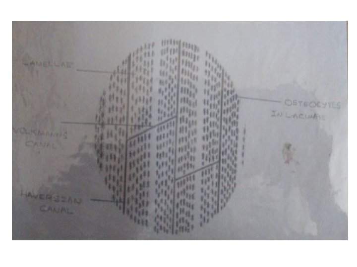

Identification points • T. S of Bone: 1. Presence of Haversian systems with central Haversian canals. 2. Presence of circumferential lamellae and interstitial lamellae. 3. Presence of outer periosteum and inner endosteum

Identification points • L. S of Bone: 1. Presence of longitudinal arrangement of lamellas on the sides of Haversian canals 2. Volkmann’s canals are visible. 3. Cement line can be seen
- Slides: 34