
Bone Diversity Dr Anderson GCIT Bone Diversity There
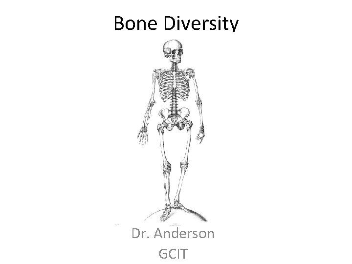
Bone Diversity Dr. Anderson GCIT
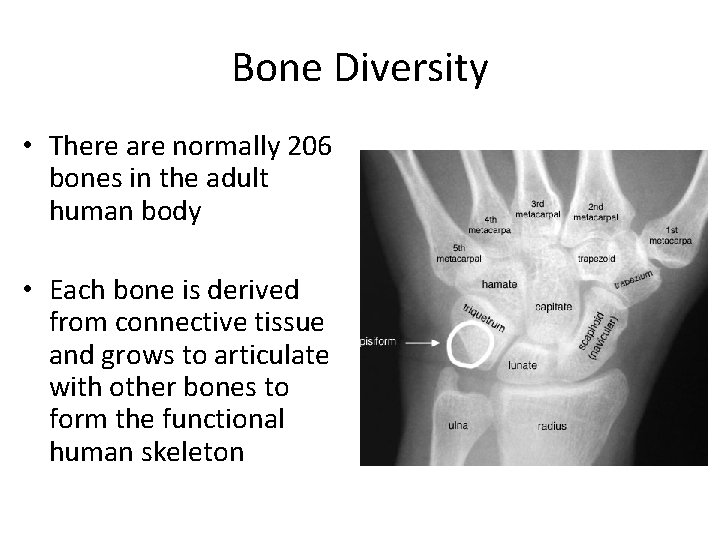
Bone Diversity • There are normally 206 bones in the adult human body • Each bone is derived from connective tissue and grows to articulate with other bones to form the functional human skeleton

The Axial Skeleton • Composed of the skull, vertebrae, ribs (thoracic cage) and coccyx

The Skull • Cranium – Made of multiple bones that are immovable (as adult) and connect in sutures, houses the brain
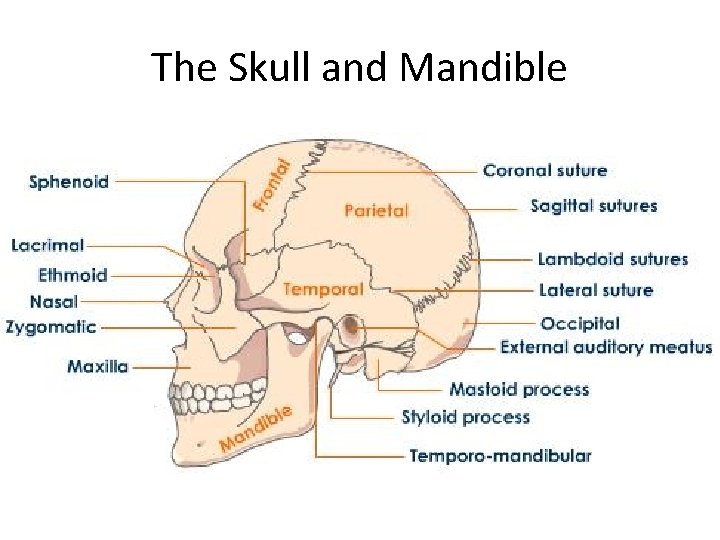
The Skull and Mandible
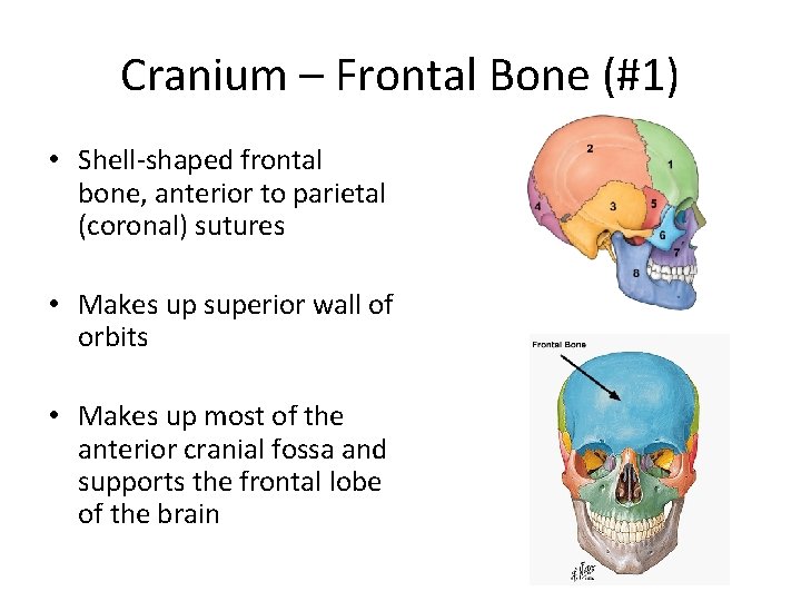
Cranium – Frontal Bone (#1) • Shell-shaped frontal bone, anterior to parietal (coronal) sutures • Makes up superior wall of orbits • Makes up most of the anterior cranial fossa and supports the frontal lobe of the brain
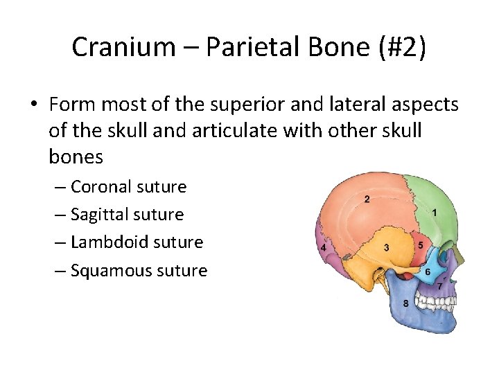
Cranium – Parietal Bone (#2) • Form most of the superior and lateral aspects of the skull and articulate with other skull bones – Coronal suture – Sagittal suture – Lambdoid suture – Squamous suture
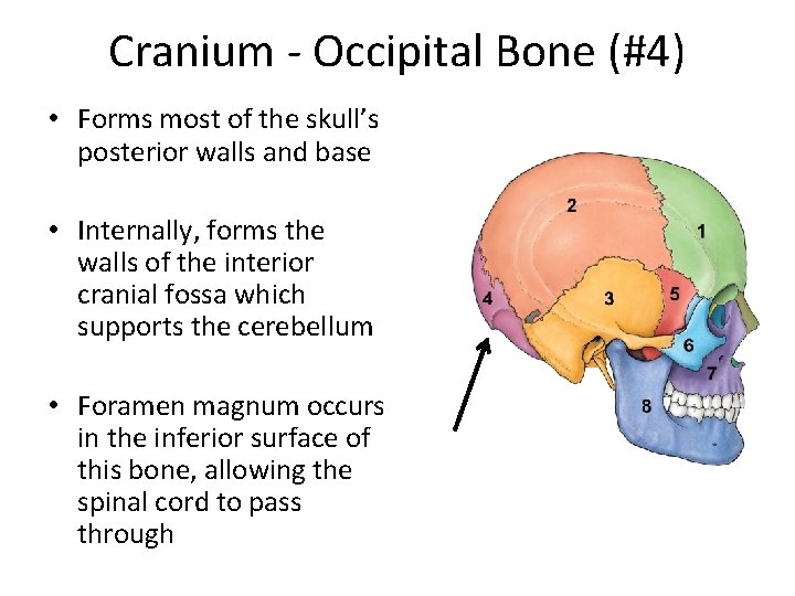
Cranium - Occipital Bone (#4) • Forms most of the skull’s posterior walls and base • Internally, forms the walls of the interior cranial fossa which supports the cerebellum • Foramen magnum occurs in the inferior surface of this bone, allowing the spinal cord to pass through
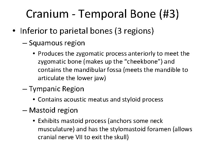
Cranium - Temporal Bone (#3) • Inferior to parietal bones (3 regions) – Squamous region • Produces the zygomatic process anteriorly to meet the zygomatic bone (makes up the “cheekbone”) and contains the mandibular fossa (meets the mandible to articulate the lower jaw) – Tympanic Region • Contains acoustic meatus and styloid process – Mastoid region • Exhibits mastoid process (anchors some neck musculature) and has the stylomastoid foramen (allows cranial nerve VII to exit the skull)
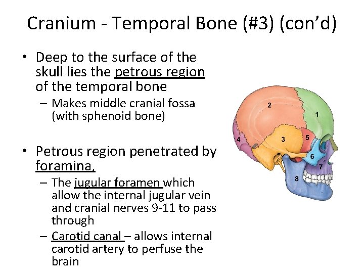
Cranium - Temporal Bone (#3) (con’d) • Deep to the surface of the skull lies the petrous region of the temporal bone – Makes middle cranial fossa (with sphenoid bone) • Petrous region penetrated by foramina, – The jugular foramen which allow the internal jugular vein and cranial nerves 9 -11 to pass through – Carotid canal – allows internal carotid artery to perfuse the brain
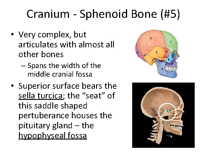
Cranium - Sphenoid Bone (#5) • Very complex, but articulates with almost all other bones – Spans the width of the middle cranial fossa • Superior surface bears the sella turcica; the “seat” of this saddle shaped pertuberance houses the pituitary gland – the hypophyseal fossa

Cranium - Sphenoid Bone (#5) (con’d) • “Batlike” wings – “Greater wings” – form the: • middle cranial fossa (along with temporal bone) • Dorsal walls of the orbits • External wall of the skull – “Lesser wings” - • Form part of the floor of the anterior cranial fossa • Pterygoid processes – anchor chewing muscles
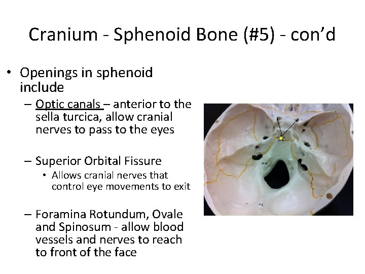
Cranium - Sphenoid Bone (#5) - con’d • Openings in sphenoid include – Optic canals – anterior to the sella turcica, allow cranial nerves to pass to the eyes – Superior Orbital Fissure • Allows cranial nerves that control eye movements to exit – Foramina Rotundum, Ovale and Spinosum - allow blood vessels and nerves to reach to front of the face
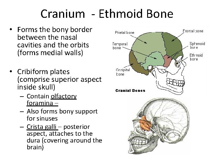
Cranium - Ethmoid Bone • Forms the bony border between the nasal cavities and the orbits (forms medial walls) • Cribiform plates (comprise superior aspect inside skull) – Contain olfactory foramina – – Also forms bony support for sinuses – Crista galli – posterior aspect, attaches to the dura (covering around the brain)

Facial Bones - Mandible • Lower Jawbone – articulates with mandibular fossa (on temporal bone) • Two main parts – Body (chin) – Ramus (vertical part)
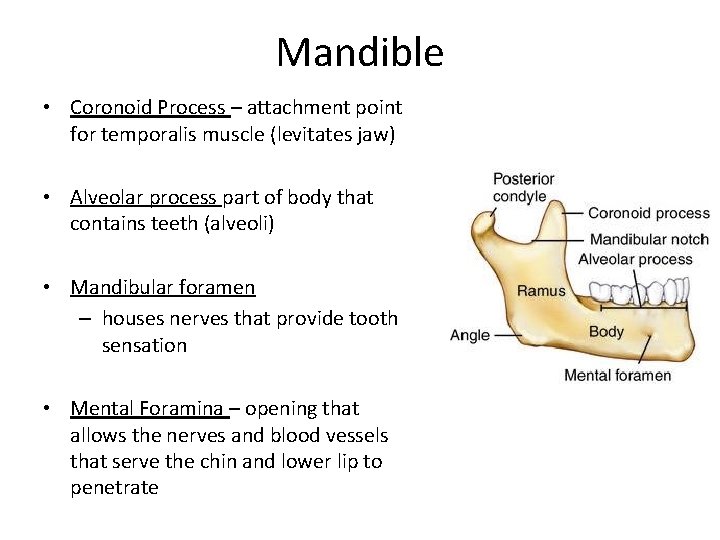
Mandible • Coronoid Process – attachment point for temporalis muscle (levitates jaw) • Alveolar process part of body that contains teeth (alveoli) • Mandibular foramen – houses nerves that provide tooth sensation • Mental Foramina – opening that allows the nerves and blood vessels that serve the chin and lower lip to penetrate
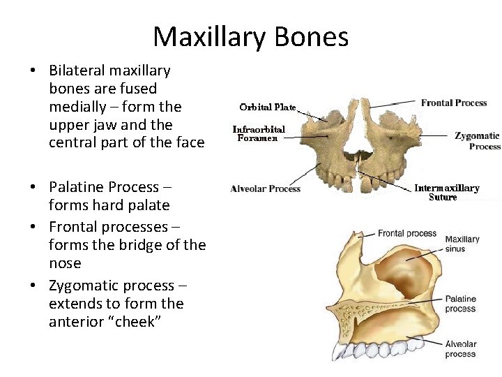
Maxillary Bones • Bilateral maxillary bones are fused medially – form the upper jaw and the central part of the face • Palatine Process – forms hard palate • Frontal processes – forms the bridge of the nose • Zygomatic process – extends to form the anterior “cheek”
Smaller Facial Bones (pg 213) • • • Zygomatic bones – form cheek bones Nasal Bones – forms bridge of nose Lacrimal Bones – medial orbit walls Palatine Bones – posterior part of hard palate Inferior nasal conchae – lateral walls of nasal cavity
All of the Aforementioned Bones are Summarized on pp. 214 -215
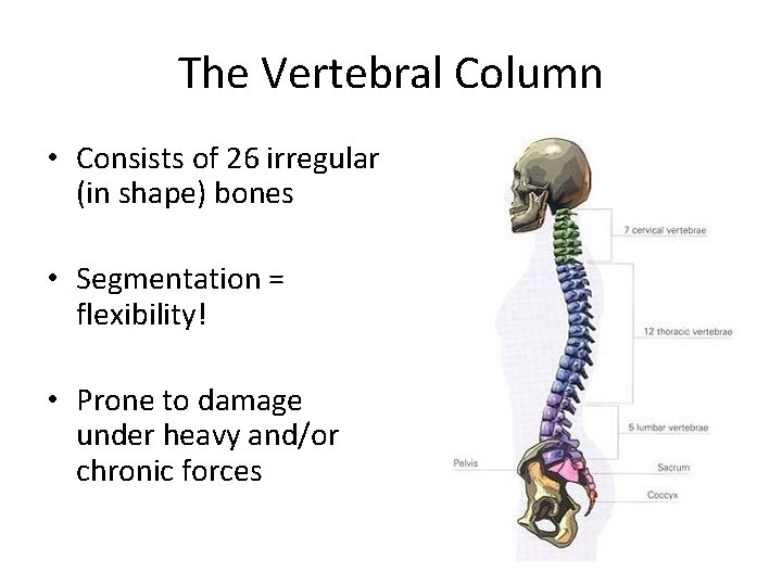
The Vertebral Column • Consists of 26 irregular (in shape) bones • Segmentation = flexibility! • Prone to damage under heavy and/or chronic forces
Vertebral Function • Transmits force from upper body to legs • Houses and protects the spinal cord • Attachment points for ribs, and for muscles/ ligaments of the back and neck
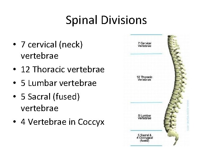
Spinal Divisions • 7 cervical (neck) vertebrae • 12 Thoracic vertebrae • 5 Lumbar vertebrae • 5 Sacral (fused) vertebrae • 4 Vertebrae in Coccyx
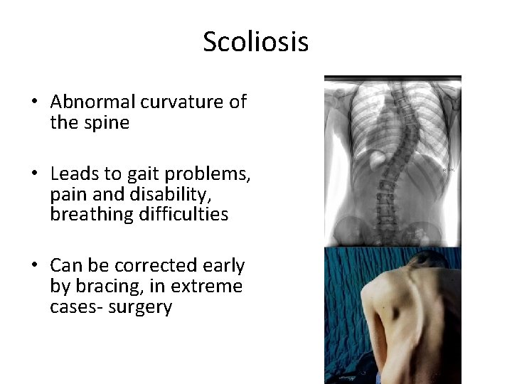
Scoliosis • Abnormal curvature of the spine • Leads to gait problems, pain and disability, breathing difficulties • Can be corrected early by bracing, in extreme cases- surgery
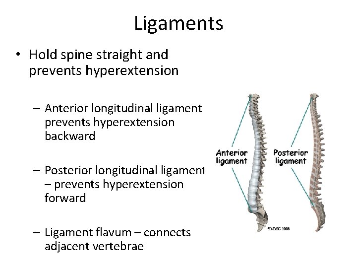
Ligaments • Hold spine straight and prevents hyperextension – Anterior longitudinal ligament – prevents hyperextension backward – Posterior longitudinal ligament – prevents hyperextension forward – Ligament flavum – connects adjacent vertebrae
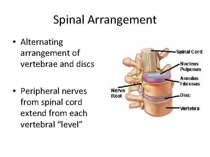
Spinal Arrangement • Alternating arrangement of vertebrae and discs • Peripheral nerves from spinal cord extend from each vertebral “level”
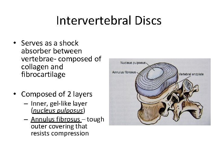
Intervertebral Discs • Serves as a shock absorber between vertebrae- composed of collagen and fibrocartilage • Composed of 2 layers – Inner, gel-like layer (nucleus pulposus) – Annulus fibrosus – tough outer covering that resists compression
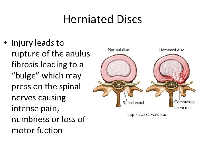
Herniated Discs • Injury leads to rupture of the anulus fibrosis leading to a “bulge” which may press on the spinal nerves causing intense pain, numbness or loss of motor fuction
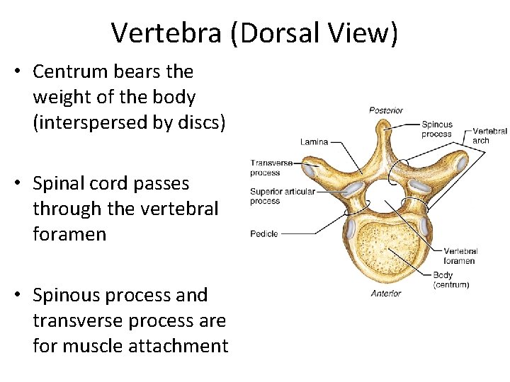
Vertebra (Dorsal View) • Centrum bears the weight of the body (interspersed by discs) • Spinal cord passes through the vertebral foramen • Spinous process and transverse process are for muscle attachment
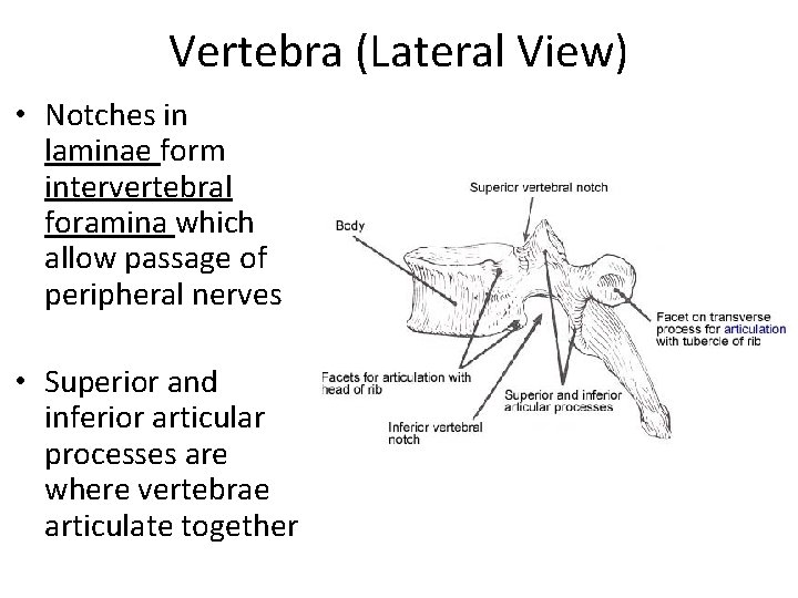
Vertebra (Lateral View) • Notches in laminae form intervertebral foramina which allow passage of peripheral nerves • Superior and inferior articular processes are where vertebrae articulate together
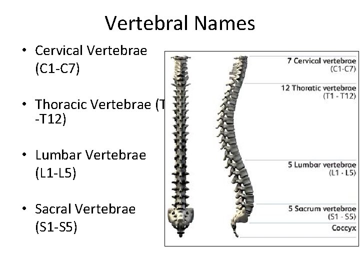
Vertebral Names • Cervical Vertebrae (C 1 -C 7) • Thoracic Vertebrae (T 1 -T 12) • Lumbar Vertebrae (L 1 -L 5) • Sacral Vertebrae (S 1 -S 5)
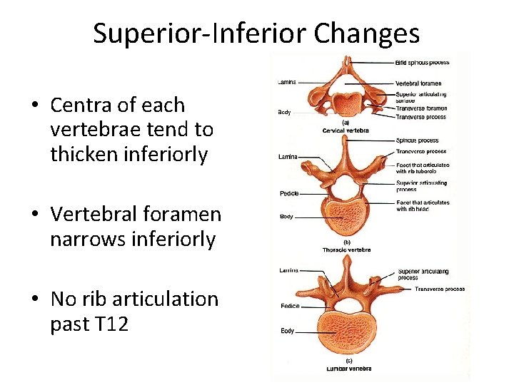
Superior-Inferior Changes • Centra of each vertebrae tend to thicken inferiorly • Vertebral foramen narrows inferiorly • No rib articulation past T 12
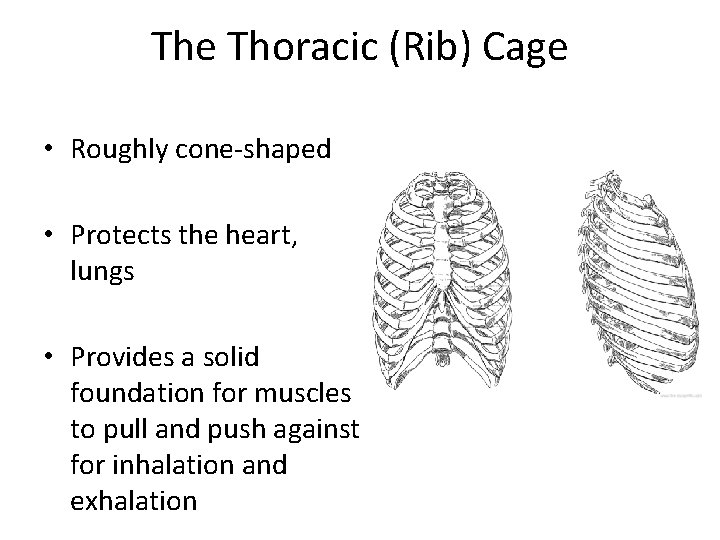
The Thoracic (Rib) Cage • Roughly cone-shaped • Protects the heart, lungs • Provides a solid foundation for muscles to pull and push against for inhalation and exhalation
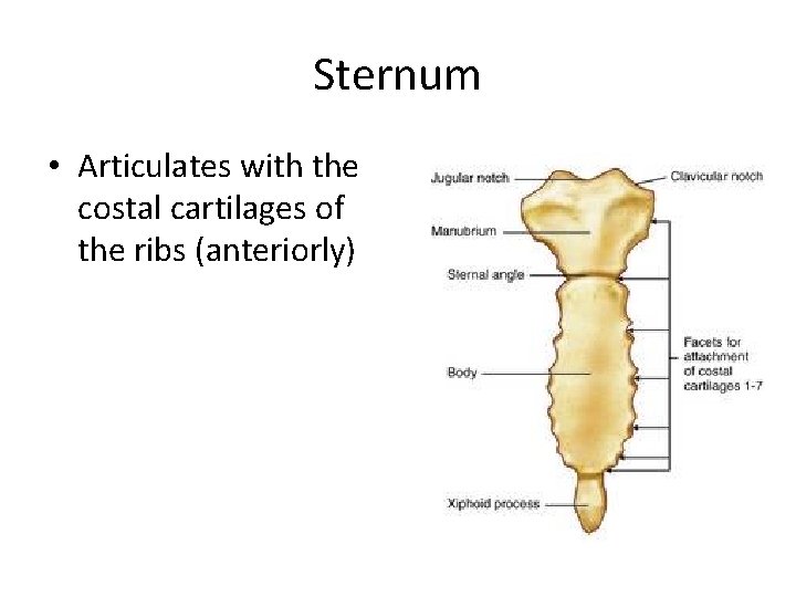
Sternum • Articulates with the costal cartilages of the ribs (anteriorly)
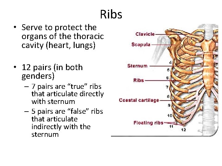
Ribs • Serve to protect the organs of the thoracic cavity (heart, lungs) • 12 pairs (in both genders) – 7 pairs are “true” ribs that articulate directly with sternum – 5 pairs are “false” ribs that articulate indirectly with the sternum

Posterior Rib Articulation
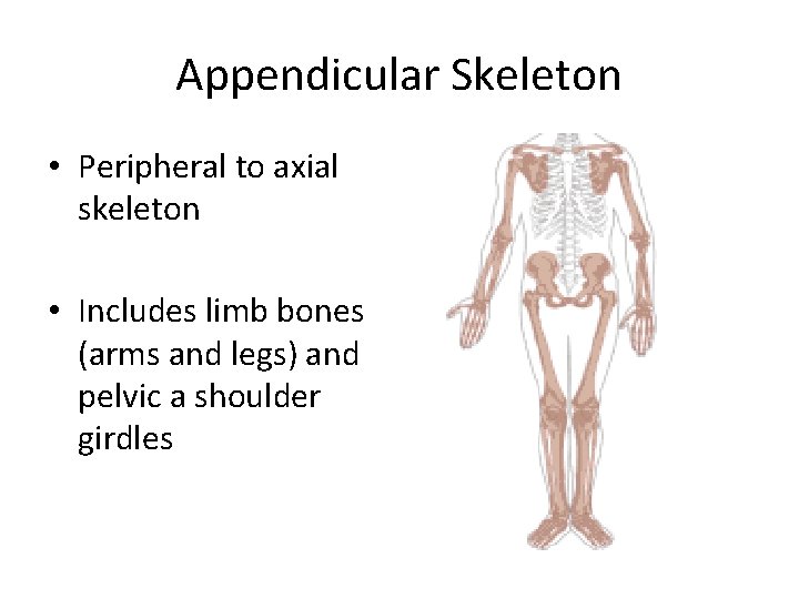
Appendicular Skeleton • Peripheral to axial skeleton • Includes limb bones (arms and legs) and pelvic a shoulder girdles
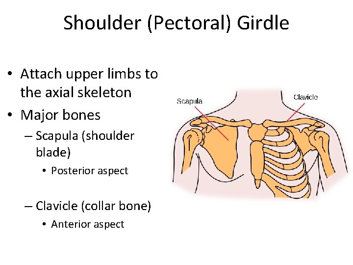
Shoulder (Pectoral) Girdle • Attach upper limbs to the axial skeleton • Major bones – Scapula (shoulder blade) • Posterior aspect – Clavicle (collar bone) • Anterior aspect
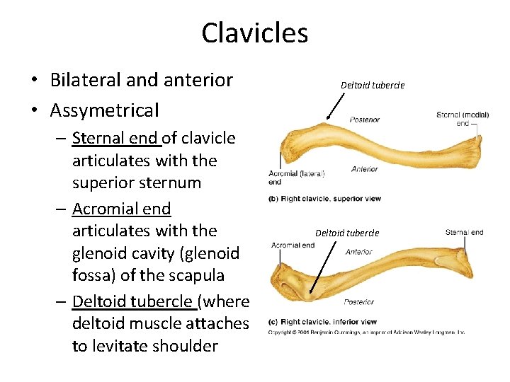
Clavicles • Bilateral and anterior • Assymetrical – Sternal end of clavicle articulates with the superior sternum – Acromial end articulates with the glenoid cavity (glenoid fossa) of the scapula – Deltoid tubercle (where deltoid muscle attaches to levitate shoulder Deltoid tubercle
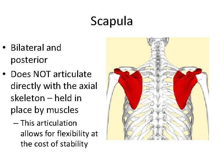
Scapula • Bilateral and posterior • Does NOT articulate directly with the axial skeleton – held in place by muscles – This articulation allows for flexibility at the cost of stability
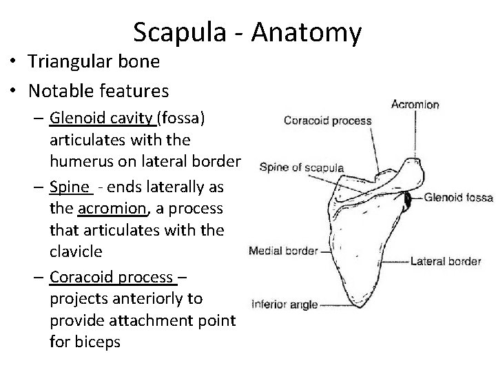
Scapula - Anatomy • Triangular bone • Notable features – Glenoid cavity (fossa) articulates with the humerus on lateral border – Spine - ends laterally as the acromion, a process that articulates with the clavicle – Coracoid process – projects anteriorly to provide attachment point for biceps
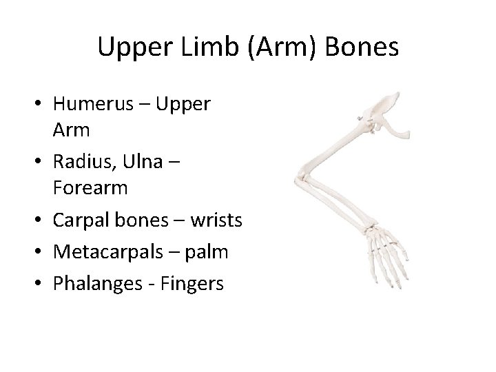
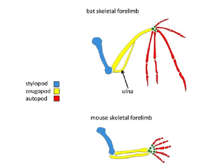
Upper Limb (Arm) Bones • Humerus – Upper Arm • Radius, Ulna – Forearm • Carpal bones – wrists • Metacarpals – palm • Phalanges - Fingers
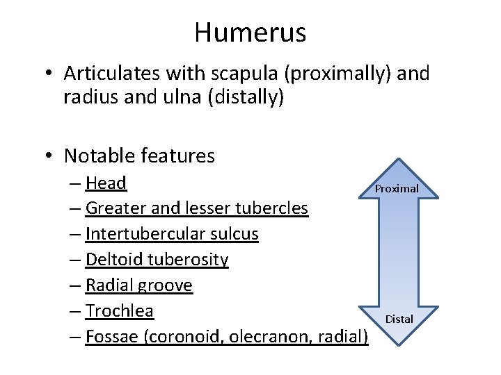
Humerus • Articulates with scapula (proximally) and radius and ulna (distally) • Notable features – Head Proximal – Greater and lesser tubercles – Intertubercular sulcus – Deltoid tuberosity – Radial groove – Trochlea Distal – Fossae (coronoid, olecranon, radial)
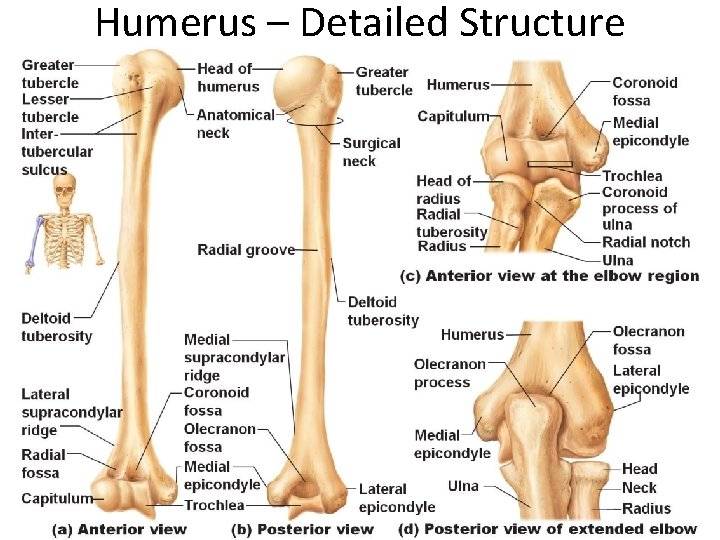
Humerus – Detailed Structure
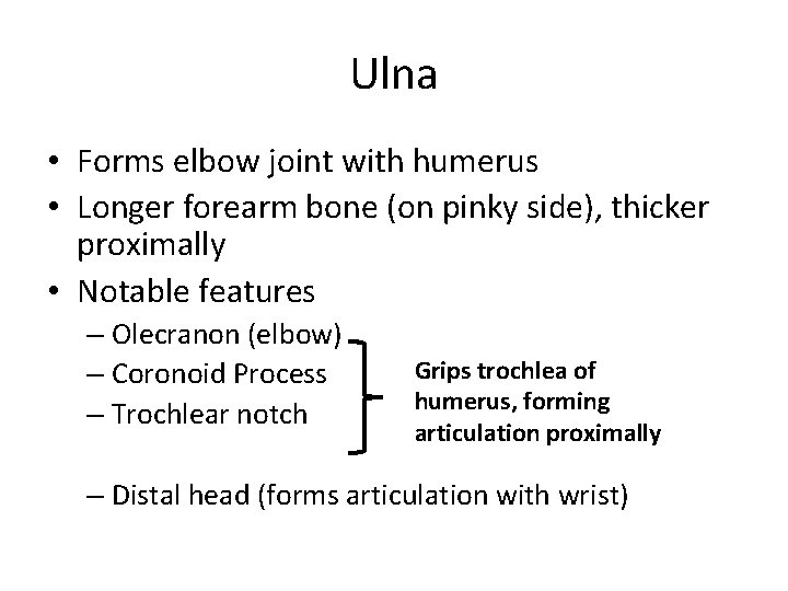
Ulna • Forms elbow joint with humerus • Longer forearm bone (on pinky side), thicker proximally • Notable features – Olecranon (elbow) – Coronoid Process – Trochlear notch Grips trochlea of humerus, forming articulation proximally – Distal head (forms articulation with wrist)
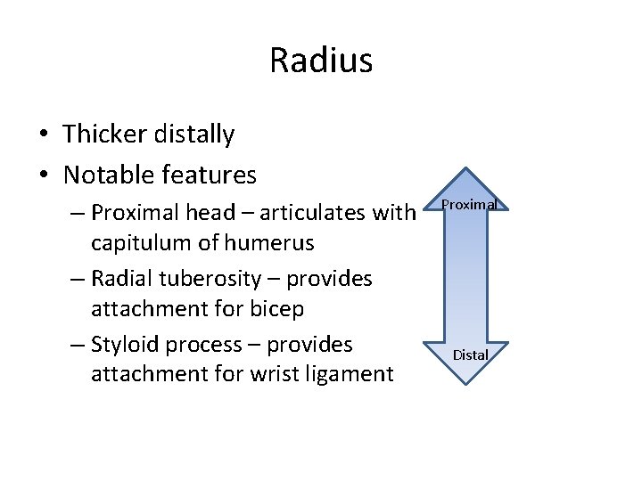
Radius • Thicker distally • Notable features – Proximal head – articulates with capitulum of humerus – Radial tuberosity – provides attachment for bicep – Styloid process – provides attachment for wrist ligament Proximal Distal
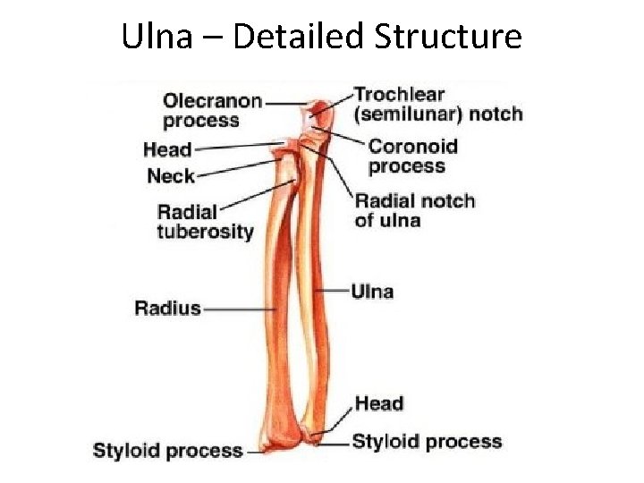
Ulna – Detailed Structure
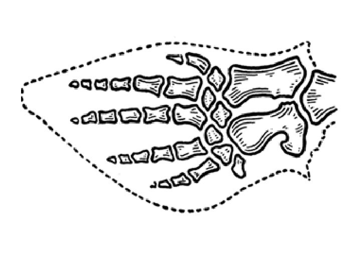
The Hand • Carpals – (8 bones total) • Metacarpals (5 bones) labeled 1 -5 (thumb to pinky) • Phalanges – multiple bones each – Two in thumb – Three in all other fingers • Distal, middle, proximal
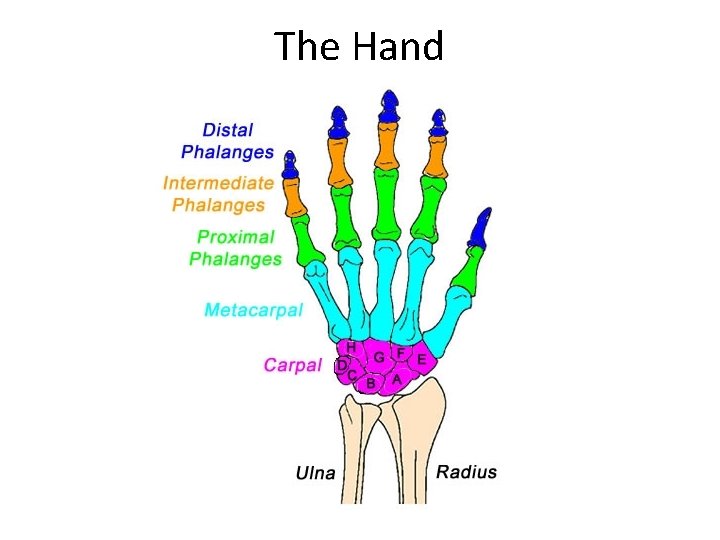
The Hand
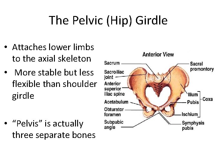
The Pelvic (Hip) Girdle • Attaches lower limbs to the axial skeleton • More stable but less flexible than shoulder girdle • “Pelvis” is actually three separate bones
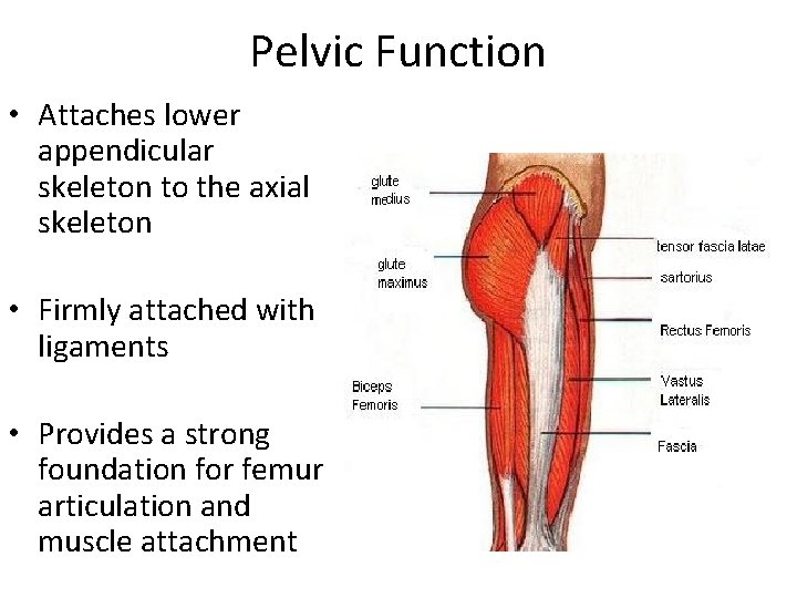
Pelvic Function • Attaches lower appendicular skeleton to the axial skeleton • Firmly attached with ligaments • Provides a strong foundation for femur articulation and muscle attachment
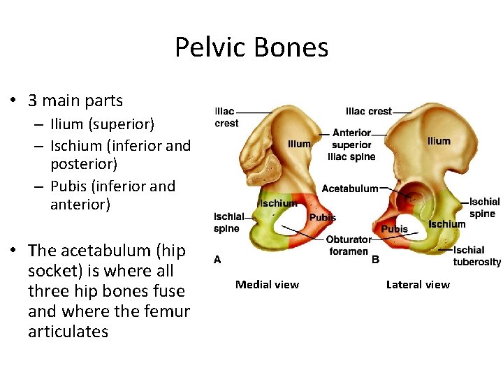
Pelvic Bones • 3 main parts – Ilium (superior) – Ischium (inferior and posterior) – Pubis (inferior and anterior) • The acetabulum (hip socket) is where all three hip bones fuse and where the femur articulates Medial view Lateral view
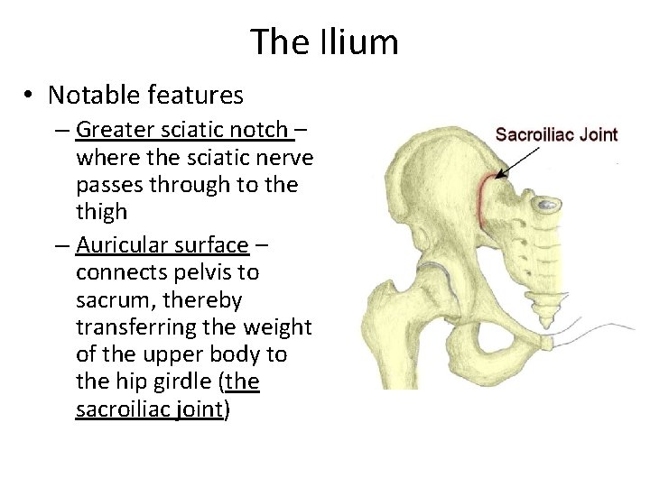
The Ilium • Notable features – Greater sciatic notch – where the sciatic nerve passes through to the thigh – Auricular surface – connects pelvis to sacrum, thereby transferring the weight of the upper body to the hip girdle (the sacroiliac joint)
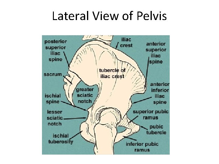
Lateral View of Pelvis
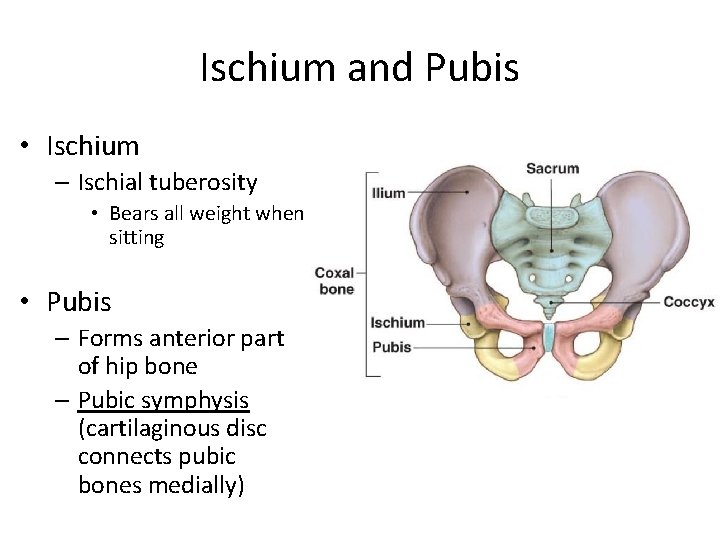
Ischium and Pubis • Ischium – Ischial tuberosity • Bears all weight when sitting • Pubis – Forms anterior part of hip bone – Pubic symphysis (cartilaginous disc connects pubic bones medially)
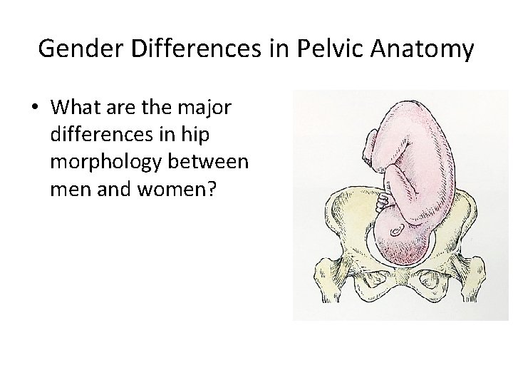
Gender Differences in Pelvic Anatomy • What are the major differences in hip morphology between men and women?
The Lower Limb (Leg) Bones • • • Femur – thigh bone Tibia – shin bone 1 Fibula – shin bone 2 Tarsals – ankle bones Metatarsals – foot bones Phalanges - toes
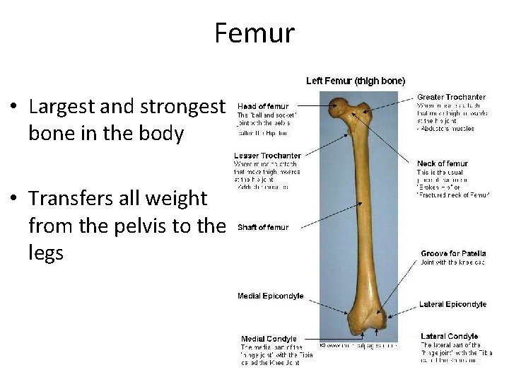
Femur • Largest and strongest bone in the body • Transfers all weight from the pelvis to the legs
Femur • Notable Features – Head – articulates with acetabulum in hip – Greater trochanter – attachment point for thigh muscles – Lesser trochanter – attachment point for gluteal muscles – Lateral/medial condyles – articulates at knee joint – Patella – knee cap
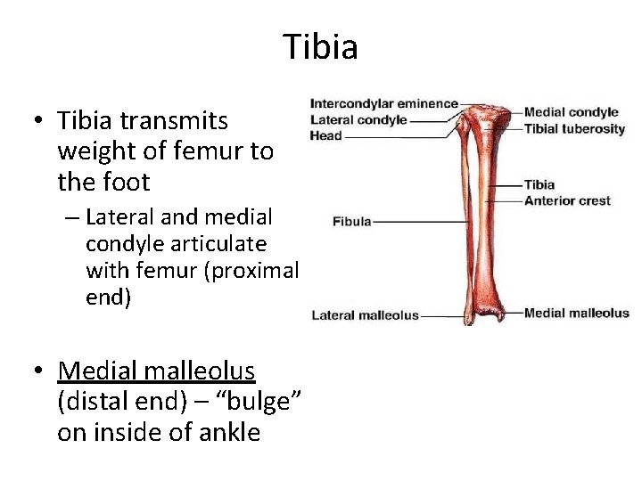
Tibia • Tibia transmits weight of femur to the foot – Lateral and medial condyle articulate with femur (proximal end) • Medial malleolus (distal end) – “bulge” on inside of ankle
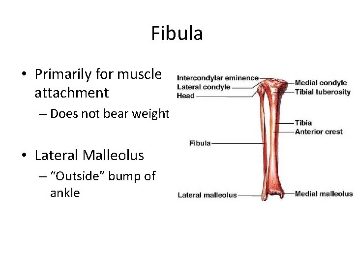
Fibula • Primarily for muscle attachment – Does not bear weight • Lateral Malleolus – “Outside” bump of ankle
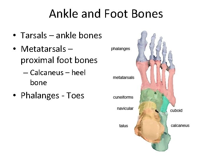
Ankle and Foot Bones • Tarsals – ankle bones • Metatarsals – proximal foot bones – Calcaneus – heel bone • Phalanges - Toes
- Slides: 63