BODY TISSUES Remembergroups of cells that are similar

BODY TISSUES • Remember…groups of cells that are similar in structure and function make TISSUES

Cell Shapes • thin, flat, angular contours • round to oval • irregular angular shapes, > 4 sides • disc shaped

Cell Shapes 2 • squarish • thick middle, tapered ends • taller than wide • long, slender Stellate • nerve cells have extensions, look starlike

• Histology: study of microscopic anatomy • Only 200 different cells types • Four primary tissue classes – epithelial tissue – connective tissue – muscular tissue – nervous tissue

EPITHELIAL TISSUE • Lining, covering, and glandular tissue of the body • Covers & lines all free body surfaces • Functions: - Protection - Absorption - Filtration - Secretion

SPECIAL CHARACTERISTICS • One or more layers of closely adhering cells- form continuous sheet and bound together – no room for blood vessels - avascular • depends on underlying connective tissue for oxygen – sits on basement membrane (connective tissue) - secreted by cells • anchors epithelium to underlying connective tissue • Always have 1 free surface – apical surface – exposed to body’s exterior or the cavity of internal organ • If well nourished, regenerate easily

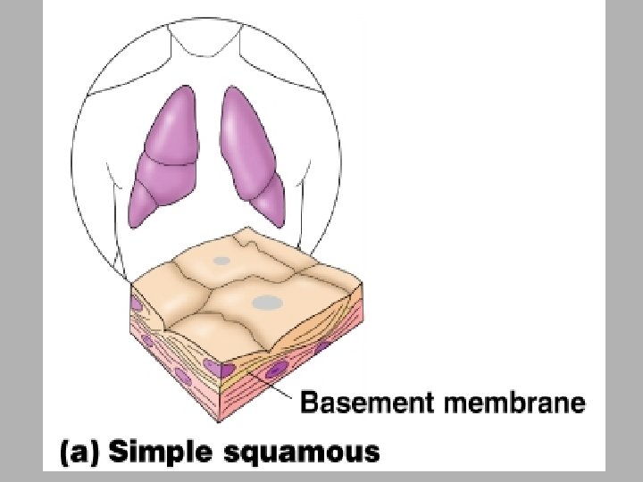
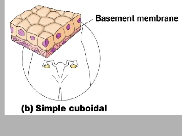
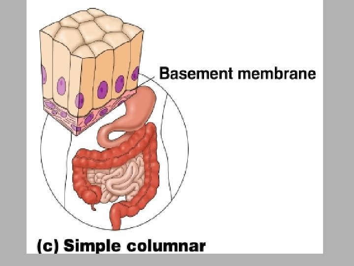
CLASSIFICATION OF EPITHELIUM • Each epithelium given 2 names 1 st – indicates number of cell layers - Simple epithelium = 1 layer - Stratified = more than 1 layer 2 nd – describes shape of cells - Squamous – flattened like fish scales - Cuboidal – cube-shaped like dice - Columnar – shaped like columns • Combine the names to fully describe epithelium

Classification of Epithelium · Shape of cells · Squamous – flattened · Cuboidal – cube-shaped · Columnar – column-like Figure 3. 16 b Copyright © 2003 Pearson Education, Inc. publishing as Benjamin Cummings Slide 3. 44 b

Simple Versus Stratified Epithelia • Simple epithelium – contains one layer of cells – named by shape of cells • Stratified epithelium – contains more than one layer – named by shape of apical cells

Simple Squamous Epithelium • Single row of flat cells on basement membrane • Forms membranes where filtration or exchange occurs • Found in alveoli (lungs), glomeruli (kidneys), capillaries,

Simple Cuboidal Epithelium • Single row of cube-shaped cells, often with microvilli • Absorption & secretion; produces mucus • Glands & bronchioles (lung tubes)

Simple Columnar Epithelium Goblet cell Microvilli • Single row of tall, narrow cells • Absorption & secretion; secretion of mucus - Goblet cells – produce lubricating mucus • Inner lining of GI tract, uterus, kidney & uterine tubes

Stratified Epithelium · Stratified squamous · Cells at the free edge are flattened · Found as a protective covering where friction is common · Locations · Skin * Weiners · Mouth · Esophagus · Vagina · Anus Copyright © 2003 Pearson Education, Inc. publishing as Benjamin Cummings Figure 3. 17 e

Stratified Squamous Epithelial layer

Stratified Epithelium · Stratified cuboidal · Two layers of cuboidal cells · Stratified columnar · Surface cells are columnar, cells underneath vary in size and shape · Stratified cuboidal and columnar · Rare in human body · Found mainly in ducts of large glands Copyright © 2003 Pearson Education, Inc. publishing as Benjamin Cummings Slide 3. 50

Stratified Cuboidal Epithelium • Two or more layers of cells; surface cells square • Stratified Columnar & Cuboidal both rare • Mostly found in ducts of glands

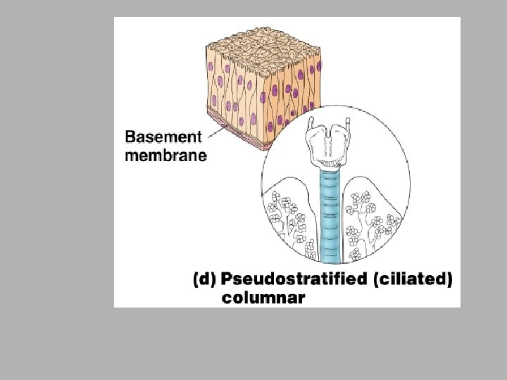
Pseudostratified Epithelium Cilia Goblet cell Basal cell • • Single row of cells, some shorter than others Absorption & secretion Secretes and propels respiratory mucus (ciliated type) Found in respiratory system

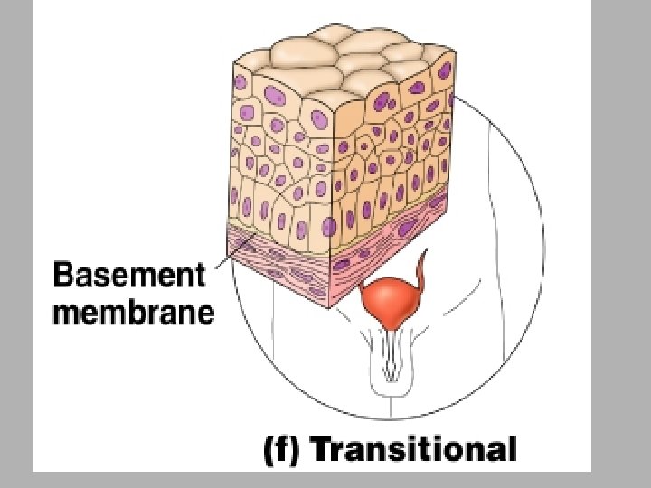
Transitional Epithelium • Multilayered epithelium with rounded surface cells that flatten when the tissue is stretched • Stretches to allow filling of urinary tract • Found in urinary tract -- kidney, ureter, bladder

Glandular Epithelium · Gland – one or more cells that secretes a particular product · Two major gland types · Endocrine gland · Ductless · Secretions are hormones · Exocrine gland · Empty through ducts to the epithelial surface · Include sweat and oil glands Copyright © 2003 Pearson Education, Inc. publishing as Benjamin Cummings Slide 3. 52

Endocrine & Exocrine Glands • Glands secrete substances for elimination or for use elsewhere in the body – composed predominantly of epithelial tissue • Exocrine glands maintain connection to surface with a duct (epithelial tube) • Endocrine glands have no ducts but secrete their products (hormones) directly into bloodstream • Mixed organs – liver secretes bile into ducts + albumin into blood – gonads release gametes + secrete hormones into blood – pancreas secretes digestive enzymes + hormones

ENDOCRINE GLANDS • • Ductless glands Secretions go directly into bloodstream All hormones Thyroid, adrenals, pituitary glands

Connective Tissue • Most abundant and variable tissue type • Cells not in direct contact since volume of extracellular matrix is greater than the volume occupied by cells • Functions – connects organs to each other – gives support & protection (physical & immune) – storage of energy & heat production – movement & transport of materials

COMMON CHARACTERISTICS OF CONNECTIVE TISSUE • Variations in blood supply - Most well vascularized - However, tendons & ligaments have poor blood supply and cartilages are avascular - Very slow to heal when injured • Extracellular maxtrix - Nonliving substance found outside the cells - Lots of different kinds - Acts as “glue”

TYPES OF CONNECTIVE TISSUE • Bone • Cartilage • Dense connective tissue • Loose connective tissue • Blood

Bone • Called osseous tissue • Bone cells sitting in cavities surrounded by rock hard matrix
 • Physical support; leverage for muscles; mineral storage • Found")
Bone Tissue (compact bone) • Physical support; leverage for muscles; mineral storage • Found in skeleton

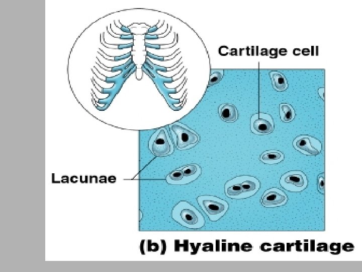
Cartilage • Less hard & more flexible than bone • Supportive connective tissue with rubbery matrix • No blood vessels so diffusion must bring in nutrients & remove wastes – injured cartilage heals slowly • Major types of cartilage depend upon fiber types – hyaline, fibrocartilage and elastic cartilage
 • Lots of collagen fibers in rubbery")
Hyaline Cartilage (most abundant type of cartilage) • Lots of collagen fibers in rubbery matrix w/ glassy, blue/white appearance • Supports airway, attaches ribs to sternum, covers ends of bones where they form joints • Fetuses have all hyaline cartilage that develops into bone by time born

FIBROCARTILAGE • Forms cushionlike disks between vertebrae • Highly compressible

Elastic Cartilage • Provides flexible, elastic support • External ear and epiglottis

DENSE CONNECTIVE TISSUE • Form strong, ropelike structures - Tendons – connect skeletal muscle to bones - Ligaments – connect bone to bone at joints - Lower layers of skin
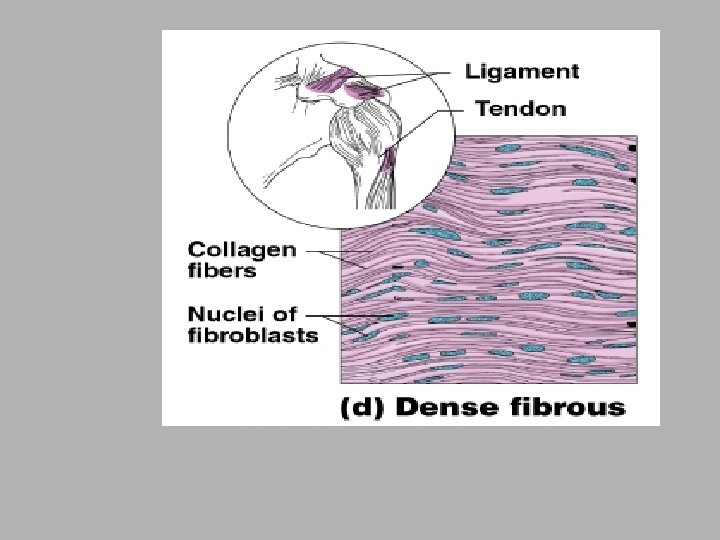

Dense Regular Connective Tissue

LOOSE CONNECTIVE TISSUE • Softer & have more cells and fewer fibers • Areolar tissue • Adipose tissue

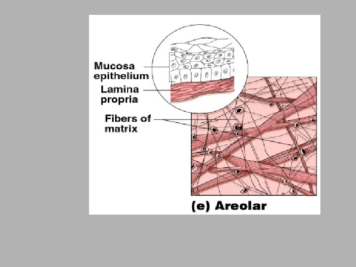
Areolar Tissue • Soft, pliable, cobwebby tissue that cushions & protects organs is wraps • Acts as universal packing tissue & connective tissue glue b/c it holds internal organs together & in proper position

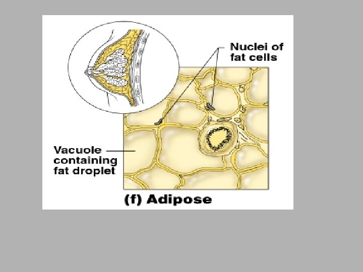
Adipose Tissue • Large, empty-looking cells with thin margins; nucleus pressed against cell membrane • Subcutaneous fat beneath skin & surrounding organs • Insulates body; protects against extreme temps; protects organs; stores fuel

Blood • Variety of cells and cell fragments; some with nuclei & some without • Nonnucleated pale pink cells or nucleated white blood cells • Found in heart and blood vessels
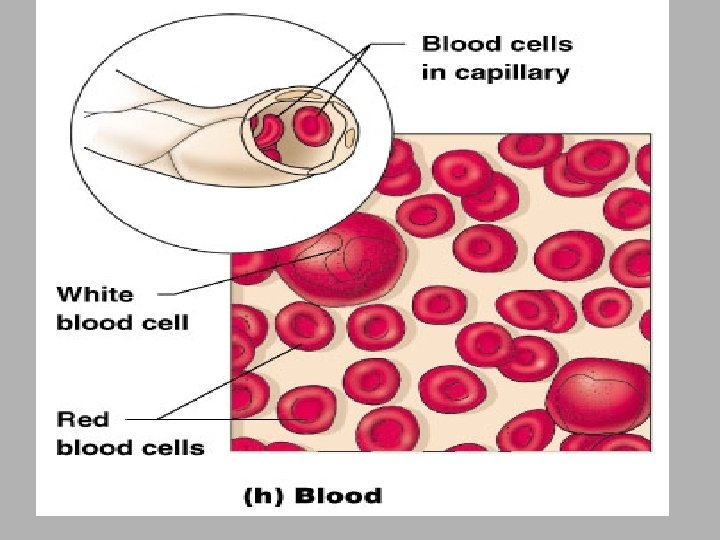

• Vascular tissue • Considered connective tissue b/c it is blood cells surrounded by nonliving, fluid matrix called blood plasma • Transport vehicle for cardiovascular system – carries nutrients, wastes, respiratory gases

MUSCLE TISSUE • Highly specialized to contract, or shorten, to produce movement • Elongated to provide long axis for contraction • Types: - Skeletal - Cardiac - Smooth
 and multiple nuclei •")
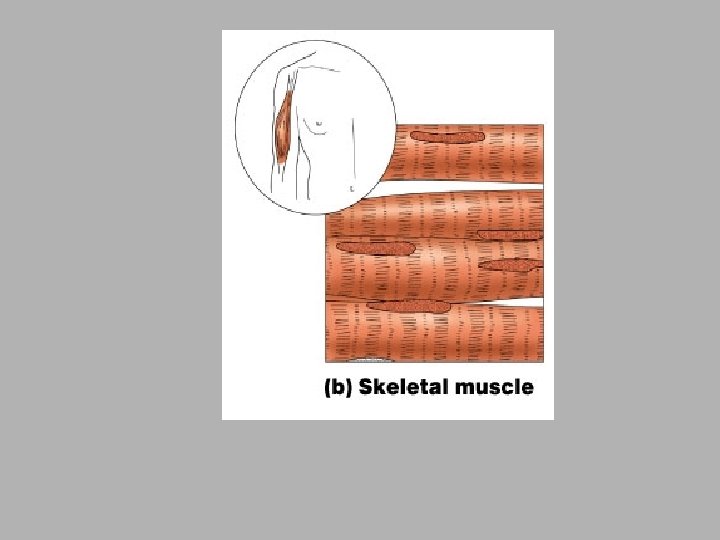
Skeletal Muscle • Long, cylindrical, unbranched cells with striations (stripes) and multiple nuclei • Movement, facial expression, posture, breathing, speech, swallowing and excretion • Skeletal muscles = voluntary

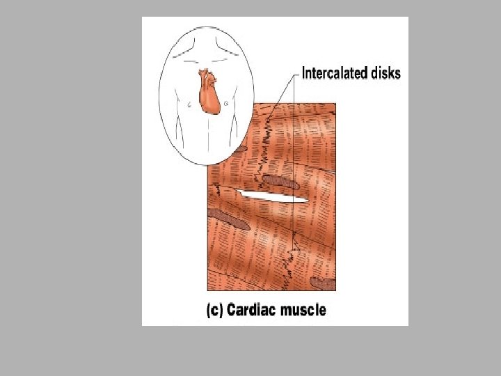
Cardiac Muscle • Short branched cells with striations and intercalated discs (junctions like clasped fingers); one central nuclei per cell • Pumping of blood • Found in the heart

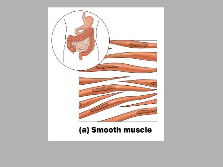
Smooth Muscle • aka visceral; no striations, single nucleus, spindle-shaped • Swallowing, GI tract functions, labor contractions, control of airflow, erection of hairs & control of pupil • Sheets of muscle in viscera; iris; hair follicles & sphincters • Peristalsis – wavelike motions

Nerve Tissue • Neurons that receive & conduct electrochemical impulses irritability & conductivity are major functions • Unique structure – cytoplasm drawn out into long extensions (>3 feet in leg) which allows single neuron to conduct impulse over long distance • Found in brain, spinal cord, nerves
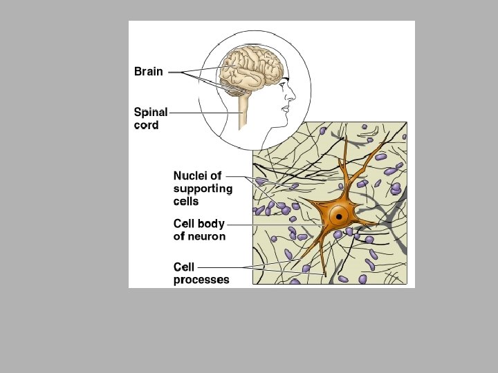

• NEUROGLIA CELLS – cells in the nervous system that support the neurons
 – secretes")
Membranes • Synovial membrane lines joints (only connective tissue comprises this layer) – secretes synovial fluid rich in hyaluronic acid • Cutaneous membrane (skin) – stratified squamous epithelium resting on layer of dermis – relatively dry layer serves protective function • Serous membrane (serosa) – simple squamous epithelium on areolar tissue covers organs and body walls & produces serous fluid • Mucous membrane (mucosae) – line passageways that open to the exterior: digestive, respiratory, urinary & reproductive – often covered with mucus secreted by goblet cells

Changes in Tissue Types • Tissues are capable of changing from one type to another • Differentiation – unspecialized tissues of embryo to specialized mature types (mesenchyme to muscle) • Metaplasia – changing from one type of mature tissue to another – simple cuboidal tissue before puberty changes to stratified squamous after puberty

Tissue Growth • Hyperplasia is tissue growth through cell multiplication • Hypertrophy is enlargement of preexisting cells – muscle grow through exercise • Neoplasia is growth of a tumor (benign or malignant) through growth of abnormal tissue

Tissue Shrinkage and Death • Atrophy is shrinkage from loss of cell size/number – senile atrophy is due to aging – disuse atrophy from lack of use (leg in a cast) • Necrosis is pathological death of tissue – gangrene is due to insufficient blood supply – gas gangrene is due to anaerobic bacterial infection – infarction is sudden death of tissue from lack of blood – decubitus ulcer is bed sore or pressure sore • Neoplasm – abnormal mass of proliferating cells

Tissue Repair • Damaged tissues are repaired in 2 ways: • Regeneration – replacement of dead or damaged cells with original cells – restores normal function – skin injuries & liver regenerate • Fibrosis – replacement of damaged cells with scar tissue (collagen) – helps hold organ together -- function is not restored – healing muscle injuries, scarring of lung tissue in TB or healing of severe cuts & burns of the skin

Regeneration of Tissues · Tissues that regenerate easily · Epithelial tissue · Fibrous connective tissue and bone · Tissues that regenerate poorly · Skeletal muscle · Tissues that are replaced largely with scar tissue · Cardiac muscle · Nervous tissue within the brain and spinal cord Copyright © 2003 Pearson Education, Inc. publishing as Benjamin Cummings Slide 3. 71

Developmental Aspects of Tissue · Epithelial tissue arises from all three primary germ layers · Muscle and connective tissue arise from the mesoderm · Nervous tissue arises from the ectoderm · With old age there is a decrease in mass and viabililty in most tissues Copyright © 2003 Pearson Education, Inc. publishing as Benjamin Cummings Slide 3. 72

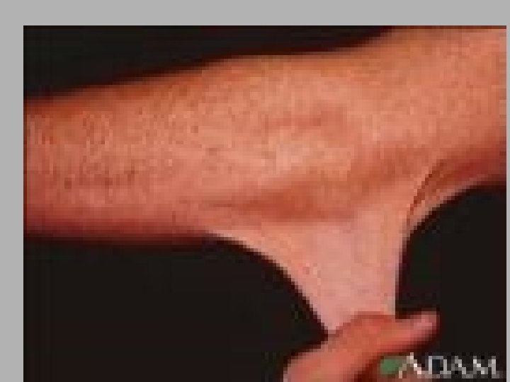
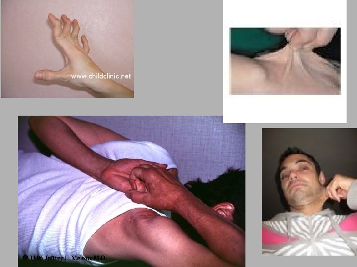
Connective Tissue Diseases • Deficiencies in collagen & elastin cause serious problems • Marfan syndrome = hereditary defect elastin fibers – tall stature, long limbs, spinal curvature & weakening of heart valves & arterial walls (rupture of aorta) • Ehlers-Danlos syndrome = hereditary defect in collagen synthesis – stretchy skin, loose joints, abnormal blood vessels, intestines and bladder • Osteogenesis imperfecta = insufficient collagen in bones making them brittle • Scurvy is lack of vitamin C needed for synthesis of 2 amino acids used to synthesize collagen – poor wound healing & subcutaneous hemorrhages
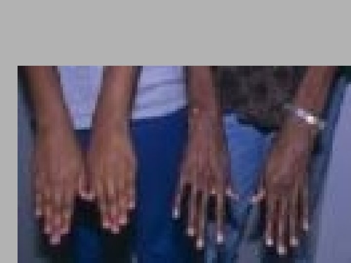

Ehlers-Danlos Syndrome

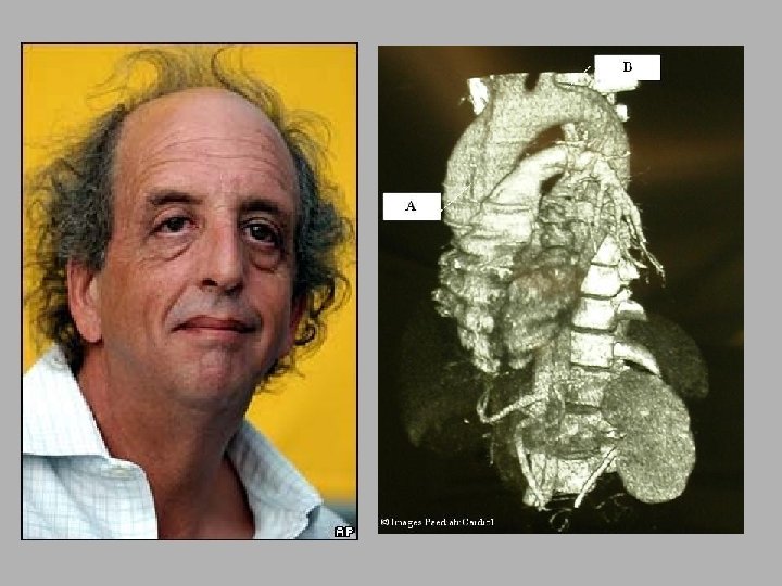
OI

Multiple fx from OI

Fatal type of OI – fx appears in utero

Wound Healing of a Laceration • Damaged vessels leak blood • Damaged cells & mast cells leak histamine – dilates blood vessels – increases blood flow – increases capillary permeability • Plasma seeps into wound carrying antibodies, clotting factors & WBCs

Wound Healing of a Laceration • Clot forms • Scab forms on surface • Macrophages start to clean up debris Scab formation & macrophage activity.

Wound Healing of a Laceration • New capillaries grow into wound • Fibroblasts deposit new collagen to replace old material • Fibroblastic phase begins in 3 -4 days & lasts up to 2 weeks Formation of granulation tissue.

Wound Healing of a Laceration • Surface epithelial cells multiply & spread beneath scab • Scab falls off • Epithelium grows thicker (regenerates) • Connective tissue forms only scar tissue (fibrosis) • Remodeling phase Epithelial regeneration & connective may last 2 years tissue fibrosis.
- Slides: 77