Body Fluids Dr Sangeetha Balakrishnan 23 February 2017

Body Fluids Dr. Sangeetha Balakrishnan 23 February, 2017

Presumptive/Preliminary/Screening/ Field/Indicative Tests for Blood 1. Luminol Test 2. Fluorescein Test 3. Phenolpthalein Test

Luminol Test at a Crime Scene 0 to 30 s

Fluorescein Test at a Crime Scene

Phenolpthalein Test • Can detect blood diluted to 1 part in 10 million. • Gives false positives for certain vegetable extracts. • How is it done? : (i) Suspected blood stain (on a cotton swab) + water + Phenolpthalein colourless (ii) Add hydrogen peroxide immediate PINK colour!
 • Both")
Confirmatory Tests for Blood 1. Teichmann Test 2. Takayama Test (Hemochromogen Test) • Both are called Microcrystal Tests. Blood + Crystallising Reagent Characteristic Shaped Crystals.

Teichmann Test • Blood + Teichmann’s Reagent hemin react with halides yellow rhomboid crystals • Microscopic Observation (heat) Hb brownish- • Teichmann’s Reagent: Potassium bromide + Potassium chloride + Potassium iodide in Acetic acid.

Teichmann Test
 • Suspected blood stain on a glass slide + heat")
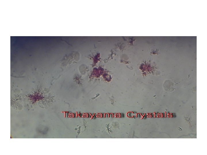
Takayama Test (Hemochromogen Test) • Suspected blood stain on a glass slide + heat + add pyridine in Sodium hydroxide + reducing sugar red feathery crystals of pyridine ferroprotoporphyrin • Very sensitive test. • Even very old blood stains give +’ve results!

Saliva • • • Saliva is produced in the mouth. Function: preliminary digestion of food. Contains: water, proteins, enzymes, salts. Alpha amylase: enzyme in saliva. Alpha amylase breaks down starch in food. It is also present in other body fluids, but in low concentrations.

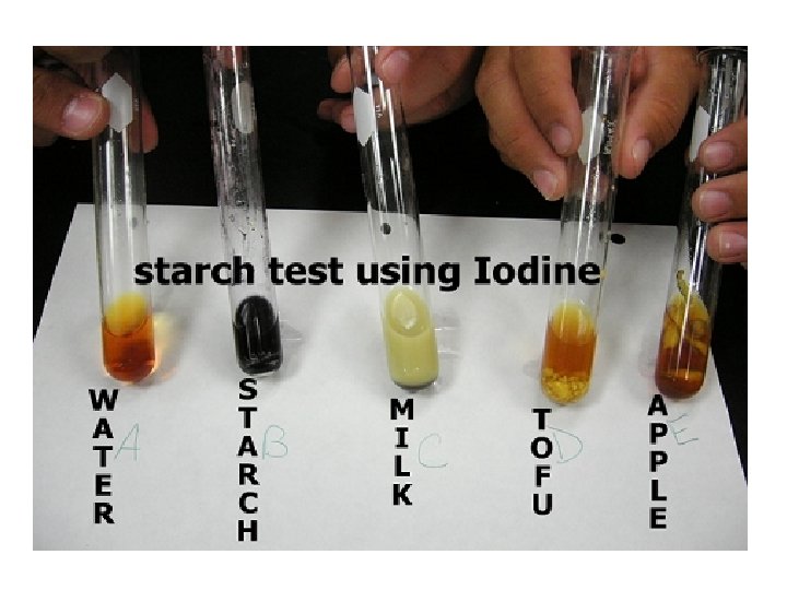
Preliminary Test - Saliva • Principle: Starch + Iodine Deep blue colour • Suspected saliva sample + water/saline incubate at body temperature. • If the suspected sample is indeed saliva, it will contain alpha amylase! • Alpha amylase will break down starch into simpler components. (digestion) • Add Iodine solution. • Absence of starch (it has been broken down) No Deep Blue colour!

Drawbacks of the Saliva Preliminary Test • Not particularly sensitive. • Not specific to saliva. • Use saliva sample for DNA testing. • Detection of saliva at scene of crime: Shine UV light fluorescence!

Saliva – Confirmatory test • Phadebas Amylase Test Developed by Pharmacia Diagnostics. Qualitative and quantitative test. Phadebas: a synthetic biochemical substrate. • The substrate has starch microspheres. • The microspheres are chemically bonded to a blue coloured dye. Phadebas substrate + suspected saliva (in water) salivary amylase digests starch microspheres break down blue dye is released!

Phadebas Amylase Test
 Creatinine (ii) Urea Presumptive Test – Urine 1. Jaffe")
Urine • Main components: (i) Creatinine (ii) Urea Presumptive Test – Urine 1. Jaffe Test • Based on the detection of creatinine • Suspected urine sample + picric acid + 5% Na. OH immediate orange colour
 Assay for Urea • Bromothymol Blue is")
Presumptive Test – Urine … cont’d 2) Assay for Urea • Bromothymol Blue is a p. H-indicator dye. • Colour: Yellow-green at p. H 6 Aqua blue at p. H > 7. 6 (i) Whatman Filter Paper + Bromothymol Blue (1 drop) allow to dry (ii) Add a drop from the suspected urine sample. (iii) Add a drop of Urease. (iv) If urea is present (meaning: urine is present), urease will degrade urea to ammonia gas. (v) This will cause the p. H to increase. (vi) Aqua blue colour!

Semen • Produced by the male sex organ. • Semen = Spermatozoa + Seminal fluid (cellular component) (fluid component) Spermatozoa (Sperm) Seminal Fluid Male reproductive cells Contains: proteins, salts, organic materials. Flavin: UV fluorescence! Consist of: head, tail and mid piece Produced in prostate gland Seminal Acid Phosphatase (SAP) Prostate Specific Antigen (p 30 protein)
. • There are")
Presumptive Test - Semen • Based on Seminal Acid Phosphatase (SAP). • There are other acid phosphatases in the body! • Brentamine Spot Test or Walker Test • Suspected Semen Sample + Brentamine Fast Blue B Intense purple colour within 2 minutes

Brentamine Fast Blue B added to suspected semen samples

Confirmatory Test - Semen 1. Christmas Tree Stain Test • Basis: Sperms analysed in lab are NOT motile. • Hence identification should be in the presence of other cellular material! • A pair of dyes is used: (i) Picroindigocarmine (PIC) (ii) Nuclear Fast Red • PIC: stains the tails green-blue-grey • Nuclear Fast Red: stains the heads (with DNA) bright crimson.

Christmas Tree Stain Test

Confirmatory Test – Semen … cont’d 2. Detection of Prostate Specific Antigen or p 30 protein: Oligospermia (low sperm count) Aspemia (no sperms) Suspected semen sample + Reagent purple. Intense

Fecal Stains • Basis: Human waste has bilirubin. • Bacteria in body, break down bilirubin to urobilinogen. Presumptive Test: Edelman’s Reagent Fecal stain bilirubin urobilinogen urobilin shine UV light + Edelman’s reagent Green fluoresence.

Confirmatory Test – Fecal Stains • Microscopy: • To look for animal and plant cells present due to digestion.

Sweat • Suspected sweat sample + Crystal Violet deep purple • Fatty acids in sweat react with the dye.

Blood Spatter Analysis

Blood Stains Passive Bloodstain Transfer Stain Eg. Clots Drops Flows Pooling Eg. Wipes Swipes Pattern Transfers Projected or Impact Stains Eg. Spatters Splashes Cast-off stains Arterial spurts or gushes

splash arterial spurts passive fall blood pool multiple drips smear

Physical Properties • Viscosity – more viscous than water – Does not spatter – smooth hard surface (tile) – Spatters – rough, hard surface (concrete) • Surface tension – blood has a high surface tension; causes a decrease in its surface area. • Blood adheres to an external surface; it will separate and spatter only when there is sufficient external force to overcome the surface tension. • Blood drops that fall on a flat surface have a spherical surface • Edges may have spikes or extensions • Satellites - small secondary droplets

• Cast off pattern: blood from a moving object coated in blood (pipe, knife) • Fine-mist spatter: high-velocity impact (gunshot) • Void: empty space; victim/attacker/object moved after attack

Blood Spatter Types 1. High velocity – gunshot wounds 2. Medium velocity – beating, stabbing 3. Low velocity – blunt object impact

Information from Blood Spatter • Used to explain events at a violent crime scene Point of origin – Angle of impact – Blood velocity (manner of death) –
 • Determine source of blood (point of origin)")
Lines of Convergence (Point of Origin) • Determine source of blood (point of origin) • Draw straight lines down long axis of blood spatter

Angle of Impact • Determined by measuring the length and width of the blood stain. arcsin A = Width/length Here A = Angle of impact.
- Slides: 36