
Body Bugs NOVA Bugs That Live on You

Body Bugs NOVA | Bugs That Live on You

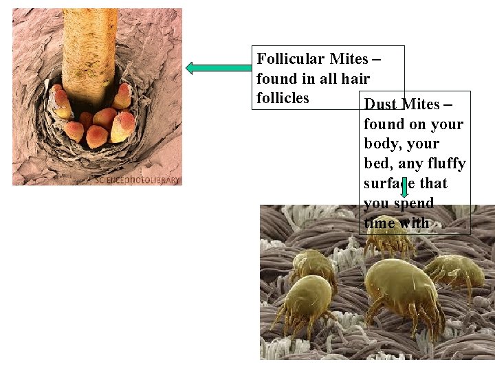
Follicular Mites – found in all hair follicles Dust Mites – found on your body, your bed, any fluffy surface that you spend time with
Essentials of Human Anatomy & Physiology Seventh Edition Elaine N. Marieb Chapter 4 Skin and Body Membranes Slides 4. 1 – 4. 32 Lecture Slides in Power. Point by Jerry L. Cook Copyright © 2003 Pearson Education, Inc. publishing as Benjamin Cummings
Skin and Body Membranes · Function of body membranes: · 1. Line or cover body surfaces · 2. Protect body surfaces · 3. Lubricate body surfaces Copyright © 2003 Pearson Education, Inc. publishing as Benjamin Cummings Slide 4. 1
2 TYPES OF MAIN MEMBRANES • 1. EPITHELIAL MEMBRANES: • Cutaneous, mucous, & serous membranes included • All of these do contain an epithelial sheet However, it is always combined with an underlying layer of connective tissue. • 2. CONNECTIVE TISSUE MEMBRANES: • Represented by Synovial Membranes = Areolar Tissue: NO Epithelial Cells
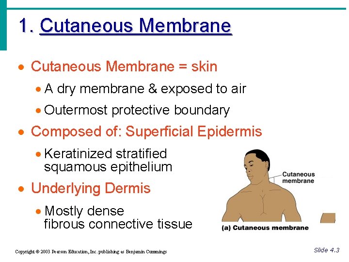
1. Cutaneous Membrane · Cutaneous Membrane = skin · A dry membrane & exposed to air · Outermost protective boundary · Composed of: Superficial Epidermis · Keratinized stratified squamous epithelium · Underlying Dermis · Mostly dense fibrous connective tissue Copyright © 2003 Pearson Education, Inc. publishing as Benjamin Cummings Figure 4. 1 a Slide 4. 3
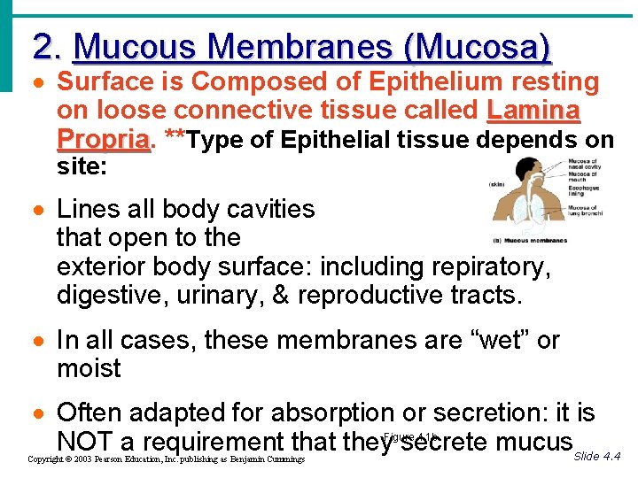
2. Mucous Membranes (Mucosa) · Surface is Composed of Epithelium resting on loose connective tissue called Lamina Propria **Type of Epithelial tissue depends on site: · Lines all body cavities that open to the exterior body surface: including repiratory, digestive, urinary, & reproductive tracts. · In all cases, these membranes are “wet” or moist · Often adapted for absorption or secretion: it is NOT a requirement that they secrete mucus Figure 4. 1 b Copyright © 2003 Pearson Education, Inc. publishing as Benjamin Cummings Slide 4. 4
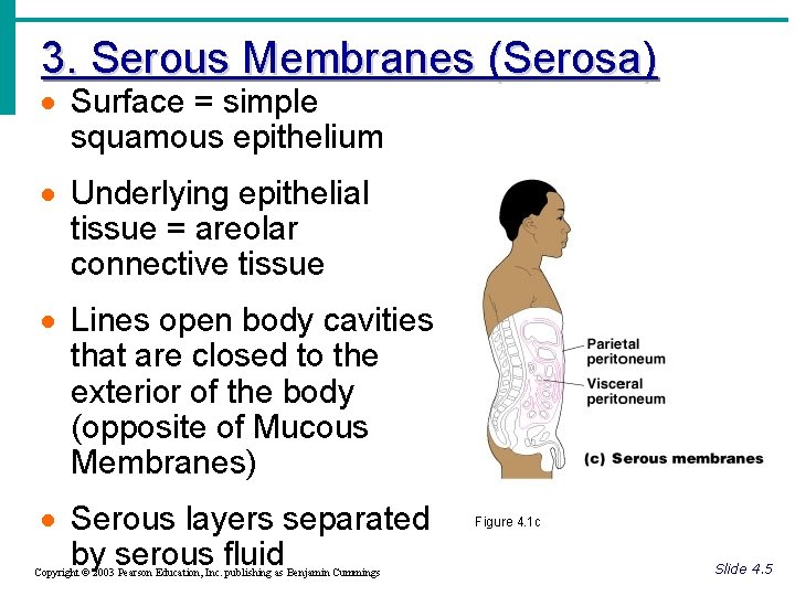
3. Serous Membranes (Serosa) · Surface = simple squamous epithelium · Underlying epithelial tissue = areolar connective tissue · Lines open body cavities that are closed to the exterior of the body (opposite of Mucous Membranes) · Serous layers separated by serous fluid Copyright © 2003 Pearson Education, Inc. publishing as Benjamin Cummings Figure 4. 1 c Slide 4. 5
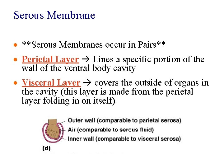
Serous Membrane · **Serous Membranes occur in Pairs** · Perietal Layer Lines a specific portion of the wall of the ventral body cavity · Visceral Layer covers the outside of organs in the cavity (this layer is made from the perietal layer folding in on itself)
Serous Membranes · Specific serous membranes: · Peritoneum · Abdominal cavity · Pleura · Around the lungs ** Good Picture to look at is on Page 97 Figure 4. 1 C · Pericardium · Around the heart Slide 4. 6
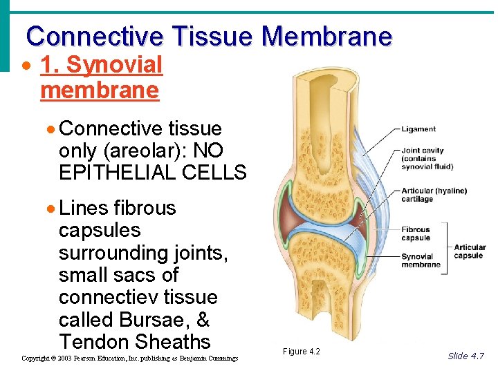
Connective Tissue Membrane · 1. Synovial membrane · Connective tissue only (areolar): NO EPITHELIAL CELLS · Lines fibrous capsules surrounding joints, small sacs of connectiev tissue called Bursae, & Tendon Sheaths Copyright © 2003 Pearson Education, Inc. publishing as Benjamin Cummings Figure 4. 2 Slide 4. 7
Integumentary System · Skin (cutaneous membrane) · Skin derivatives: · Sweat glands · Oil glands · Hairs · Nails Copyright © 2003 Pearson Education, Inc. publishing as Benjamin Cummings Slide 4. 8

Integumentary Functions
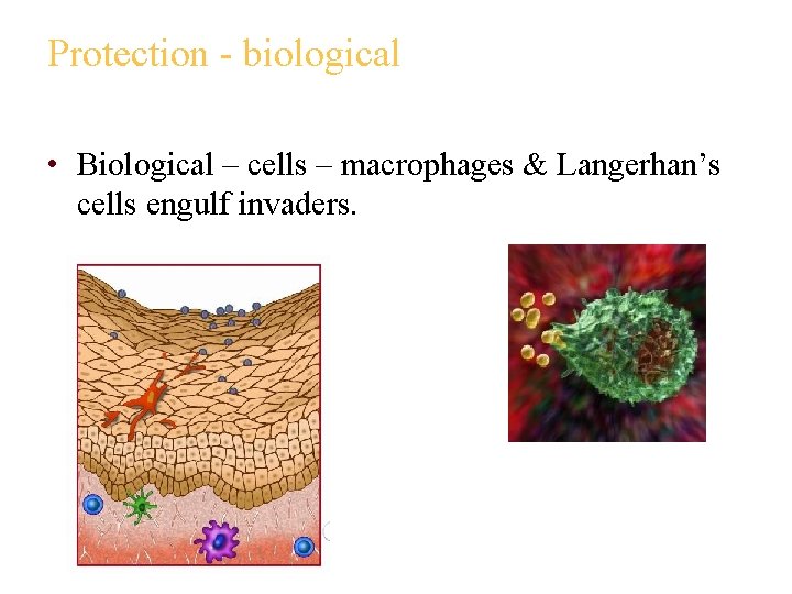
Protection - biological • Biological – cells – macrophages & Langerhan’s cells engulf invaders.
Protection - mechanical • Mechanical – physical • Continuity – skin is made impermeable through the tight junctions & desmosomes • Keratin – physically blocks the passage of water and water soluble substances • FYI: Molecules that can pass through your skin: • Lipid soluble – O 2, CO 2, ADEK, steroids, nicotine, & other transdermal medications • Oleoresins – poison ivy & poison oak, etc – you have less than 10 min to wash it off!
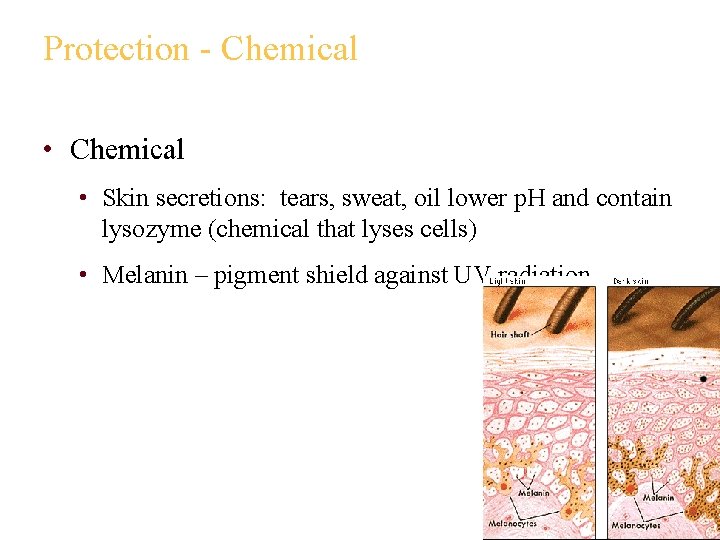
Protection - Chemical • Skin secretions: tears, sweat, oil lower p. H and contain lysozyme (chemical that lyses cells) • Melanin – pigment shield against UV radiation
Excretion • Limited amounts of nitrogen containing wastes (most is excreted in urine unless you have a kidney disorder in which case urea will be excreted in small amounts through skin) • Sodium chloride through sweat • Water through sweat • Heat through sweat
Homeostasis of Body Temperature • Heat production – chief determinant is muscle activity • Heat loss/gain • 80% of heat transfer is through the skin – the rest is through the mucosa (dogs are opposite!) • Regulated by vasoconstriction & vasodilation since heat is carried primarily in the water content of the blood (reminder: on page 1 of notes)
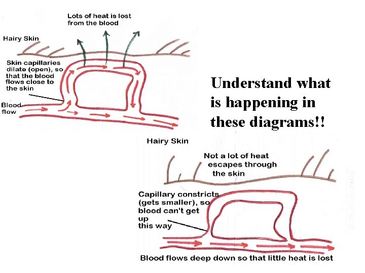
Understand what is happening in these diagrams!!
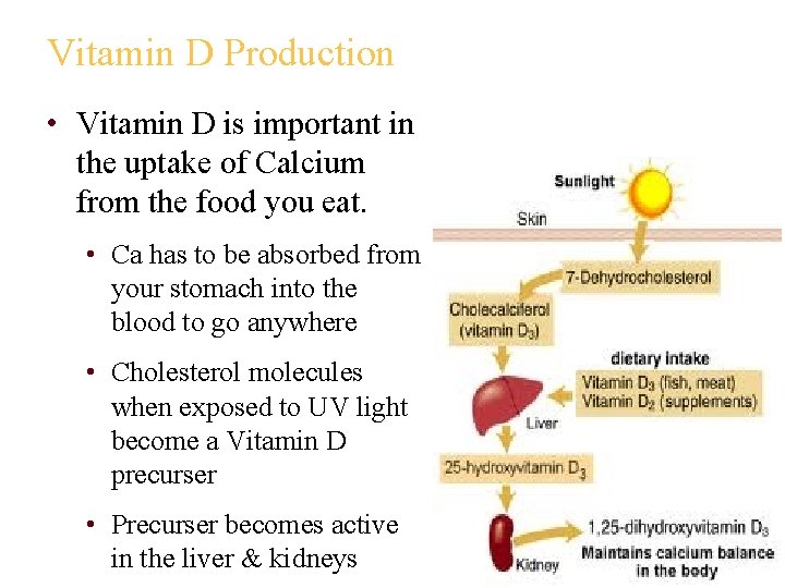
Vitamin D Production • Vitamin D is important in the uptake of Calcium from the food you eat. • Ca has to be absorbed from your stomach into the blood to go anywhere • Cholesterol molecules when exposed to UV light become a Vitamin D precurser • Precurser becomes active in the liver & kidneys
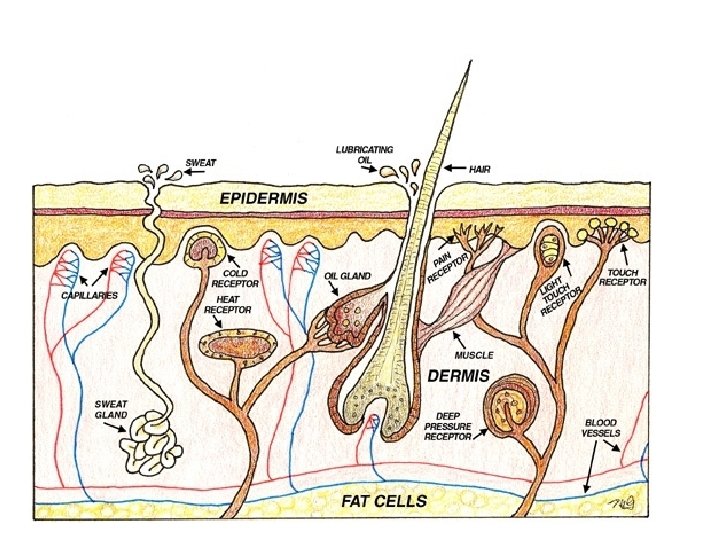
Sensation – skin receptors Don’t need to memorize • Free nerve ending = pain • Meissner’s corpuscles = discriminating touch, light pressure (think fly on your arm) • Krause’s – cold • Ruffini’s – heat • Merkel Disc – Medium pressure • Pacinian corpuscle – Deep pressure See diagram in notes
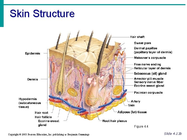
Skin Structure · Epidermis – outer layer: · Stratified Squamous epithelium · Often keratinized (tough & hardened by keratin) · Dermis: · Dense connective tissue Copyright © 2003 Pearson Education, Inc. publishing as Benjamin Cummings Slide 4. 10 a
Skin Structure · Deep to dermis is the hypodermis (Subcutaneous Tissue) · Not part of the skin · Anchors skin to underlying organs · Composed mostly of adipose tissue · Responsible for “curves” on your figure · *Serve as a shock absorber and insulates deeper tissues Copyright © 2003 Pearson Education, Inc. publishing as Benjamin Cummings Slide 4. 10 b
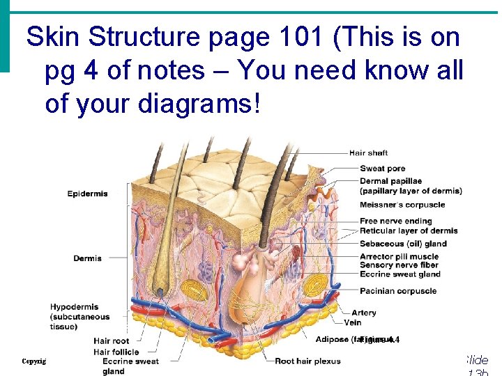
Skin Structure page 101 (This is on pg 4 of notes – You need know all of your diagrams! Figure 4. 4 Copyright © 2003 Pearson Education, Inc. publishing as Benjamin Cummings Slide
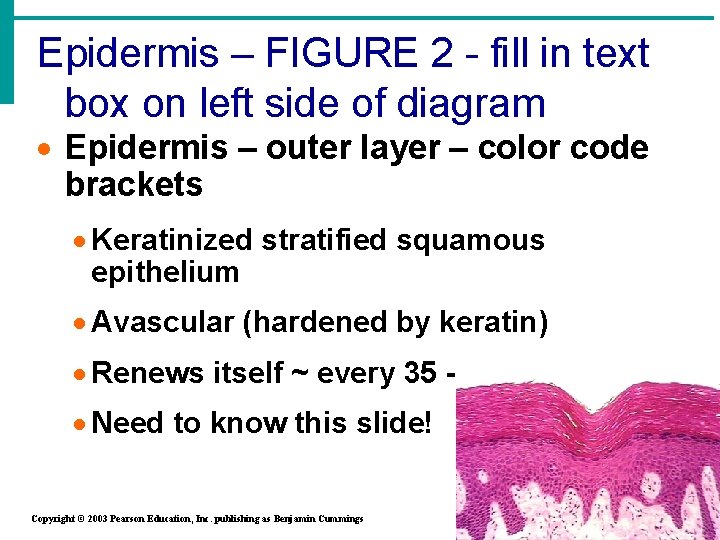
Epidermis – FIGURE 2 - fill in text box on left side of diagram · Epidermis – outer layer – color code brackets · Keratinized stratified squamous epithelium · Avascular (hardened by keratin) · Renews itself ~ every 35 - 45 days · Need to know this slide! Copyright © 2003 Pearson Education, Inc. publishing as Benjamin Cummings Slide
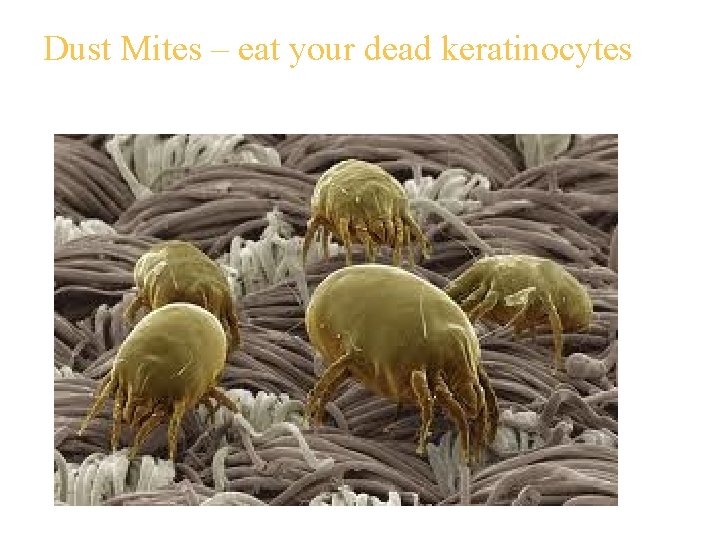
Dust Mites – eat your dead keratinocytes
Epidermis – cell types – Go to FIGURE 1 of notes & fill in cell info (upper right) · Keratinocytes · produce keratin – waterproofing protein ·Why important? To keep you from gaining/losing water · Originate in deeper layers & get pushed to surface – becomes keratin filled & dies · Reminder: Connected to each other by desmosomes & tight junctions · Cell production & keratinization are accelerated in areas of friction ·Callus – thickened skin for protection Slide Copyright © 2003 Pearson Education, Inc. publishing as Benjamin Cummings
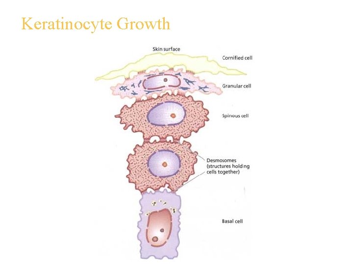
Keratinocyte Growth
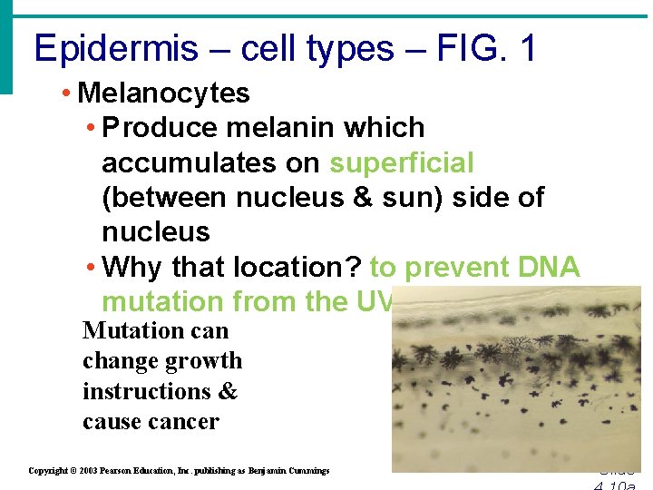
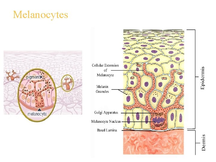
Epidermis – cell types – FIG. 1 • Melanocytes • Produce melanin which accumulates on superficial (between nucleus & sun) side of nucleus • Why that location? to prevent DNA mutation from the UV radiation Mutation can change growth instructions & cause cancer Copyright © 2003 Pearson Education, Inc. publishing as Benjamin Cummings Slide
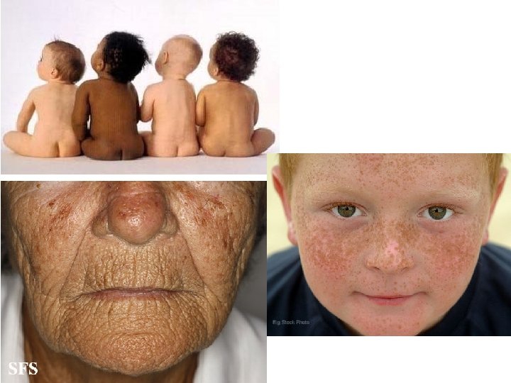
Side Note: • Freckles and moles = areas where melanin is concentrated in one spot. • Despite protection of melanin, over time the exposure eventually damages the skin permanently: • Elastic fibers begin to clump = leathery skin • Depresses the Immune System • Skin Cancer
Melanocytes
Label the 5 strata on FIGURE 1 left side
Reflection • Through research it has been found that darker skinned individuals seldom have skin cancer. • Why do you think that is?
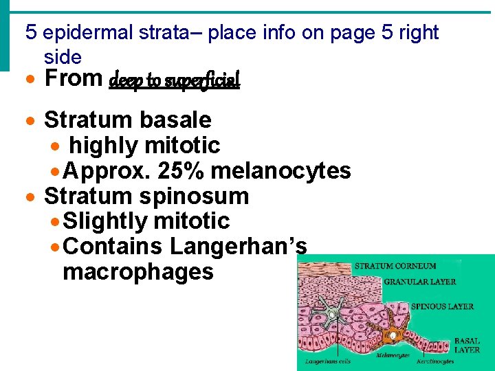
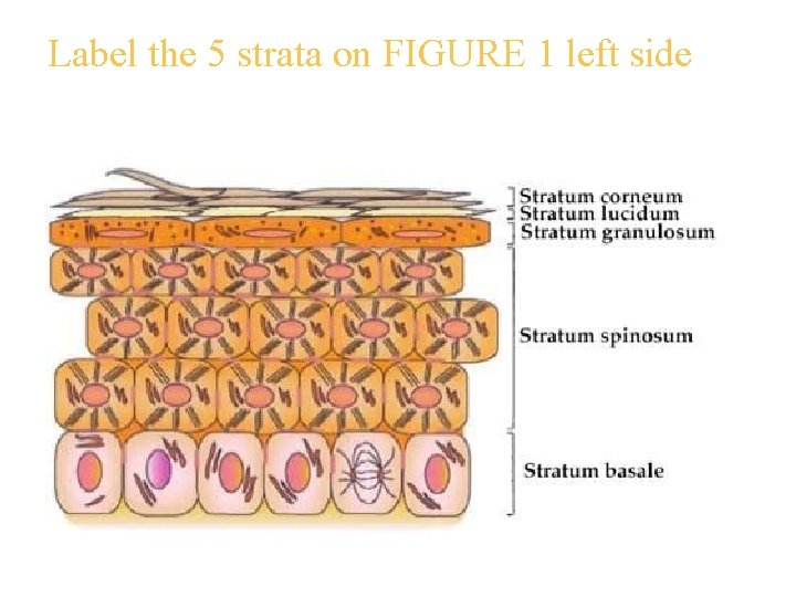
5 epidermal strata– place info on page 5 right side · From deep to superficial · Stratum basale · highly mitotic · Approx. 25% melanocytes · Stratum spinosum · Slightly mitotic · Contains Langerhan’s macrophages Slide
5 strata of the Epidermis · Stratum granulosum · Also contains Langerhans cell · contains keratin granules · Stratum lucidum · found in thicker epidermis – palms, soles, callus · Completely keratinized (and dead!) Copyright © 2003 Pearson Education, Inc. publishing as Benjamin Cummings Slide
5 strata of the Epidermis · Stratum corneum · Also completely keratinized (& dead) · Tough, waterproofing protection · AGAIN: Why is waterproofing important? Copyright © 2003 Pearson Education, Inc. publishing as Benjamin Cummings Slide
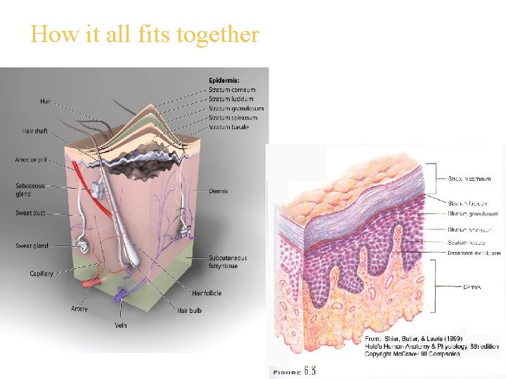
How it all fits together
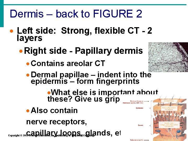
Dermis – back to FIGURE 2 · Left side: Strong, flexible CT - 2 layers · Right side - Papillary dermis · Contains areolar CT · Dermal papillae – indent into the epidermis – form fingerprints ·What else is important about these? Give us grip ! · Also contain nerve receptors, capillary loops, glands, etc. Copyright © 2003 Pearson Education, Inc. publishing as Benjamin Cummings Slide
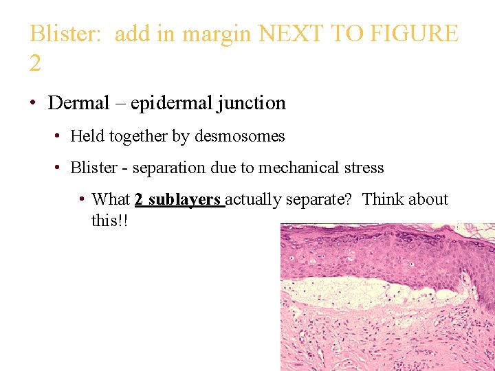
Blister: add in margin NEXT TO FIGURE 2 • Dermal – epidermal junction • Held together by desmosomes • Blister - separation due to mechanical stress • What 2 sublayers actually separate? Think about this!!
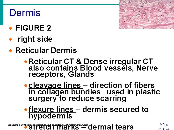
Dermis · FIGURE 2 · right side · Reticular Dermis · Reticular CT & Dense irregular CT – also contains Blood vessels, Nerve receptors, Glands · cleavage lines – direction of fibers in collagen bundles – used in plastic surgery to reduce scarring · flexure lines – dermis secured to hypodermis Slide · stretch marks – dermal tears Copyright © 2003 Pearson Education, Inc. publishing as Benjamin Cummings
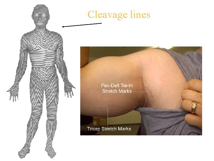
Cleavage lines
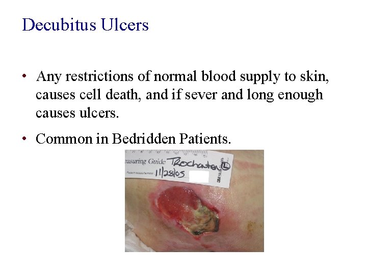
Decubitus Ulcers • Any restrictions of normal blood supply to skin, causes cell death, and if sever and long enough causes ulcers. • Common in Bedridden Patients.
DERMIS • Both Collagen & Elastic Fibers are found throughout the dermis. • *Collagen helps with toughness & hydration. • *Elastic Fibers help with elasticity when we are young • As we age the amount of these fibers decrease skin begins to sag and wrinkle
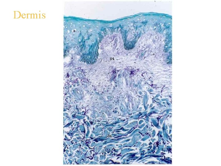
Dermis
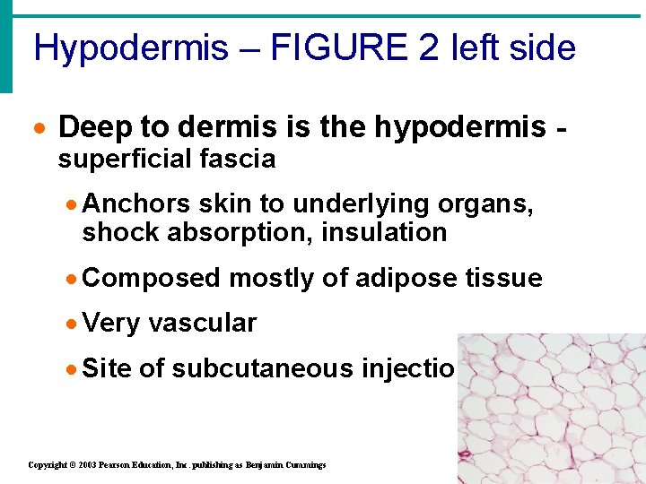
Hypodermis – FIGURE 2 left side · Deep to dermis is the hypodermis superficial fascia · Anchors skin to underlying organs, shock absorption, insulation · Composed mostly of adipose tissue · Very vascular · Site of subcutaneous injections Copyright © 2003 Pearson Education, Inc. publishing as Benjamin Cummings Slide
5 Tissues • There have been 5 types of tissues during the previous slides • You will need to MEMORIZE the location/function/slide of these 5 for this unit test
Skin Structure Figure 4. 4 Copyright © 2003 Pearson Education, Inc. publishing as Benjamin Cummings Slide 4. 13 b
Skin color
Normal Skin Color Determinants · Determined by a combo of: · Types of pigments present · Blood circulation · Stratum corneum thickness Copyright © 2003 Pearson Education, Inc. publishing as Benjamin Cummings Slide 4. 14
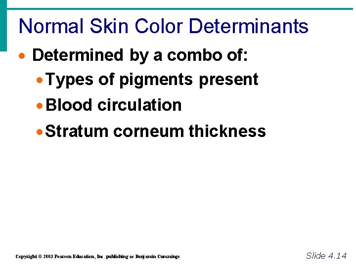
Normal Skin Color Determinants · Melanin · Yellow, brown or black pigments produced in melanocytes found in stratum basale – transferred to keratinocytes · Local accumulations form freckles & pigmented moles · Amount of melanin produced depends upon genetic and exposure to sunlight · Solar elastosis – clumping of elastin fibers = leathery looking skin Slide 4. 14 Copyright © 2003 Pearson Education, Inc. publishing as Benjamin Cummings
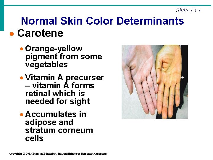
Slide 4. 14 Normal Skin Color Determinants · Carotene · Orange-yellow pigment from some vegetables · Vitamin A precurser – vitamin A forms retinal which is needed for sight · Accumulates in adipose and stratum corneum cells Copyright © 2003 Pearson Education, Inc. publishing as Benjamin Cummings
Normal Skin Color Determinants · Hemoglobin & blood circulation ·Red coloring from oxygenated blood cells in dermis capillaries ·Oxygen content determines the extent of red coloring ·More obvious in fair Copyright © 2003 Pearson Education, Inc. publishing as Benjamin Cummings Slide 4. 14
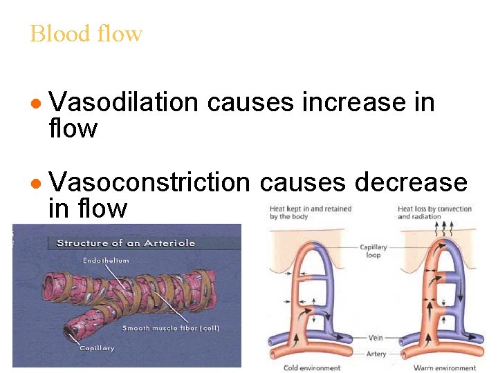
Blood flow · Vasodilation causes increase in flow · Vasoconstriction causes decrease in flow
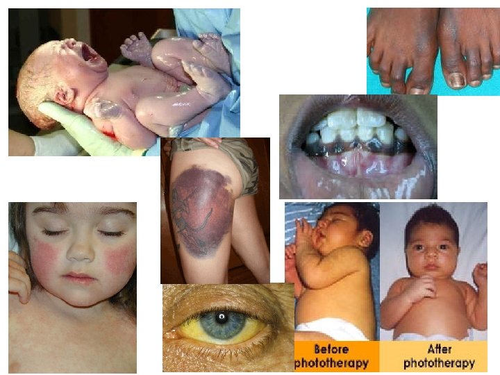
Skin as a Diagnostic · Skin color is influenced by emotional & disease states: You should know the states that cause these. · Cyanosis – bluish color - lack of oxygen · Erythema – redness – heat, inflammation, fever · Add this -Pallor – paleness – lack of blood flow · Jaundice – yellowish color – liver damage · Bronzing – bronze (tan) – Addison’s disease · Hematomas – black & blue – blood under skin Copyright © 2003 Pearson Education, Inc. publishing as Benjamin Cummings Slide 4. 14
Skin Derivatives
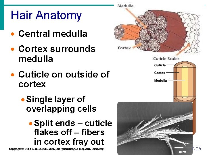
Hair Anatomy · Central medulla · Cortex surrounds medulla · Cuticle on outside of cortex · Single layer of overlapping cells · Split ends – cuticle flakes off – fibers in cortex fray out Copyright © 2003 Pearson Education, Inc. publishing as Benjamin Cummings Figure 4. 7 b Slide 4. 19
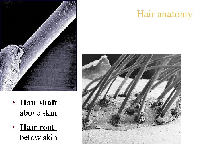
Hair anatomy • Hair shaft – above skin • Hair root – below skin
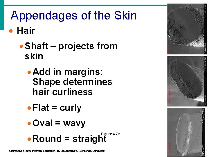
Appendages of the Skin · Hair · Shaft – projects from skin · Add in margins: Shape determines hair curliness · Flat = curly · Oval = wavy Figure 4. 7 c · Round = straight Copyright © 2003 Pearson Education, Inc. publishing as Benjamin Cummings Slide 4. 18
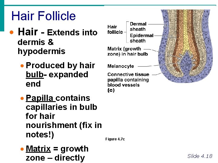
Hair Follicle · Hair - Extends into dermis & hypodermis · Produced by hair bulb- expanded end · Papilla contains capillaries in bulb for hair nourishment (fix in notes!) · Matrix = growth zone – directly Figure 4. 7 c Slide 4. 18
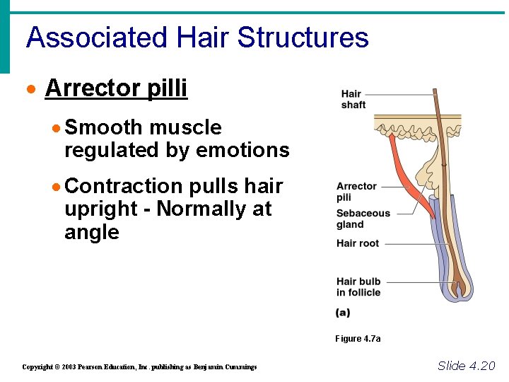
Associated Hair Structures · Arrector pilli · Smooth muscle regulated by emotions · Contraction pulls hair upright - Normally at angle Figure 4. 7 a Copyright © 2003 Pearson Education, Inc. publishing as Benjamin Cummings Slide 4. 20
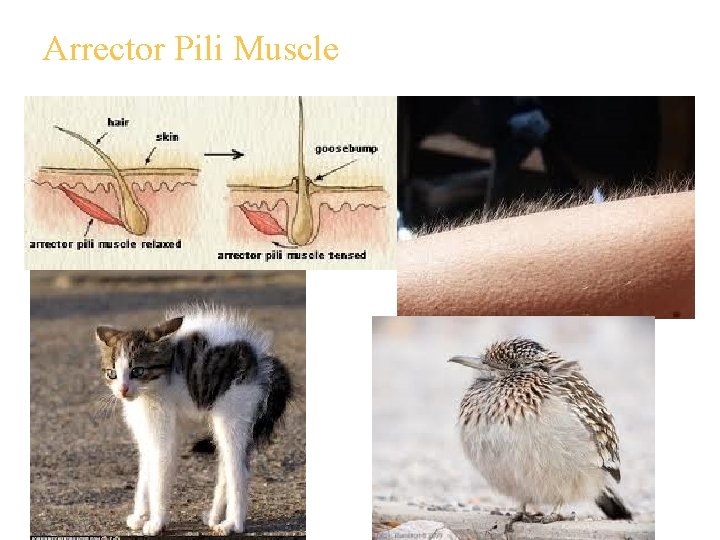
Arrector Pili Muscle
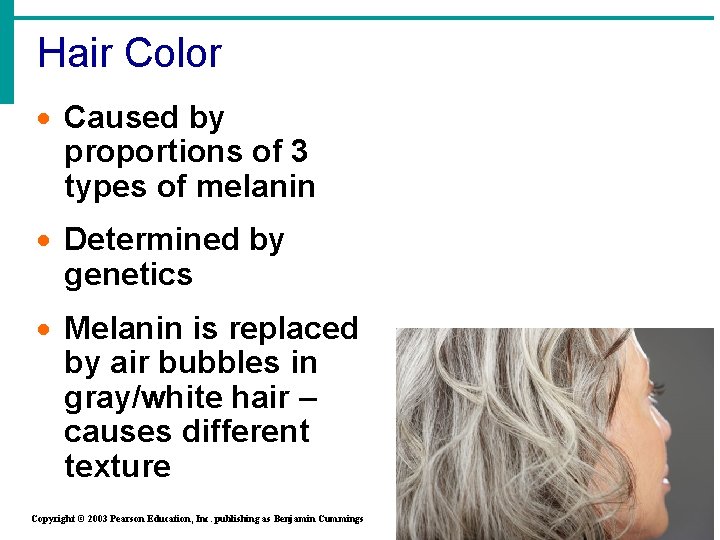
Hair Color · Caused by proportions of 3 types of melanin · Determined by genetics · Melanin is replaced by air bubbles in gray/white hair – causes different texture Copyright © 2003 Pearson Education, Inc. publishing as Benjamin Cummings Figure 4. 7 b Slide 4. 19
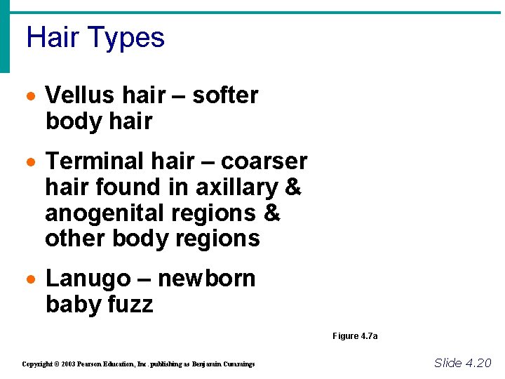
Hair Types · Vellus hair – softer body hair · Terminal hair – coarser hair found in axillary & anogenital regions & other body regions · Lanugo – newborn baby fuzz Figure 4. 7 a Copyright © 2003 Pearson Education, Inc. publishing as Benjamin Cummings Slide 4. 20
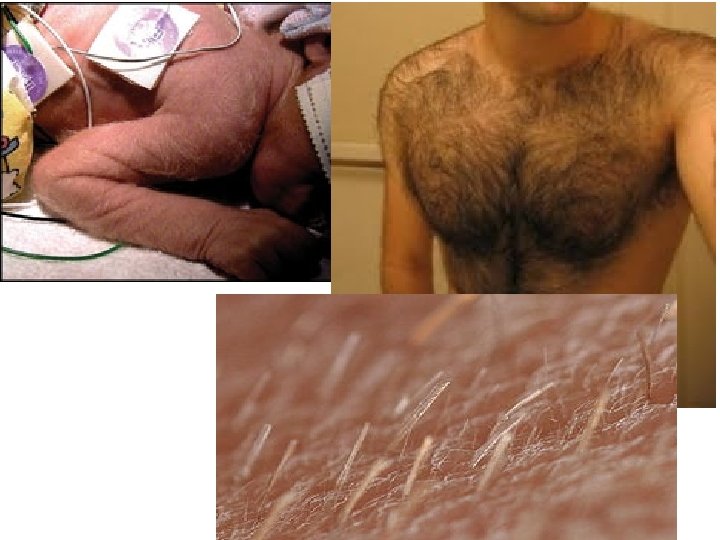
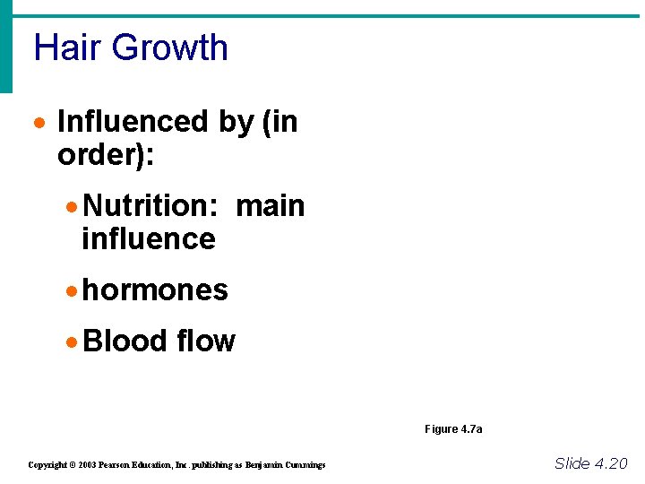
Hair Growth · Influenced by (in order): · Nutrition: main influence · hormones · Blood flow Figure 4. 7 a Copyright © 2003 Pearson Education, Inc. publishing as Benjamin Cummings Slide 4. 20
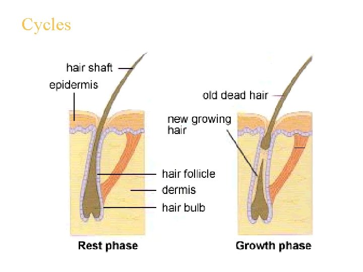
Cycles
Thought Question: know the answers to these questions!! • Why is the hair on your head longer than the hair in your eyebrows? • Why does some hair fall out every day? • Approx 100 hairs per day are lost from your head! That is why your sink is stopped up!
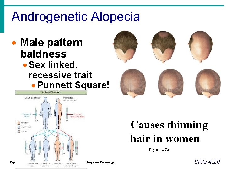
Androgenetic Alopecia · Male pattern baldness · Sex linked, recessive trait · Punnett Square! Causes thinning hair in women Figure 4. 7 a Copyright © 2003 Pearson Education, Inc. publishing as Benjamin Cummings Slide 4. 20
Androgenetic Alopecia q Increased activity in androgen receptors causes hair loss & increases other health risks such as coronary heart disease and prostate cancer in men and polycystic ovary syndrome in women q. Terminal hair replaced by vellus hair -progresses posteriorly q. Treatment: drugs that inhibit testosterone production (Rogaine, Propecia)
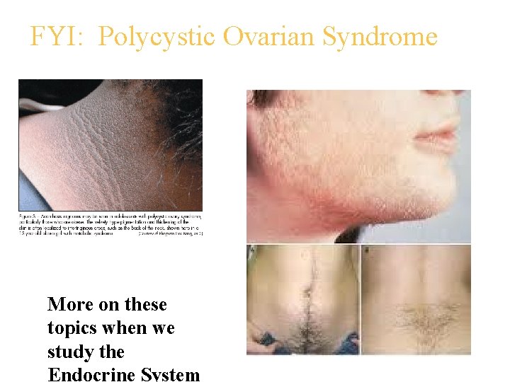
FYI: Polycystic Ovarian Syndrome More on these topics when we study the Endocrine System
Imbalance: FYI • Thinning hair can be caused by an abundance of factors. • Heterozygous traits • Nutrition • Medications • Stress (affects nutrition & hormones) • Hormones • Physical factors
Appendages of the Skin · Nails: Just know diagram & these imp. facts – add to diagram: · Scale-like modifications of the epidermis · Heavily keratinized · Stratum basale extends beneath the nail bed to form nail matrix · Which is responsible for growth · Lack of pigment makes them colorless Copyright © 2003 Pearson Education, Inc. publishing as Benjamin Cummings Slide 4. 21
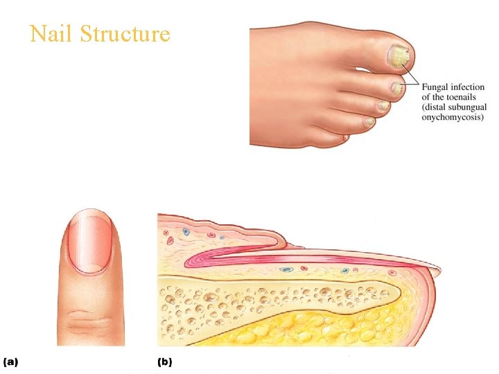
Nail Structure
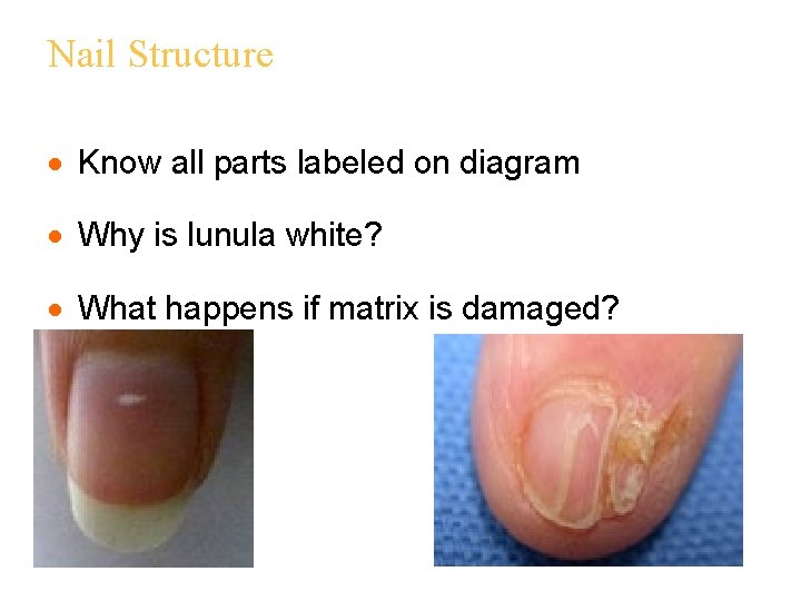
Nail Structure · Know all parts labeled on diagram · Why is lunula white? · What happens if matrix is damaged?
Cutaneous Glands • Are ALL Exocrine Glands they release secretions to the surface using DUCTS. Reside in the Dermis. (Made in the Stratum Basale) • These fall into 2 groups: • 1. Sebaceous Glands • 2. Sweat Glands
Appendages of the Skin · 1. Sebaceous glands found all over skin except palms and soles of feet. · Produce oil called Sebum. · Lubricant for skin · This sebum also kills bacteria · Most with ducts that empty into hair follicles · Glands are activated at puberty · If these glands are blocked causes acne if material oxidizes and dries, get blackheads. Copyright © 2003 Pearson Education, Inc. publishing as Benjamin Cummings Slide 4. 15
Sweat Glands · Sudoriferous (sweat) glands · Three types · Eccrine glands · Widely distributed in skin: abundant on palms, soles, forehead · Open via duct to pore on skin surface · Sweat composition: mostly water with a slightly acidic 4 -6 p. H · Function: thermoregulation Copyright © 2003 Pearson Education, Inc. publishing as Benjamin Cummings Slide 4. 16
Sweat Glands • Apocrine glands (also sudoriferous) · Ducts empty into hair follicles · Found mainly in anogenital & axillary region · Begin to function at puberty due to hormones · Organic contents: Fatty acids and proteins – can have a yellowish color that stains clothes · Odor is from associated bacteria Copyright © 2003 Pearson Education, Inc. publishing as Benjamin Cummings Slide 4. 17
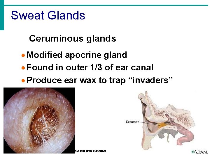
Sweat Glands Ceruminous glands · Modified apocrine gland · Found in outer 1/3 of ear canal · Produce ear wax to trap “invaders” Copyright © 2003 Pearson Education, Inc. publishing as Benjamin Cummings Slide 4. 17
Appendages of the Skin · Sebaceous glands (all over except palms and soles of feet) · Produce oil for waterproofing · Lubricant for skin & kills bacteria · Most with ducts that empty into hair follicles · Glands are activated at puberty: stimulated by hormones Copyright © 2003 Pearson Education, Inc. publishing as Benjamin Cummings Slide 4. 15
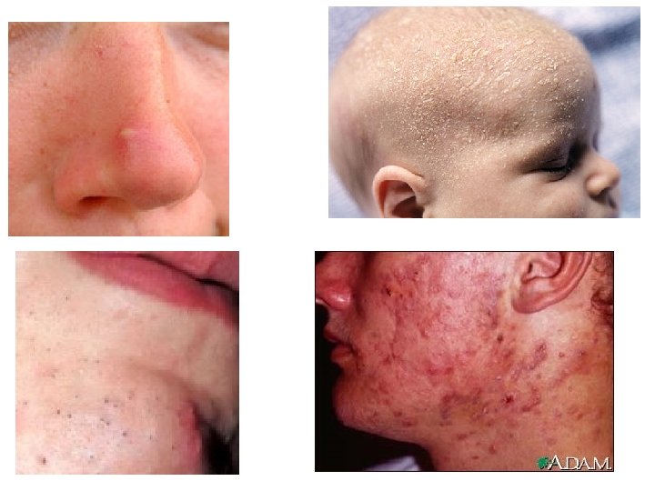
Imbalances • Upper right hand box in notes; • Acne – active infection of sebaceous glands • Whitehead - Sebaceous gland duct blocked by sebum • Blackhead – sebum oxidizes, dries, and darkens • Seborrhea – “cradle cap” – Over activity of sebaceous glands in infants
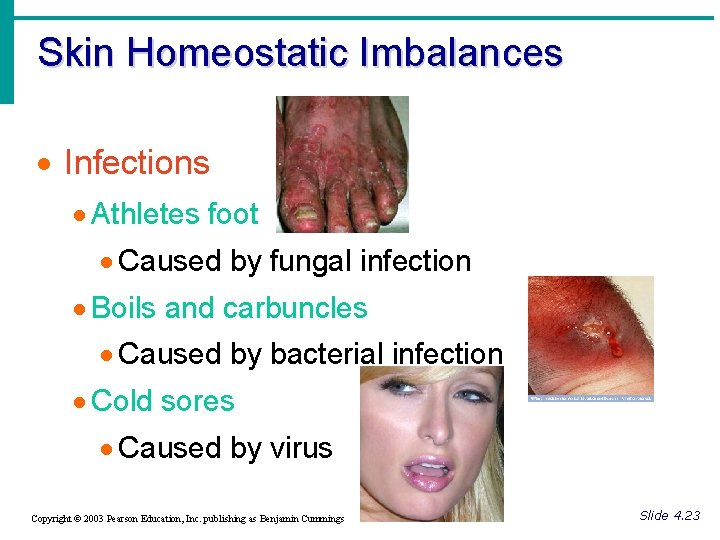
Skin Homeostatic Imbalances · Infections · Athletes foot · Caused by fungal infection · Boils and carbuncles · Caused by bacterial infection · Cold sores · Caused by virus Copyright © 2003 Pearson Education, Inc. publishing as Benjamin Cummings Slide 4. 23
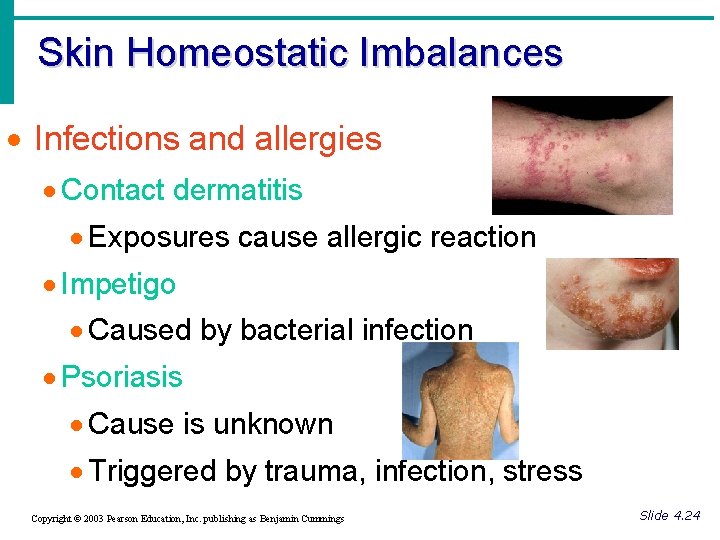
Skin Homeostatic Imbalances · Infections and allergies · Contact dermatitis · Exposures cause allergic reaction · Impetigo · Caused by bacterial infection · Psoriasis · Cause is unknown · Triggered by trauma, infection, stress Copyright © 2003 Pearson Education, Inc. publishing as Benjamin Cummings Slide 4. 24

Skin Homeostatic Imbalances · Burns · Tissue damage and cell death caused by heat, electricity, UV radiation, or chemicals · Associated dangers · Dehydration · Electrolyte imbalance · Circulatory shock Copyright © 2003 Pearson Education, Inc. publishing as Benjamin Cummings Slide 4. 25
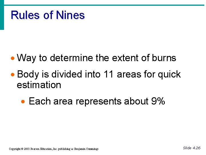
Rules of Nines · Way to determine the extent of burns · Body is divided into 11 areas for quick estimation · Each area represents about 9% Copyright © 2003 Pearson Education, Inc. publishing as Benjamin Cummings Slide 4. 26
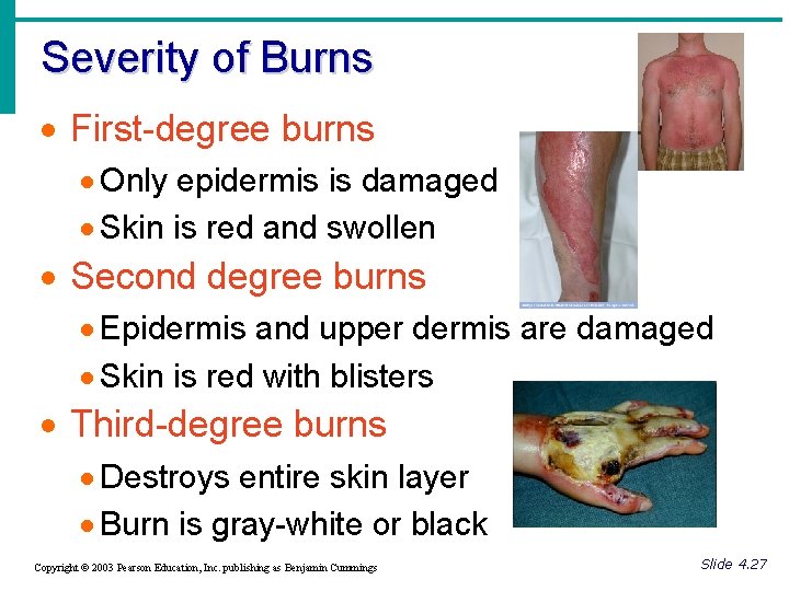
Severity of Burns · First-degree burns · Only epidermis is damaged · Skin is red and swollen · Second degree burns · Epidermis and upper dermis are damaged · Skin is red with blisters · Third-degree burns · Destroys entire skin layer · Burn is gray-white or black Copyright © 2003 Pearson Education, Inc. publishing as Benjamin Cummings Slide 4. 27
Critical Burns · Burns are considered critical if: · Over 25% of body has second degree burns · Over 10% of the body has third degree burns · There are third degree burns of the face, hands, or feet Copyright © 2003 Pearson Education, Inc. publishing as Benjamin Cummings Slide 4. 28
Skin Cancer · Cancer – abnormal cell mass · Two types · 1. Benign · Does not spread (encapsulated) · 2. Malignant · Metastasized (moves) to other parts of the body · Skin cancer is the most common type of cancer Copyright © 2003 Pearson Education, Inc. publishing as Benjamin Cummings Slide 4. 29
Skin Cancer Types · Basal cell carcinoma · Least malignant · Most common type · Arises from statum basale · Squamous cell carcinoma · Arises from stratum spinosum · Metastasizes to lymph nodes · Early removal allows a good chance of cure Copyright © 2003 Pearson Education, Inc. publishing as Benjamin Cummings Slide 4. 30
Skin Cancer Types · Malignant melanoma · Most deadly of skin cancers · Cancer of melanocytes · Metastasizes rapidly to lymph and blood vessels · Detection uses ABCD rule Copyright © 2003 Pearson Education, Inc. publishing as Benjamin Cummings Slide 4. 31
ABCD Rule · A = Asymmetry · Two sides of pigmented mole do not match · B = Border irregularity · Borders of mole are not smooth · C = Color · Different colors in pigmented area · D = Diameter · Spot is larger then 6 mm in diameter Copyright © 2003 Pearson Education, Inc. publishing as Benjamin Cummings Slide 4. 32
- Slides: 97