Blood vessels The pulp organ is extensively vascularized

Blood vessels: The pulp organ is extensively vascularized. It • is known that the blood vessels of both the pulp and the periodontium arise from the inferior or superior alveolar artery and also drain by the same veins in both the mandibular and maxillary regions. The communication of the vessels of the pulp with the periodontium, in addition to the apical connections, is further enhanced by connections through the accessory canals.

These relationships are of considerable clinical • significance in the event of a potential pathologic condition in either the periodontium or the pulp, because the infection has a potential to spread through the accessory and apical canals. Along their course they give off numerous branches in the radicular pulp that pass peripherally to form a plexus in the odontogenic region. Pulpal blood flow is more rapid than in most areas of the body and pulpal pressure is among the highest of body tissues.

Vascular Supply to the Pulp:

The endothelial cells of these vessels contain • numerous micropinocytotic vesicles, which function in transendothelial fluid movement. Occasionally a fibroblast or pericyte lies on the surface of these vessels. Pericytes are capillaryassociated fibroblasts. They are present partially encircling the • capillaries. They have contractile properties and they are • capable of reducing the size of the capillary lumen.

Both venous-venous anastomosis and arteriole-venous • anastomosis occur in the pulp. The arteriole-venous shunts may have an important role in regulation of pulpal blood flow. Frequently arteriole or precapillary loops with capillaries are found under- lying the odontogenic zone in the coronal pulp. A few peripheral capillaries found among the odonto- • blasts have fenestrations in the endothelial cells. These pores are located in the thin part of the capillary wall and are spanned only by the thin diaphragm of contacting inner and outer plasma membranes of endothelial cells.
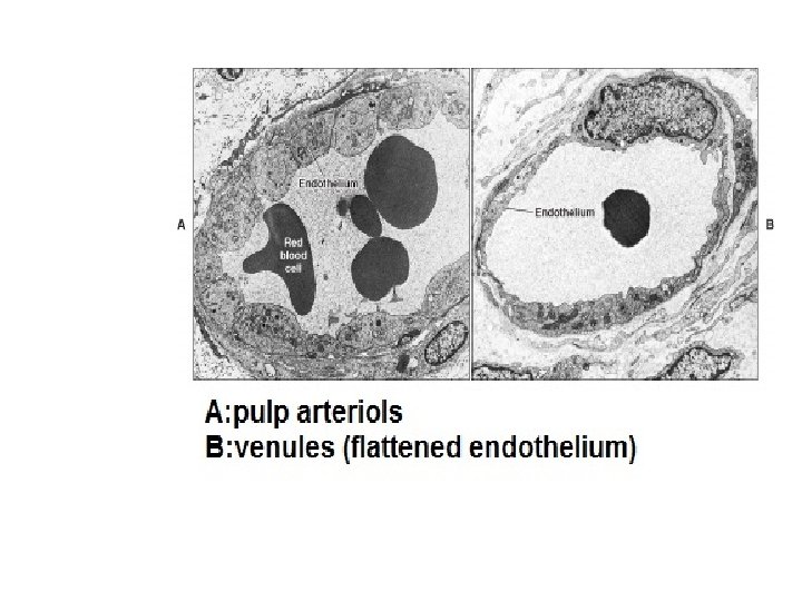

These fenestrated capillaries are assumed to be • involved in rapid transport of metabolites at a time when the odontoblasts are active in the process of dentinal matrix formation and its subsequent calcification. Both fenestrated and continuous terminal capillaries are found in the odontogenic region. During active dentinogenesis capillaries appear among • the odontoblasts adjacent to the predentin Later, after the teeth have reached occlusion and dentinogenesis slows down, these vessels usually retreat to a subodontoblastic position.

Lymph vessels: Lymph capillaries are described as • endothelium-lined tubes that join thin-walled lymph venules or veins in the central pulp. The lymphatic capillaries have thin walls. The lymphatic vessels were more numerous in the central part of the pulp than in the peripheral areas.

In inflamed pulps, due to increased • interstitial fluid pressure, gap junction develops between the endothelial cells of the dilated lymph capillaries. Lymph vessels draining the pulp and • periodontal ligament have a common outlet. Those draining the anterior teeth pass to • the submental lymph nodes; those of the posterior teeth pass to the

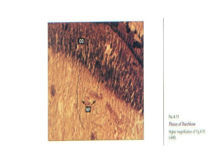
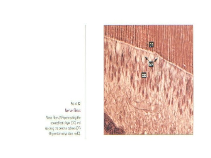
Nerves The abundant nerve supply in the pulp follows • the distribution of the blood vessels. The majority of the nerves that enter the pulp are nonmyelinated are sympathetic in nature. They have terminals on the muscle cells of the larger vessels and function in vasoconstriction. Thick nerve bundles enter the apical foramen • and pass along the radicular pulp to the coronal pulp where their fibers separate and


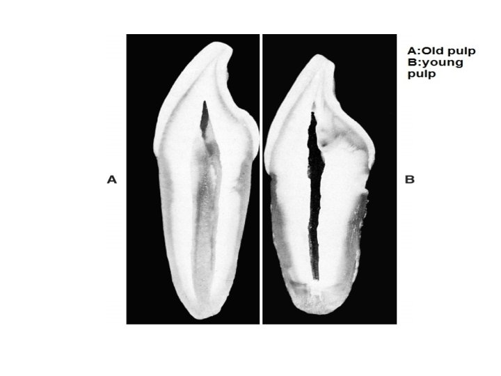
Age-Related and Pathologic Changes in the Pulp • • Specific changes occur in dental pulp with age. Cell death results in a decreased number of cells. The surviving fibroblasts respond by producing more fibrous matrix (increased type I over type II collagen) but less ground substance that contains less water. So with age the pulp becomes: a) less cellular b) more fibrous c) overall reduction in volume due to the continued deposition of dentin (secondary/reactive)

Stages in Pulp Aging • Some attrition of the pulp as the result of normal aging as well as trauma from wearing of the enamel at the cusp ; the pulp horn is not as well defined due to responsive ingrowth of secondary dentin below the worn cusp. • • The pulp horn continues to be reduced in response to increased wearing of the overlying enamel. An overall reduction in pulp cavity dimensions through the continued deposition of normal secondary dentin has occurred. Histology of the pulp reveals a decreased cellularity coupled with increased fibrosis. Cementum deposition continues and the apical foramen subsequently has undergone a reduction in diameter. •

• Aging decreases the ability of dental pulp to respond to injury and repair itself. The fact that the pulp is surrounded by mineralized dentin makes relatively minor pathologic events like inflammation, that cause swelling elsewhere, lead to a compression of the pulp leading to intense pain. This generally results in the death of the pulp.
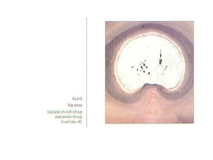
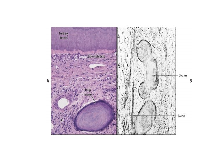
 • Small calcified bodies are present in")
Calcified Bodies in the Pulp (Pulp Stones) • Small calcified bodies are present in up to 50% of the pulp of newly erupted teeth and in over 90% of older teeth. These calcified bodies are generally found loose within the pulp but may eventually grow large enough to encroach on adjacent dentin and become attached. These bodies are classified by either their development or histology

Causes : • 1 -Epithelio-Mesenchymal Interactions. Small groups of epithelial cells become isolated from the epithelial root sheath during development and end up in the dental papilla. Here they interact with mesenchymal cells resulting in their differentiation into odontoblasts. They form small dentinal structures within the pulp

2 -Calcific Degenerations. • Spontaneous calcification of pulp components (collagen fibers, ground substance, cell debris, etc. ) may expand or induce pulpal cells into osteoblasts. These cells then produce concentric layers of calcifying matrix on the surface of the mass - but no cells become entrapped. • Diffuse Calcification. A variation of the above whereby seriously degenerated pulp undergoes calcification in a number of locations. These bodies resemble calcific degenerations except for their smaller size and increased number.

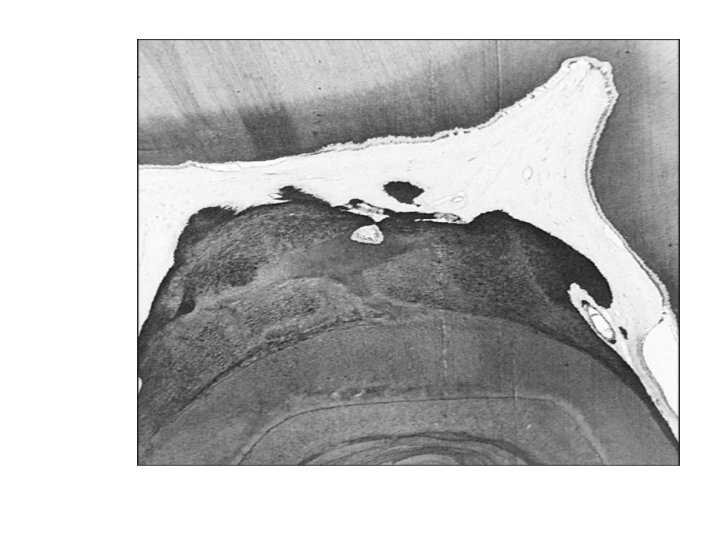
• The pulp cavity in old age. Continued wearing of the enamel on the cusp has resulted in the formation of dead tracts of dentin. It has also stimulated the formation of reactive secondary dentin that has obliterated the pulp horn and now grows into the coronal pulp cavity. • • Aging decreases the ability of dental pulp to respond to injury and repair itself. The fact that the pulp is surrounded by mineralized dentin makes relatively minor pathologic events like inflammation, that cause swelling elsewhere, lead to a compression of the pulp leading to intense pain. This generally results in the death of the pulp.
 • Small calcified bodies are present in")
Calcified Bodies in the Pulp (Pulp Stones) • Small calcified bodies are present in up to 50% of the pulp of newly erupted teeth and in over 90% of older teeth. These calcified bodies are generally found loose within the pulp but may eventually grow large enough to encroach on adjacent dentin and become attached. These bodies are classified by either their development or histology.

Causes : • 1 -Epithelio-Mesenchymal Interactions. Small groups of epithelial cells become isolated from the epithelial root sheath during development and end up in the dental papilla. Here they interact with mesenchymal cells resulting in their differentiation into odontoblasts. They form small dentinal structures within the pulp. • 2 -Calcific Degenerations. Spontaneous calcification of pulp components (collagen fibers, ground substance, cell debris, etc. ) may

Diffuse Calcification. • A subtype of pulp stone whereby seriously degenerated pulp undergoes calcification in a number of locations. These bodies resemble calcific degenerations except for their smaller size and increased number. • •

2. Histology • Calcified bodies in the pulp may be composed of dentin, irregularly calcified tissue, or both. A calcified body containing tubular dentin is referred to as a "true" pulp stone or denticle. • True pulp stones exhibit radiating striations reminiscent of dentinal tubules. Usually those bodies formed by an epithelio- mesenchymal interaction, are true pulp stones. •
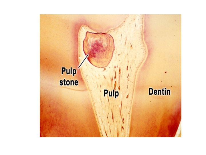

Irregularly calcified tissue generally does not bear much resemblance to any known tissue and as such is referred to as a "false" pulp stone or denticle. False pulp stones generally exhibit either a hyaline-like homogeneous morphology or appear to be composed of concentric lamellae.
- Slides: 31