Blood vessels of the head and the neck

Blood vessels of the head and the neck ED II. Additional images to the lecture Dr. Ágota Ádám

External carotid artery Maxillary a. !!!

Lingual artery 2 nd branch of ECA – at the level of lesser horn of the hyoid bone Deep lingual a. Sublingual a. Medial lingual sulcus
 and Béclard Borders")
How to locate the lingual artery? The triangles of Pirogov (Pirogoff) and Béclard Borders of the Pirogov trigone: -Intermediate tendon of digastric m. Fig. 1 Schematic drawing of - post. Border of left side Beclard’s, Lesser’s, and mylohyoid m. Pirogoff’s triangles -N. XII. 54 Surg Radiol Anat (2011) 33: 53– 57 Borders of the Béclard 123 trigone: -Post. border of hyoglossus m. - post. belly of digastric m. -Greater horn of hyoid bone R. Shane Tubbs • Mark Rasmussen • Marios Loukas • Mohammadali M. Shoja • Aaron A. Cohen-Gadol, Surg Radiol Anat (2011) 33: 53– 57 DOI 10. 1007/s 00276 -010 -0697 -2

Facial artery 3 rd branch of ECA - submental a. - inf. and sup. labial branches - lat. nasal a. - angular a. (end branch)

Ascending pharyngeal artery Originates between ECA and ICA - Pharyngeal branches → wall of the pharynx - Inferior tympanic a. → tympanic canalicule → tympanic cavity - Post meningeal a. →jugular foramen → dura
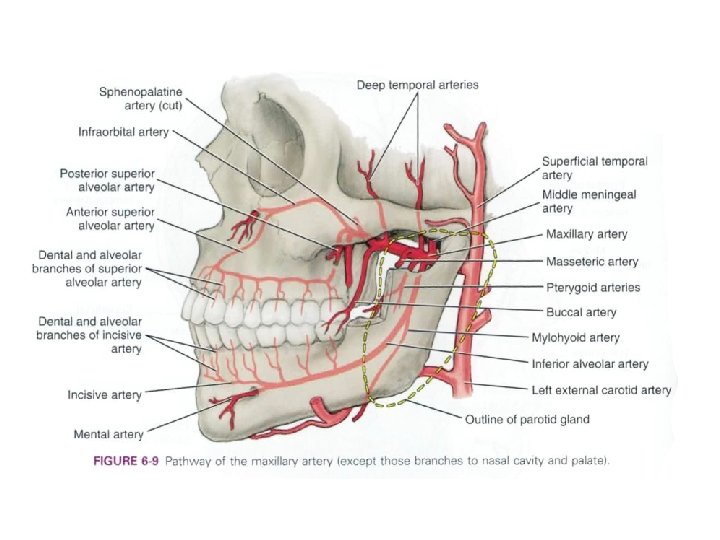

Maxillary artery

Carotid sinus and carotid body The common carotid artery has two specialized organs near its bifurcation: the carotid sinus and the carotid body ⇒ information concerning pressure and chemical composition of the arterial blood Innervation: glossopharyngeal nerve, with small contributions from the cervical sympathetic trunk and the vagus nerve. carotid sinus: baroreceptor; usually appears as a dilation of the lower end of the internal carotid body: chemoreceptor; a reddish-brown, oval structure, 5– 7 mm in height and 2. 5– 4 mm in width. It lies either posterior to the carotid bifurcation or between its branches, and is attached to, or sometimes partly embedded in, their adventitia

Blood supply of the gingiva From lingual and maxillary arteries Upper gingiva (around the maxillary cheek): - The buccal gingivae around the maxillary cheek teeth are supplied by gingival and perforating branches from the posterior superior alveolar artery and by the buccal branch of the maxillary artery. - The labial gingivae of anterior teeth are supplied by labial branches of the infraorbital artery and by perforating branches of the anterior superior alveolar artery. -The palatal gingivae are supplied primarily by branches of the greater palatine artery. Lower gingiva (around the mandibular cheek): - The buccal gingivae associated with the mandibular cheek teeth are supplied by the buccal branch of the maxillary artery and by perforating branches from the inferior alveolar artery. -The labial gingivae around the anterior teeth are supplied by the mental artery and by perforating branches of the incisive artery. -The lingual gingivae are supplied by perforating branches from the inferior alveolar artery and by its lingual branch, and by the main lingual artery, a branch of the external carotid artery.

Blood supply of the palate principally from the greater palatine artery The greater palatine artery descends with its accompanying nerve in the palatine canal, where it gives off two or three lesser palatine arteries, which are transmitted through the lesser palatine canals and foramina to supply the soft palate and tonsil, and anastomose with the ascending palatine branch of the facial artery. The greater palatine artery emerges on to the oral surface of the palate at the greater palatine foramen adjacent to the second maxillary molar and runs in a curved groove near the alveolar border of the hard palate to the incisive canal. It ascends this canal and anastomoses with septal branches of the nasopalatine artery to supply the gingivae, palatine glands and mucous membrane.

Veins of the neck

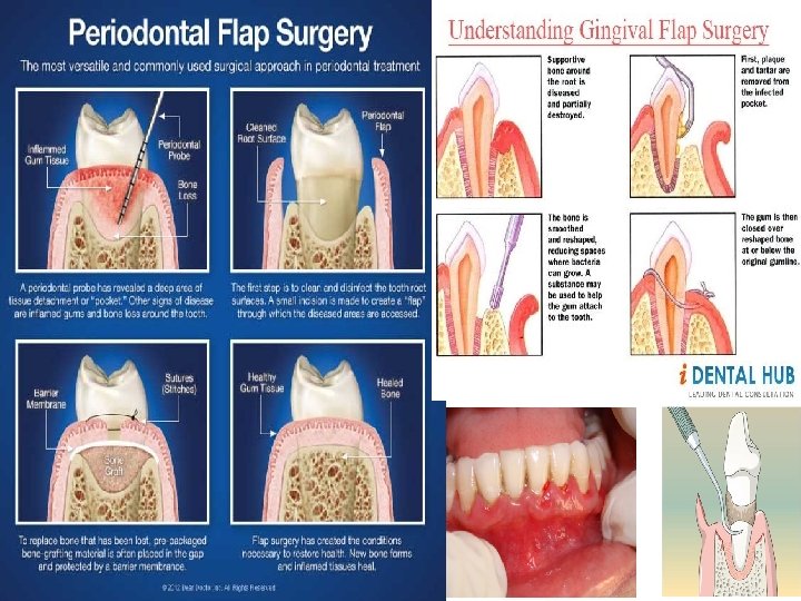
Clinical relevances in dentistry

Anastomosis between the greater palatine artery and nasopalatine arteries Vascular corrosion casting with rasin; by Dr. Arvin Shahbazi

Latex milk injection; by Dr. Arvin Shahbazi

Common complications after local anaesthesia Hematoma after dental anaesthesia A severe hematoma on the anterior floor of the mouth after implant placement in the anterior mandible. (from: "Implant Dentistry - A Rapidly Evolving Practice", book edited by Ilser Turkyilmaz)
 Ultrasound examination of the carotid artery")
Other clinical relevances (general medicine) Ultrasound examination of the carotid artery

Pterion: is the region where the frontal, parietal, temporal, and sphenoid bones join together weakest part of the skull The anterior division of the middle meningeal artery runs underneath the pterion ⇒ a traumatic blow to the pterion may rupture the middle meningeal artery causing an epidural haematoma

Thank you for you attention!
- Slides: 20