Blood Vessels Arteries Veins and Capillaries Cycles Blood

Blood Vessels: Arteries, Veins and Capillaries

Cycles • Blood vessels are organized into three primary cycles 1. Cardiac Circulation: route taken by blood within the heart. 2. Pulmonary Circulation: pathway of the blood from the heart to the lungs and back. 3. Systemic Circulation: pathway of blood from the heart to the rest of the body, includes all blood vessels other than those associated with the lungs.

Arteries • Carry oxygen-rich* blood AWAY from the heart. • Able to stretch and recoil • Thick-walled, with three layers: • Outer: connective tissue (tissue between organs) • Middle: muscle and elastic connective tissue • Inner: connective tissue *Exception: Pulmonary Arteries carry oxygen-poor blood

Arterioles • Smaller arteries • Blood flows from large arteries into arterioles • Middle layer: elastic fibers and smooth muscle

Capillaries • Very narrow blood vessels. • Blood flows into capillaries from arterioles. • Regulated by sphincters • Sphincters only open when new blood needed. – e. g. open in brain all the time, not always in muscle

Capillaries • Single layer of cells, no muscle – Easily ruptured, causes bruising • Site of GAS and FLUID EXCHANGE between blood and body cells (lose O 2, pick up CO 2)

Gas Exchange

Venules • Capillaries merge to form small veins which carry the oxygen-poor blood • Have a thin muscle layer • Venules merge to form veins

Veins • Return oxygen-poor* blood TO the heart • Lack the ability to contract. • Low blood pressure. – Far away from heart. – Loss of fluids to tissues in the capillaries. • Veins can prevent blood from flowing backward: – One-way (uni-directional) valves – Skeletal muscle of the surrounding area helps push blood through veins *Exception: Pulmonary Veins carry oxygen-rich blood

Arteries vs. Veins

Blood

Plasma • Fluid portion of the blood that carries blood cells. • Made up of 90% water, the other 10% made up of blood proteins, glucose, vitamins, minerals, dissolved gases, waste products of cell metabolism. • Also transports CO 2.

Red Blood Cells • Erythrocytes • Make up 44% of blood. • Specialized for transport of O 2. Without them plasma could only carry 2% of the oxygen that normally travels through our bodies. • Shape: biconcave disk to increase surface area. • No nucleus, lifespan of 120 days, constantly reproduced. • Males ~ 5. 5 billion RBC/m. L blood; Females ~ 4. 5 billion.

Red Blood Cells • Packed with 280 million molecules of hemoglobin, an ironcontaining molecule that binds with oxygen. • Hemoglobin has 4 globular protein molecules (globin) and 1 iron molecule (protein) – High affinity for oxygen – Hemoglobin + oxygen = oxy-hemoglobin • RBC lose their nucleus when they enter the blood stream in order to carry more hemoglobin.

White Blood Cells • • Make up about 1% of blood's volume. Produced in bone marrow. White blood cells contain nuclei and appear colourless. They play many roles in fighting off infection and protecting the body from pathogens. – The number of WBC may increase by double when you are fighting off an infection. – Pus: fragments of remaining protein of the WBC and the invader.

Leukocytes and Lymphocytes • Two of the most important disease-fighting white blood cells are leukocytes and lymphocytes. • Leukocytes (macrophages) engulf and digest pathogens. – Innate immune response (generalized response of the body to infection). – Can pass through the wall of the capillaries.
. – Recognize")
Leukocytes and Lymphocytes • Lymphocytes – Acquired immune response (specific immune response). – Recognize and remember specific pathogens and fend them off if they attack again.

Platelets • Are not cells. • Fragments of larger cells that broke apart in the bone marrow. • They contain no nucleus and break down relatively quickly. • They help the blood to clot and protect the body from excessive blood loss after an injury.

Blood Pressure • Force of the blood on the walls of the arteries. • Normal BP 120/80 mm Hg; decreases as you move away from the heart. – Stroke Volume: volume of blood leaving heart (L) – Heart Rate: number of beats (contractions) per minute (bpm)
: amount of blood pumped")
Blood Pressure Two factors determine BP: 1. Cardiac Output (CO): amount of blood pumped from the heart each minute = Heart Rate (HR) x Stroke Volume (SV) – ⇡ CO = ⇡ BP – increase CO by ⇡HR or ⇡ Stroke Volume (stronger heart) 2. Arteriolar resistance: diameter of the arteriole determines the amount of blood flow – ⇡ diameter = ⇣ BP

Blood Pressure Regulation • Diameter of blood vessels regulated by the medulla oblongata. • Vasoconstriction: nerve impulses cause muscle to contract, reducing diameter of vessel, reduces flow to tissue, increases pressure • Vasodilation: nerve impulses cause muscles to relax, increasing diameter of vessel, increases flow to tissue, decreases pressure
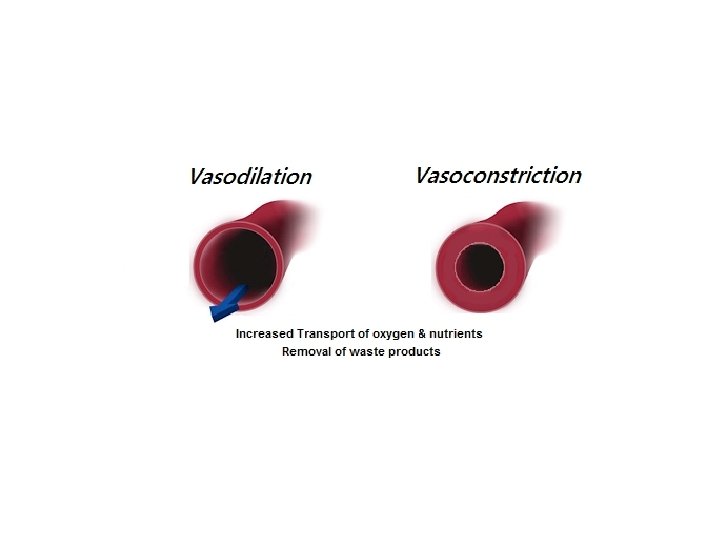

Benefits of Vasodilation • lowering blood pressure • assists in eliminating excess metabolic produced heat • enhances clotting factor & leukocyte entry into damage tissue • increases delivery of oxygen & nutrients during energy consuming activities Benefits of Vasoconstriction • retain heat in cold climates • reduce excessive blood loss

5 Temperature Regulation The external temperature varies during the day and from season to season, sometimes by as much as 40 o. C, but the human body temperature stays at about 37 o. C This is achieved by sweating, vasodilation, vasoconstriction, and shivering If the body temperature rises, the sweat glands in the skin are activated and secrete sweat on to the surface of the skin When the sweat evaporates, it takes heat from the body and cools it down

6 Section through skin The sweat gland extracts sweat from the blood and passes it up the duct to the skin surface where it evaporates 0. 25 mm Sweat gland evaporation sweat pore epidermis sweat duct sweat gland blood vessel

Vasodilation much heat lost If the body temperature rises, the blood vessels in the skin dilate (become wider) and allow more blood to flow near the surface. The heat loss from the blood through the skin helps cool the circulating blood Vasoconstriction If the body temperature falls. The blood vessels in the skin constrict. Less warm blood flows near the surface so less heat is lost Vasoconstriction & dilation little heat lost 7

5 Vasodilators The natural signals for causing vessels to dilate are referred to as vasodilators: • Nerve impulses • Hormones • Nitrates (drugs to treat hypertension, congestive heart failure) • Alcohol

5 Vasoconstrictors • Prostaglandins • Serotonin, epinephrine • Drugs: • Cocaine • Stimulants • Decongestants • Amphetamines • Antihistamines • caffeine
- Slides: 28