Blood the Cardiovascular System Honors Anatomy Physiology 2007

Blood & the Cardiovascular System Honors Anatomy & Physiology 2007

Homeostatic Functions of Blood n Transportation: transports needed nutrients and gases to cells and transports waste products away from cells n Protection: from microscopic organisms through phagocytosis and antibodies; clots to protect against blood loss n Regulation: of acid-base balance by neutralizing levels of CO 2, lactic acid, etc. ; regulates body temp. ; controls volume of blood flow to different areas
: due to oxygenation of the hemoglobin protein")
General Characteristics Red in color (NOT BLUE): due to oxygenation of the hemoglobin protein on RBC’s n Viscous: thicker and stickier than water n Slightly alkaline: p. H = 7. 35 -7. 45 n – Acidosis: too acidic (too many H+) – Alkalosis: too basic (too few H+) n Volume: avg. male = 5 -6 liters avg. female = 4 -5 liters

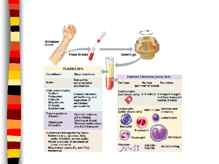
Blood Plasma n Liquid portion of blood n 92% water and 8% dissolved elements – Formed elements, gases, metabolic wastes, nutrients & hormones – Components of plasma vary based on cell’s needs – Adjustments made in order to maintain homeostasis
 n Anucleate cells; few organelles n")
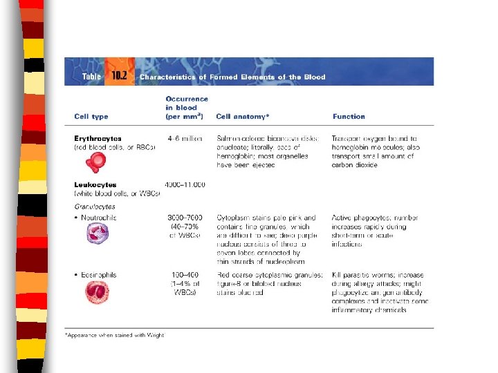
Formed Elements Erythrocytes aka Red Blood Cells (RBCs) n Anucleate cells; few organelles n Hematocrit: % of RBC’s to total blood volume n – 95% of formed elements – 1000 RBC: 1 WBC n Function: to transport O 2 & CO 2 – Hemoglobin: iron containing protein on RBC’s; carries O 2

RBC Life Cycle n Before birth: made in yolk sac, spleen & liver n After birth: made in red bone marrow n Life expectancy: 120 days because of no nucleus or organelles

Regulation of RBC Formation Liver & kidneys monitor counts n Erythropoietin excreted from kidneys when counts are low—regulates hematopoiesis n 10 billions die each HOUR n – Macrophages eat toxins & debris – Iron recycled by red marrow – Hgb: broken down into bilirubin released as bile pigment by liver

Control of Erythrocyte Production
 n Make up less than 1%")
Formed Elements Leukocytes aka White Blood Cells (WBCs) n Make up less than 1% of blood volume n Function: used in body’s defense n – Diapedesis: the ability to exit the blood stream to conduct an immune response – Positive chemotaxis: ability to locate bodily infection by responding to chemical signals

Types of Leukocytes · Granules in their cytoplasm can be stained · Include neutrophils, eosinophils, and basophils

Formed Elements n Thrombocytes aka Platelets n Fragments of complete cells n 1/10 size of RBC; shaped like an oval disc n Function: used in clotting process when blood cells are ruptured
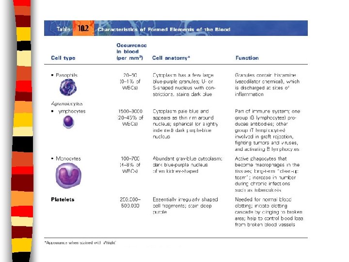

Hemostasis n Mechanism to stop bleeding – Blood vessel spasm: smooth muscle is stimulated to contract to bring broken ends of vessel together; lasts less than 30 minutes – Platelet Plug Formation: platelets become large and sticky and form a PLUG – Coagulation: series of events forming a blood clot

Blood Disorders Thrombus: blood clot in vein or artery n Embolus: free floating thrombus; can be trapped in vessel causing stroke or heart attack n Hemophilia: inability to clot n Anemia: decreased O 2 carrying capacity n – Low RBC’s – Low hemoglobin

Blood Disorders n Sickle Cell Anemia: n Polycythemia: excessive RBC’s sickle shaped RBC’s causing increased that clog vessels viscosity & and impairs blood decreased blood flow n Leukemia: increased numbers of WBC’s; function abnormally

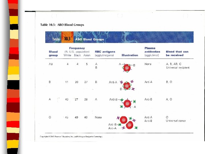
Blood Groups and Transfusions n Antigens: genetically determined proteins; the body recognizes them as “self” or “foreign”; stimulates immune system to release a defense (antibodies) against “foreigners” n Antibodies: recognizes antigens and decides if they are “foreign” or “self”

Blood Groups & Transfusions n Agglutination: clumping of RBC’s when antigen and antibody don’t match; leads to clogging of small vessels n Transfusion Reactions: when blood types don’t match causing: kidney failure, fever, vomiting, chills and nausea
: person having Rh antigens on RBC’s n Rh(-): person")
Rh Blood Type n Rh(+): person having Rh antigens on RBC’s n Rh(-): person without Rh antigens on RBC’s n Rh sensitization: process by which an Rh(-) person develops anti-Rh antibodies due to exposure to Rh(+) blood
 blood from")
Rh Blood Type n Example: pregnant mother with Rhblood; baby with Rh(+) blood from father – First baby: anti-Rh antibodies produced but no effect on baby – Second baby: anti-Rh antibodies enter fetal circulation causing hypoxia and anemia – Disorder called: erythroblastosis fetalis

The Heart n Function: transportation of nutrients and wastes via blood n Lies between the lungs in the thoracic cavity n Approximately the size of a fist n Weighs less than a pound

Anatomy of the Heart n Apex: pointed, inferior aspect of the heart n Base: broad, superior aspect of the heart

Serous Membrane n Pericardium AKA pericardial sac: the parietal peritoneum – Holds and protects the heart; makes pericardial fluid n Epicardium: outer layer of heart wall – Serves to protect the heart – Separated from pericardium by pericardial cavity

Walls and Coverings: 3 layers Epicardium n Myocardium: ring-like arrangements of cardiac muscle n – the part that contracts n Endocardium: white membrane – lines inside of heart chambers & blood vessels – reduces friction between wall and blood flow

Heart Chambers n Two atria – Receiving chambers – Auricles: extensions that allow for increase in volume – Do not aid in pumping; myocardium very thin – Filled with blood from veins and acts to fill ventricles with blood – Separated by interatrial septum – Fossa Ovalis: remains of fetal heart opening
 – Propels blood into")
Heart Chambers n Two Ventricles – Discharging chambers (actual pumps) – Propels blood into circulation – Thicker walls than atria – Left thicker than right – Separated by interventricular septum

Anatomy of the Heart

Internal Anatomy of the Heart
 – Located between atria and")
Heart Valves: Atrioventricular Valves n Atrioventricular Valves (AV valves) – Located between atria and ventricles – Anchored to papillary muscles via chordae tendinae – Tricuspid Valve: lies between R atrium and ventricle; has 3 cusps – Bicuspid Valve (mitral valve): lies between L atrium and ventricle; has 2 cusps

Atrioventricular Valves Continued n Function: permits one-way flow of blood from atria to ventricles ONLY upon atrial contraction n Ventricular contraction forces the AV valve flaps up, causing them to close off the opening to the atria and directing the flow of blood out of the heart

Heart Valves: Semilunar Valves n Semilunar Valves – Located between the ventricles and their major arteries – Made of 3 -half moon shaped cusps each 1. Pulmonary valve: between R ventricle and pulmonary artery 2. Aortic valve: between L ventricle and aorta

Semilunar Valves Continued n Function: to direct one-way flow of blood from the ventricles to the pulmonary trunk and aorta n Ventricular contraction forces SL valves open (& AV valves shut) – All due to pressure increases n Ventricular relaxation causes closure of SL valves, preventing backflow

Heart Valves
 Blood enters the R atrium and exits")
Blood Flow Through The Heart 1. ) Blood enters the R atrium and exits the heart via the L ventricle. In that time it leaves the heart to exchange gas in the lungs. 2. )Trace the path of blood flow through the heart naming all of the chambers, valves and vessels it passes through along the way!

Blood Flow Through the Heart

Supplying Blood to the Heart Coronary Circulation: the flow of blood to the heart itself n The nutrients found in the blood cannot be absorbed from the internal chamber walls in order to nourish the myocardium n R & L Coronary Arteries: found at base of aorta; supplies heart with nourishing blood n Coronary Sinus: “waste” blood brought back to the R atrium n

Circulation n Pulmonary Circulation: the route of blood between the lungs and heart n Systemic Circulation: the route of blood transport between the heart and all organs and tissues (except the lungs)

Vessel Walls n 3 layers in arteries and veins n Tunica Intima: surrounds lumen – Very elastic n Tunica Media: smooth muscle and elastic fibers n Tunica Adventitia: thin layer – Anchors artery to neighboring structures

Circulation n Arteries: transports blood away from the heart – Highly elastic – Lumen becomes progressively smaller as the arteries branch – Blood pressure HIGH n Arterioles: same function as arteries – Thinner walls – Smaller lumens n Capillaries: microscopic vessels where exchange takes place – Walls made of squamos epithelium

Circulation n Venules: returns “wasted” blood to heart – Larger in diameter than capillaries – Increase in size en route to heart – Formed from the union of capillaries n Veins: returns “wasted” blood to heart – Larger lumens than venules – Distensible: walls able to distend/stretch – LOW blood pressure – Blood flow variations • Muscular and Respiratory pumps • One-way valves

Blood Vessels

Blood Vessels

Circulation Irregularities n Varicose Veins: weakened valves allows blood to pool and permanently stretch vein walls n Hemorrhoids: varicosities in veins surrounding anal canal n Murmurs: defective valve – May shunt blood to organs

Heart Physiology: Cardiac Cycle n The cyclic contraction of both atria and ventricles n These events make up 1 heartbeat n Beat rhythmically: atria beat, then ventricles; atria beat, then ventricles ETC. n Blood moves due to pressure changes

Heart Physiology: Cardiac Cycle n Pressure changes: – Systole: state of contraction; high pressure within that specific chamber • Allows blood to exit chamber – Diastole: state of relaxation; low pressure within that specific chamber • Allows blood to enter chamber

Heart Physiology: Heart Sounds n “Lub-Dup, pause, lub-dup, pause, lubdup, pause” n Cause: vibrational sounds of heart valve closures n Lub = closing of AV valves with ventricular systole n Dup = closing of SL valves with ventricular diastole

Heart Physiology: Heart Conduction System n Heart receives impulses from autonomic system, but can beat without nervous stimulation n Cardiac cells INHERENTLY contract n Conduction system: patches of pacesetting cardiac cells
 Node: located in upper wall of")
Heart Physiology: Heart Conduction System n Sinoatrial (SA) Node: located in upper wall of R atrium – aka “Pacemaker” – Initiates each cardiac cycle • Generates electrical impulses causing atrial contraction • Stimulates AV node
 Node: located on the R, inferior")
Heart Physiology: Heart Conduction System n Atrioventricular (AV) Node: located on the R, inferior portion of the interatrial septum – Activated by SA Node – Generates impulses down conducting fibers: AV Bundle (Bundle of His) – AV Bundle follows interventricular septum and sends impulses left and right as it travels

Heart Physiology: Heart Conduction System n AV Node Continued – At apex of heart, AV Bundle forms branches called Purkinje Fibers – Purkinje Fibers pass deep into myocardium and initiate ventricular systole – Contraction occurs upward: a wringing motion from apex towards base of heart

Heart Conduction You Tube Cardiac Conduction System Electrical System of the Heart Animation - MADE EASY
: an instrument that records the electrical changes in")
Electrocardiogram n Electrocardiogram (ECG or EKG): an instrument that records the electrical changes in the myocardium – Flat baseline: myocardium polarized – P wave: SA node fires; atria depolarized – QRS complex: AV node fires; ventricles depolarized – T wave: ventricular repolarization
 n. A measurable value that represents heart activity n Heart Rate")
Cardiac Output (CO) n. A measurable value that represents heart activity n Heart Rate (HR): number of heart beats (cardiac cycles) per minute – Avg. adult 75 beats/min. n Stroke Volume (SV): volume of blood ejected by ventricles per heart beat – Avg. adult 70 ml/min.

Cardiac Output CO = HR X SV Avg. resting adult: 75/min X 70 ml. /min. = 5250 ml. /min -OR 5. 2 liters of blood pumped through the body EACH MINUTE at rest!

Starlings Law of the Heart n How far cardiac cells are stretched (how full the chambers get), the stronger the heart will contract
 n The force exerted by blood against the inner walls of")
Blood Pressure (BP) n The force exerted by blood against the inner walls of vessels n Highest pressure in aorta, lowest in veins n Blood moves from high to low pressure n Read using a sphygmomanometer n Systole/diastole: avg. adult = 120/80
 Cardiac Output: increase in CO = increase in")
Factors Affecting Blood Pressure 1. ) Cardiac Output: increase in CO = increase in BP 2. ) Peripheral Resistance (PR): the friction or drag produces when blood passes over vessel walls • smaller lumens = high PR • greater blood viscosity = high PR • high PR = high BP
 Blood Volume: a measure of the amount of")
Factors Affecting Blood Pressure 3. ) Blood Volume: a measure of the amount of blood plasma & formed elements present in the cardiovascular system • Blood volume drops = BP drops

Blood Pressure
- Slides: 61