Blood supply to the brain The cerebrospinal fluid
 Mark Kozsurek, M. D. ,")
Blood supply to the brain The cerebrospinal fluid (CSF) Mark Kozsurek, M. D. , Ph. D. mark@kozsurek. hu 21/09/2017, EM II.

Extremly high demand for oxygen and nutrients: human brain represents 2% of the body weight, but receives 15% of the cardiac output, 20% of total body oxygen consumption and 25% of total body glucose utilization. Cerebrovascular deseases and stroke are among the major causes of death.

I. Arteries supplying the brain

2 sources of blood: ICA and VA

Vertebro-basilar system atlas laterally axis upward backward C 6 CTA: CT angiography

C 7 C 6 C 5 cavernous sinus carotid canal C 4 C 3 ant. clinoid proc. C 2 foramen lacerum C 1 X-ray angiogram
 C 2")
C 6 C 7 C 5 C 4 cavernous sinus (C 3) C 2

Another classification: Majority of these branches will never be seen and is not necessary to note them!

Circle of Willis

Circle of Willis pituitary stalk optic chiasm oculomotor n. mamillary bodies abducens n.

1. Circle of Willis encloses the optic chiasm, pituitary stalk and mamillary bodies. 2. Oculomotor nerve exits between the post. cerebral and sup. cerebellar arteries. 3. Vertebral arteries of the two sides unite to form the basilar artery at the ponto-medullary junction. The root of the abducens nerve and initial segment of the ant. inf. cerebellar artery can also be found here.

so m ar g in al br . pa rie to su oc lc cip us it al ca llo pericallosal br. A 3 A 2 A 1 ant. communicating

Heubner’s

pa rie to su oc lc cip us it al ACA PCA MCA

oculomotor n. PCA sca BA aica VA pica sca: superior cerebellar aica: anterior inferior cerebellar pica: posterior inferior cerebellar

Clinical considerations • Atherosclerosis – brain infarctions • Subarachnoidal hemorrhage

Ant. cerebral artery Weakness/paralysis of muscles and loss of sensory functions on the lower limbs of the contralateral side. Middle cerebral artery Paralysis and sensory disfunction involving head and neck and the upper limbs of the contralateral side. In case of damage of the dominant hemisphere speech disorders are also present. Post. cerebral artery Visual field defficiencies or blindness. Vertebro-basillar system Eye movement (gaze) disorders, double vision Anisocoria (pupils are different in size) Vertigo, loss of balance Dysphagia and dysphonia (disorder of swallowing and phonation) Drowsiness or unconsciousness

Subarachnoid hemorrhage

Aneurism clipping

Endovascular coiling
 The extracellular fluid of the CNS is separated from the blood")
Blood-brain barrier (BBB) The extracellular fluid of the CNS is separated from the blood by the BBB ensuring strictly controlled and mainly carrier protein assisted transport of macromolecules. Is formed by endothelial cells attached to one other by tight junctions, basement membrane, astrocytic endfeet. Protects the CNS from possibly toxic agents but makes development of medicines acting on the CNS difficult (e. g. antibiotics in infections).

Life outside the BBB: the circumventricular organs „Circumventricular” = around the ventricles Incomplet or missing BBB Highly capillarized structure Secretion of neurohormons or detection of hormons, glucose, ions, etc.
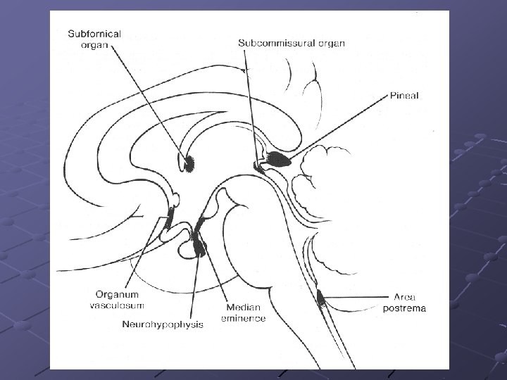

Subfornical organ sensory fluid regulation Organum vasculosum sensory, secretory detects peptides, fluid regulation Median eminence secretory regulates the anterior pituitary through the release of neurohormones Neurohypophysis secretory store and secretes the hormones oxytocin and ADH into the blood, but does not synthesize either hormone Subcommissural organ secretory secretes certain proteins into the cerebrospinal fluid, its specific function is as yet unknown. Pineal gland secretory stimulated by darkness to secrete melatonin and is associated with circadian rhythms Area postrema sensory the vomiting centre of the brain (can detect noxious substances in the blood and stimulate vomiting in order to rid the body of these toxic chemicals)

II. Veins drainig the brain

superior sagittal sinus SUPERFICIAL VEINS TROLARD’S VEIN LABBE’S VEIN cavernous sinus transverse sinus

midd le ce r. sa l great cerebral vein thala m o s triate ral oid chor ba inter nal c ereb deep v. of septum pell. ant. cerebral DEEP VEINS

Almost the total volume of veinous blood collected from the brain leaves the skull through the jugular foramen and the internal jugular vein. If the jugular foramen and/or the internal jugular vein is getting occluded, blood may escape through the diploic and emissary veins connecting the dural sinuses with the veins of the scalp skin.
: form a network between the external")
Diploic veins (frontal, anterior and posterior temporal, occipital): form a network between the external and internal compact bony layers of the skull and connect dural sinuses with the external veins.
: pearce the skull directly and connect")
emissary diploic Emissary veins (occipital, parietal, condylar, mastoid): pearce the skull directly and connect dural sinuses with external veins.
 Provides mechanical protection for the brain and the spinal")
III. The cerebrospinal fluid (CSF) Provides mechanical protection for the brain and the spinal cord. When floating in the CSF brain weights only 50 g (!) according to the Archimedes’ principle.

internal and external CSF spaces internal = ventricles external = subarachnoidal space

Choroid plexus of fourth ventricle Surface of a choroid plexus

post. choroidal from PCA ant. choroidal from ICA or MCA choroidal a. of the 4 th ventricle from pica
 cystern 2 lateral apertures of Luschka")
1 median aperture of Magendi cerebellomedullary (or great) cystern 2 lateral apertures of Luschka lateral pontine (or pontocerebellar) cystern

Site of CSF resorption: arachnoid granulations in the superior sagittal sinus and lateral lacunae.

Hydrocephalus Increased volume and/or pressure of CSF due to accelerated synthesis, blocked circulation or insufficient resorption of liquor.

anterior cerebral middle cerebral posterior cerebral

Thank You !!!
- Slides: 39