BLOOD SMEAR EXAMINATION Making Blood smear I PREPARATION

BLOOD SMEAR EXAMINATION Making Blood smear

I- PREPARATION OF BLOOD SMEAR There are three types of blood smears: 1. The cover glass smear. 2. The wedge smear. 3. The spun smear. The are two additional types of blood smear used for specific purposes 1. Buffy coat smear for WBCs < 1. 0× 109/L 2. Thick blood smears for blood parasites.

WEDGE BLOOD SMEAR Specimen : EDTA blood within 2 to 3 hours & collected to the mark on tube. Not's : May change RBCs morphology such as Spiculated (crenated) cells if : 1. Excessive amount of anticoagulant to specimen 2. Old blood - long standing. 3. Warm environment (room temperature) may hasten changes.

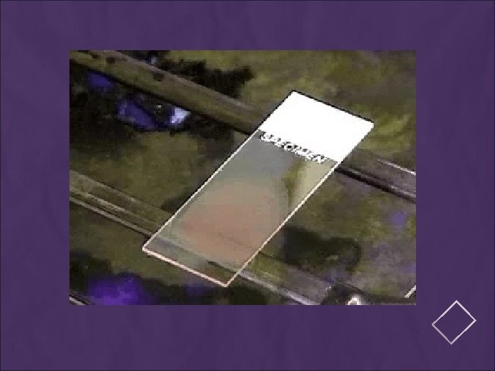
PROCEDURE placing a drop of blood from mixed sample on a clean glass slide. Spreader slide using another clean glass slide at 30 -40 degree angle. Control thickness of the smear by changing the angle of spreader slide Allow the blood film to air-dry completely before staining. (Do not blow to dry. The moisture from your breath will cause RBC artifact. )

STEPS FOR BLOOD FILM

THE THICKNESS OF THE SPREAD Notes: 1. If the hematocrit is increased, the angle of the s preader slide should be decreased. 2. If the hematocrit is decreased, the angle of the spreader slide should be increased.

high HCT small angle low HCT large angle

CHARACTERISTICS OF A GOOD SMEAR 1. Thick at one end, thinning out to a smooth rounded feather edge. 2. Should occupy 2/3 of the total slide area. 3. Should not touch any edge of the slide. 4. Should be margin free, except for point of application. Note: As soon as the drop of blood is placed on the glass slide, the smear should be made without delay. Any delay results in an abnormal distribution of the white blood cells, with many of the large white cells accumulating at the thin edge of the smear.

COMMON CAUSES OF A POOR BLOOD SMEAR 1. Drop of blood too large or too small. 2. Spreader slide pushed across the slide in a jerky manner. 3. Failure to keep the entire edge of the spreader slide against the slide while making the smear. 4. Failure to keep the spreader slide at a 30° angle with the slide. 5. Failure to push the spreader slide completely across the slide. 6. Irregular spread with ridges and long tail: Edge of spreader dirty or chipped; dusty slide 7. Holes in film: Slide contaminated with fat or grease 8. Cellular degenerative changes: delay in fixing, inadequate fixing time or methanol contaminated with water.

EXAMPLES OF UNACCEPTABLE SMEARS A: Blood film with jagged tail made from a spreader with achipped end. B: Film which is too thick C: Film which is too long, too wide, uneven thickness and made on a greasy slide. D: A well-made blood film.

EXAMPLES OF UNACCEPTABLE SMEARS

BIOLOGIC CAUSES OF A POOR SMEAR 1. Cold agglutinin - RBCs will clump together. Warm the blood at 37° C for 5 minutes, and then remake the smear. 2. Lipemia - holes will appear in the smear. There is nothing you can do to correct this. 3. Rouleaux - RBC’s will form into stacks resembling coins. There is nothing you can do to correct this

SLIDE FIXATION & STAINING

II- FIXING THE FILMS To preserve the morphology of the cells, films must be fixed as soon as possible after they have dried. It is important to prevent contact with water before fixation is complete. Methyl alcohol (methanol) is the choice, although ethyl alcohol ("absolute alcohol") can be used. Methylated spirit (95% ethanol) must not be used as it contains water. To fix the films, place them in a covered staining jar or tray containing the alcohol for 2 -3 minutes. In humid climates it might be necessary to replace the methanol 2 -3 times per day; the old portions can be used for storing clean slides.

III. STAINING THE FILM Romanowsky staining: Romanowsky stains are universally employed for staining blood films and are generally very satisfactory. There a number of different combinations of these dyes, which vary, in their staining characteristics. 1. May-Grunwald-Giemsa is a good method for routine work. 2. Giemsa stain is thought to produce more delicate staining characteristics. 3 - Wright's stain is a simpler method. 4. Leishman's is also a simple method, which is especially suitable when a stained blood film is required urgently or the routine stain is not available (e. g. at night). 5. Field's stain is a rapid stain used primarily on thin films for malarial parasites.

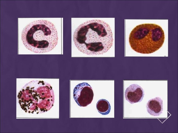
PRINCIPLE The main components of a Romanowsky stain are: … A cationic or basic dye (methylene blue or its oxidation products such as azure B), which binds to anionic sites and gives a blue-grey color to nucleic acids (DNA or RNA), nucleoproteins, granules of basophils and weakly to granules of neutrophils … An anionic or acidic dye such as eosin Y or eosin B, which binds to cationic sites on proteins and gives an orange-red color to hemoglobin and eosinophil granules. … p. H value of phosphate buffer is very important.
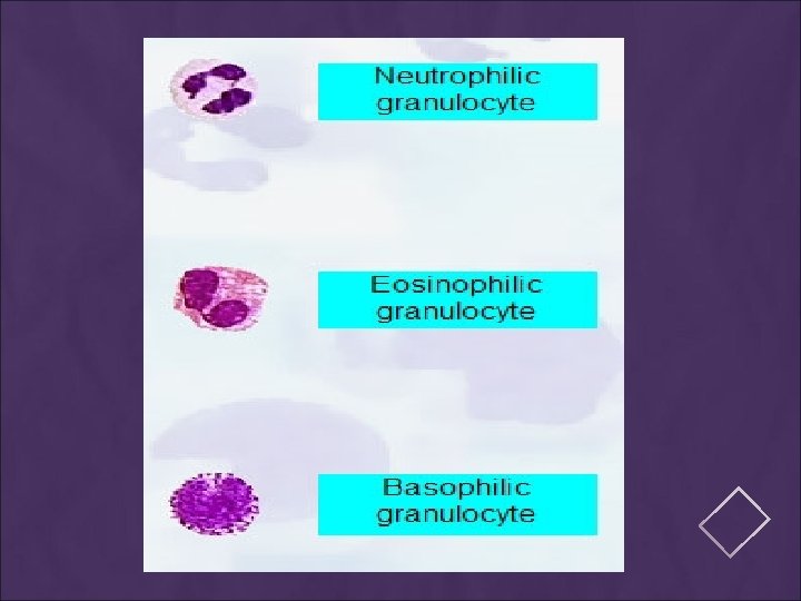

Eosinophilic granules Blue nucleus Basophilic granules

PLATELETS

MANUAL DIFFERENTIAL COUNTS These counts are done in the same area as WBC and platelet estimates with the red cells barely touching. This takes place under × 100 (oil) using the zigzag method. Count 100 WBCs including all cell lines from immature to mature. Reporting results Absolute number of cells/µl = % of cell type in differential x white cell count

Observing direction: Observe one field and record the number of WBC according to the different type then turn to another field in the snake-liked direction *avoid repeat or miss some cells

NORMAL PERIPHERAL BLOOD SMEAR

LEUKOCYTOSIS Leukocytosis, a WBC above 10, 000 is usually due to an increase in one of the five types of white blood cells and is given the name of the cell that shows the primary increase. 1. Neutrophilic leukocytosis = neutrophilia 2. Lymphocytic leukocytosis = lymphocytosis 3. Eosinophilic leukocytosis = eosinophilia 4. Monocytic leukocytosis =monocytosis 5. Basophilic leukocytosis = basophilia

tail body head

- Slides: 27