
BLOOD Provides a mechanism for rapid transport of
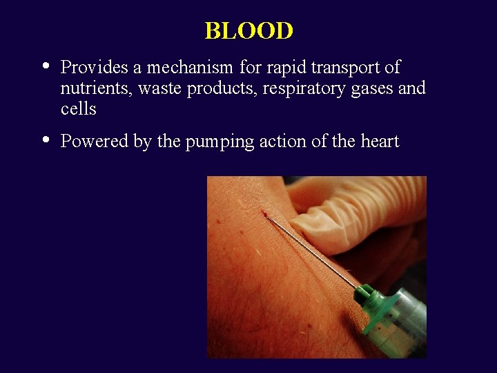
BLOOD • Provides a mechanism for rapid transport of nutrients, waste products, respiratory gases and cells • Powered by the pumping action of the heart
Introduction • Cardiovascular System • • • System made up of blood vessels, blood and heart. Major function is to transport nutrients, gases and hormones to the cells and pick up wastes from cells to transport them to areas of body where they are excreted Lymphatic System • Network of vessels that return the fluid escaped from blood vessels back to the bloodstream • Includes lymphocytes, lymphoid tissue and lymphoid organs which fight infections and give immunity to disease Circulatory System • Together the cardiovascular system and lymphatic system make up the circulatory system
Functions Of Blood • Transportation - the blood transports dissolved gases, nutrients, hormones and metabolic wastes. • Protection - the blood restricts fluid losses through damaged vessels. Platelets in the blood and clotting proteins minimize blood loss when a blood vessel is damaged. • Regulation • Blood regulates the p. H and electrolyte composition of the interstitial fluids. • Blood regulates body temperature.
Composition Of Blood • • Contains cellular and liquid components A specialized connective tissue • • • Plasma – fluid portion and fibrinogen Blood volume • • • Blood cells – formed elements Males: 5 – 6 liters Females: 4 – 5 liters The p. H of blood is about 7. 35 -7. 45
Formed Elements • Blood cells • Erythrocytes, leukocytes, and platelets • Staining of blood cells • Acidic dye – eosin – stains pink • Basic dye – methylene blue – stains blue and purple

Blood Plasma • • • Straw-colored, sticky fluid portion of blood Approximately 90% water Contains: • • Ions – Na+ and Cl. Nutrients – sugars, amino acids, lipids, cholesterol, vitamins and trace elements • Three main proteins - Albumin (60%), globulin (35%), fibrinogen (4%) • • Dissolved Gasses – including O 2 and CO 2 Waste Products – other protein wastes such as urea and bilirubin
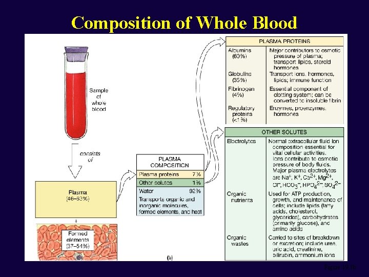
Composition of Whole Blood Figure 19. 1 b
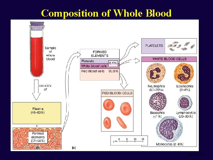
Composition of Whole Blood Figure 19. 1 c
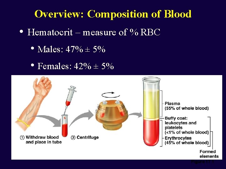
Overview: Composition of Blood • Hematocrit – measure of % RBC • Males: 47% ± 5% • Females: 42% ± 5% Figure 17. 1

Wright’s Stain Figure 17. 2 b
Erythrocytes – Red Blood Cells (RBCs) • Oxygen-transporting cells • • 7. 5 µm in diameter (diameter of capillary 8 – 10µm) Most numerous of the formed elements • • Females: 4. 3 – 5. 2 million cells/cubic millimeter Males: 5. 2 – 5. 8 million cells/cubic millimeter • Made in the red bone marrow in long bones, cranial bones, ribs, sternum, and vertebrae • Average lifespan 100 – 120 days
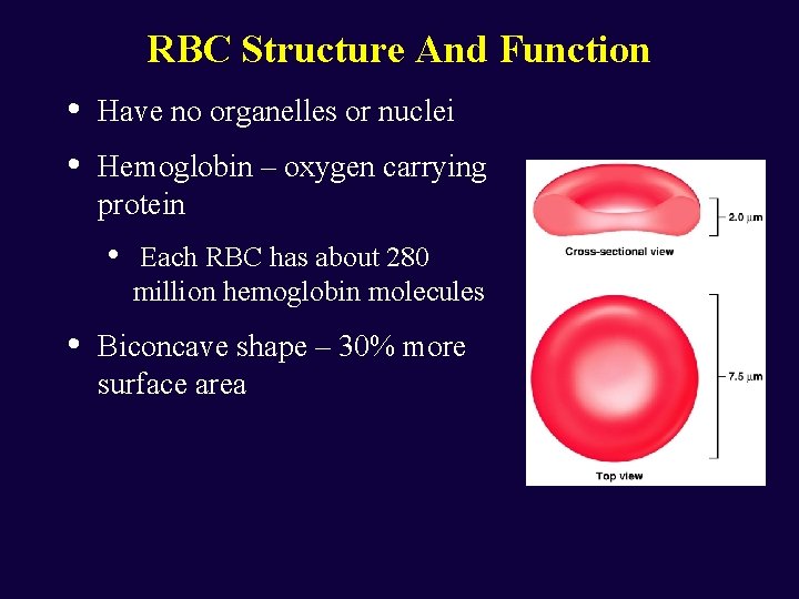
RBC Structure And Function • • Have no organelles or nuclei Hemoglobin – oxygen carrying protein • • Each RBC has about 280 million hemoglobin molecules Biconcave shape – 30% more surface area
Leukocytes – White Blood Cells (WBCs) • Protect the body from infectious microorganisms • 4, 800 – 11, 000/cubic millimeter • Function outside the bloodstream in loose connective tissue • Diapedesis – circulating leukocytes leave the capillaries • • • WBCs have a nucleus and are larger than RBCs Most produced in bone marrow Lifespan of 12 hours to several years
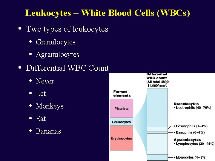
Leukocytes – White Blood Cells (WBCs) • Two types of leukocytes • • • Granulocytes Agranulocytes Differential WBC Count • • • Never Let Monkeys Eat Bananas Figure 17. 5
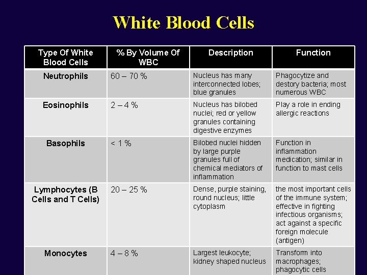
White Blood Cells Type Of White Blood Cells % By Volume Of WBC Description Function Neutrophils 60 – 70 % Nucleus has many interconnected lobes; blue granules Phagocytize and destory bacteria; most numerous WBC Eosinophils 2– 4% Nucleus has bilobed nuclei; red or yellow granules containing digestive enzymes Play a role in ending allergic reactions <1% Bilobed nuclei hidden by large purple granules full of chemical mediators of inflammation Function in inflammation medication; similar in function to mast cells 20 – 25 % Dense, purple staining, round nucleus; little cytoplasm the most important cells of the immune system; effective in fighting infectious organisms; act against a specific foreign molecule (antigen) 4– 8% Largest leukocyte; kidney shaped nucleus Transform into macrophages; phagocytic cells Basophils Lymphocytes (B Cells and T Cells) Monocytes
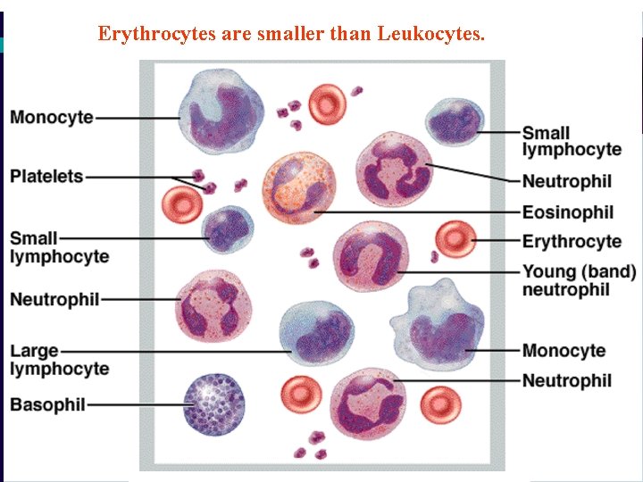
Erythrocytes are smaller than Leukocytes.
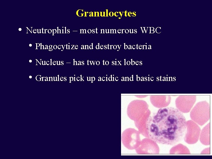
Granulocytes • Neutrophils – most numerous WBC • Phagocytize and destroy bacteria • Nucleus – has two to six lobes • Granules pick up acidic and basic stains Figure 17. 4 a
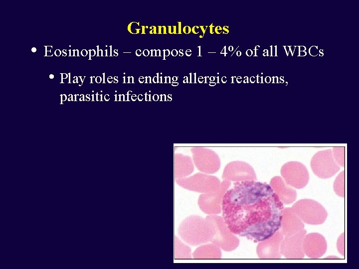
Granulocytes • Eosinophils – compose 1 – 4% of all WBCs • Play roles in ending allergic reactions, parasitic infections Figure 17. 4 b
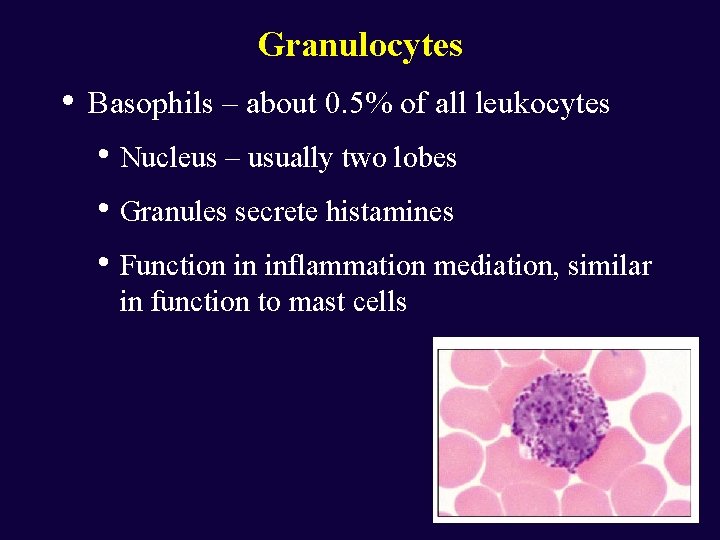
Granulocytes • Basophils – about 0. 5% of all leukocytes • Nucleus – usually two lobes • Granules secrete histamines • Function in inflammation mediation, similar in function to mast cells
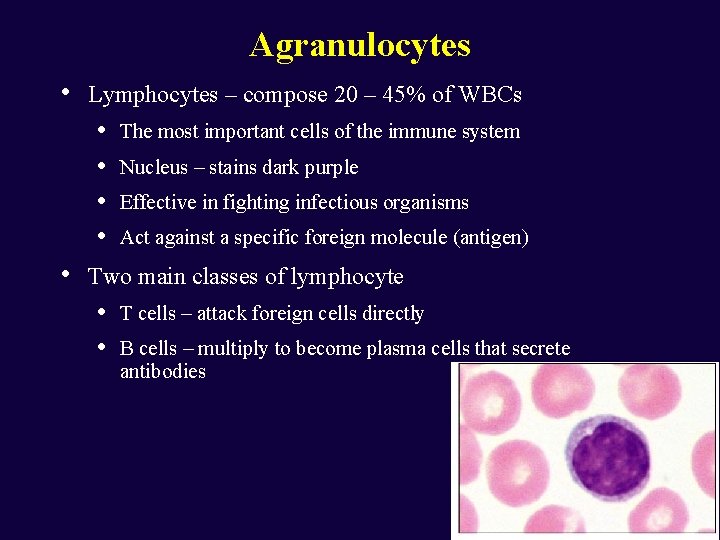
Agranulocytes • Lymphocytes – compose 20 – 45% of WBCs • • • The most important cells of the immune system Nucleus – stains dark purple Effective in fighting infectious organisms Act against a specific foreign molecule (antigen) Two main classes of lymphocyte • • T cells – attack foreign cells directly B cells – multiply to become plasma cells that secrete antibodies Figure 17. 4 d
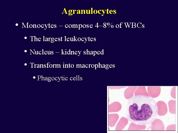
Agranulocytes • Monocytes – compose 4– 8% of WBCs • The largest leukocytes • Nucleus – kidney shaped • Transform into macrophages • Phagocytic cells Figure 17. 4 e
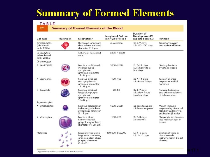
Summary of Formed Elements Table 17. 1
Platelets • • Structure • Small cellular fragments; originate in bone marrow from giant cell megakaryocyte • Contain several clotting factors – calcium ions, ADP, serotonin Function • Involved in stopping bleeding when a blood vessel is damaged; Process is called hemostasis
Blood Cell Formation • • • Hematopoiesis – process by which blood cells are formed 100 billion new blood cells formed each day Takes place in the red bone marrow of the humerus, femur, sternum, ribs, vertebra and pelvis • Red marrow – actively generates new blood cells • Contains immature erythrocytes • Remains in epiphyses, girdles, and axial skeleton • Yellow marrow – dormant • Contains many fat cells • Located in the long bones of adults • Tissue framework for red marrow • Reticular connective tissue
Cell Lines in Blood Cell Formation • • All blood cells originate in bone marrow All originate from one cell type • Blood stem cell (pluripotential hematopoeitic stem cell) • Lymphoid stem cells - give rise to lymphocytes • Myeloid stem cells - give rise to all other blood cells
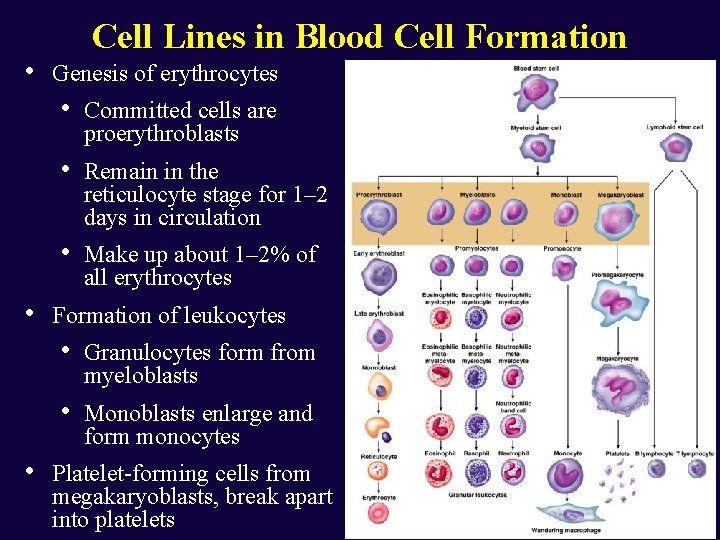
• • • Cell Lines in Blood Cell Formation Genesis of erythrocytes • Committed cells are proerythroblasts • Remain in the reticulocyte stage for 1– 2 days in circulation • Make up about 1– 2% of all erythrocytes Formation of leukocytes • Granulocytes form from myeloblasts • Monoblasts enlarge and form monocytes Platelet-forming cells from megakaryoblasts, break apart into platelets
The Blood Throughout Life • First blood cells develop with the earliest blood vessels • • Mesenchyme cells cluster into blood islands • Bone marrow becomes major hematopoietic organ at month 7 Late in the second month the liver and spleen take over blood formation
RBC life span and circulation • Replaced at a rate of approximately 3 million new blood cells entering the circulation per second • Damaged or dead RBCs are recycled by phagocytes • Components of hemoglobin individually recycled • • Heme stripped of iron and converted to biliverdin, then bilirubin Iron is recycled by being stored in phagocytes, or transported throughout the blood stream bound to transferrin
Feedback Regulation of Erythropoiesis - regulated by renal oxygen content. - Erythropoietin, a glycoprotein hormone, is produced by renal cells in response to a decreased renal blood O 2 content. - Erythropoietin stimulates erythrocyte production in the red bone marrow.
A drop in renal blood oxygen level can result from: 1) reduced numbers of red blood cells due to hemorrhage or excess RBC destruction. 2) reduced availability of oxygen to the blood, as might occur at high altitudes or during pneumonia. 3) increased demands for oxygen (common in those who are engaged in aerobic exercise).
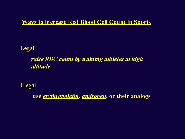
Ways to increase Red Blood Cell Count in Sports Legal raise RBC count by training athletes at high altitude Illegal use erythropoietin, androgen, or their analogs
Dietary Requirements for Erythropoiesis Iron vitamin B 12 folic acid More important to women due to the loss of blood during menstruation
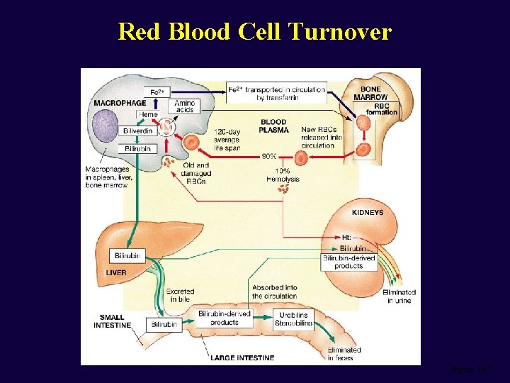
Red Blood Cell Turnover Figure 19. 5
Human Blood Groups
Human Blood Groups - were learned from tragedies (death) caused by mismatch during transfusion in ancient time. - ABO blood types were identified in 1900 by Karl Landstein (1930 Nobel laureate). - Other blood types were identified later.
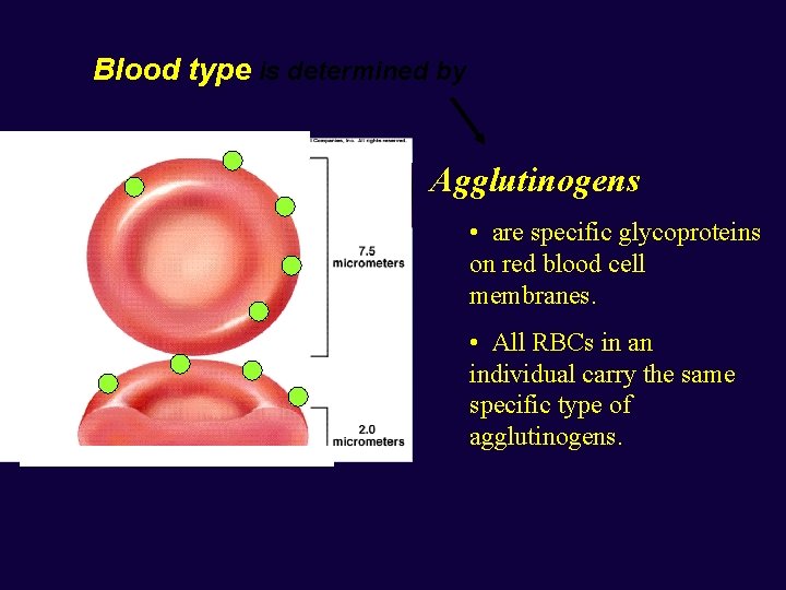
Blood type is determined by Agglutinogens • are specific glycoproteins on red blood cell membranes. • All RBCs in an individual carry the same specific type of agglutinogens.
ABO Blood Groups Type A: RBCs carry agglutinogen A. Type B: RBCs carry agglutinogen B. Type O: RBCs carry no A nor B agglutinogens. Type AB: RBCs carry both A and B agglutinogens.
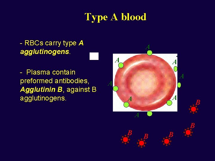
Type A blood - RBCs carry type A agglutinogens. A A - Plasma contain preformed antibodies, Agglutinin B, against B agglutinogens. A A A B B B B
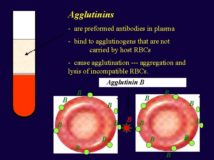
Agglutinins - are preformed antibodies in plasma - bind to agglutinogens that are not carried by host RBCs - cause agglutination --- aggregation and lysis of incompatible RBCs. Agglutinin B B B B B
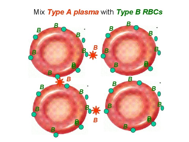
Mix Type A plasma with Type B RBCs B B B B B B B B B
Type B recipient
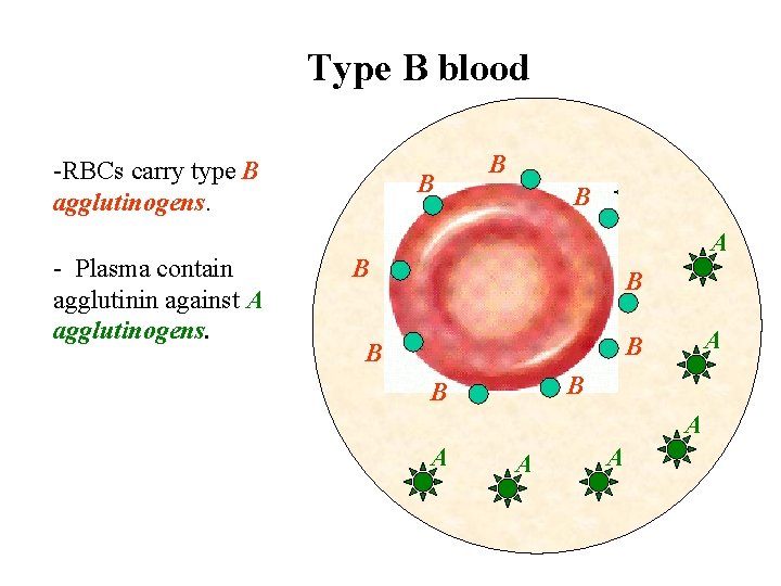
Type B blood -RBCs carry type B agglutinogens. - Plasma contain agglutinin against A agglutinogens. B B B A A
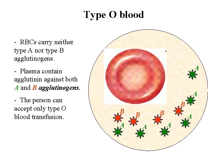
Type O blood - RBCs carry neither type A nor type B agglutinogens. A - Plasma contain agglutinin against both A and B agglutinogens. - The person can accept only type O blood transfusion. A B B B A A A
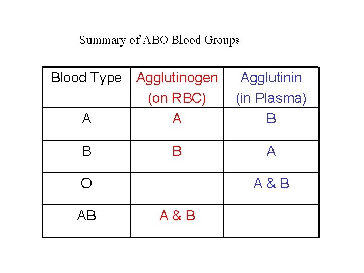
Summary of ABO Blood Groups Blood Type Agglutinogen (on RBC) A A B B O AB Agglutinin (in Plasma) B A A&B
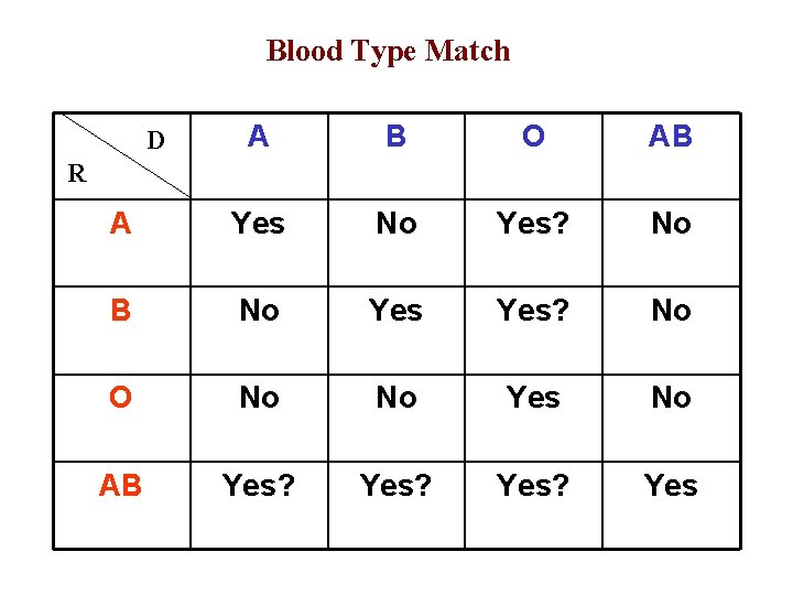
Blood Type Match A B O AB A Yes No Yes? No B No Yes? No O No No Yes No AB Yes? Yes D R
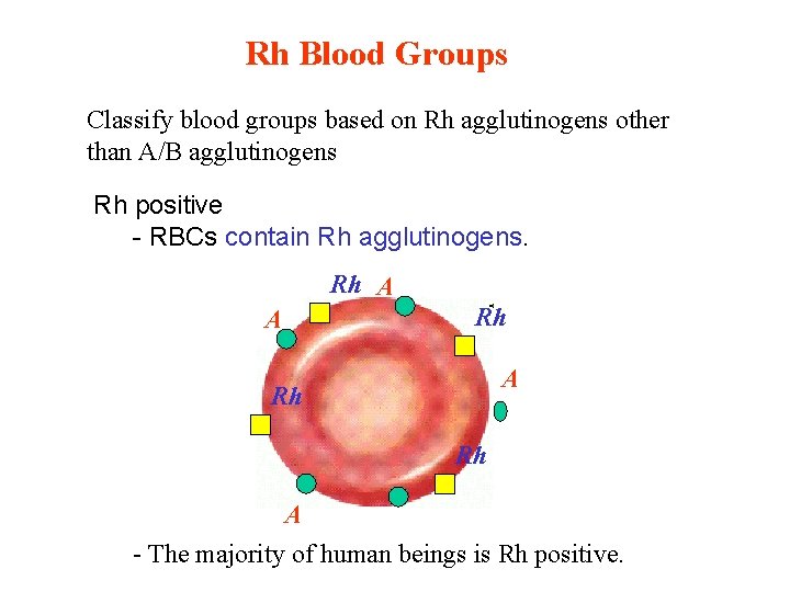
Rh Blood Groups Classify blood groups based on Rh agglutinogens other than A/B agglutinogens Rh positive - RBCs contain Rh agglutinogens. Rh A A Rh Rh A - The majority of human beings is Rh positive.
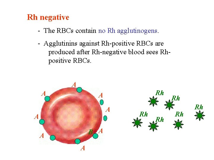
Rh negative - The RBCs contain no Rh agglutinogens. - Agglutinins against Rh-positive RBCs are produced after Rh-negative blood sees Rhpositive RBCs. A A Rh A A A B A A A Rh Rh Rh
The problem with a Rh-negative mother and her Rh-positive fetus.
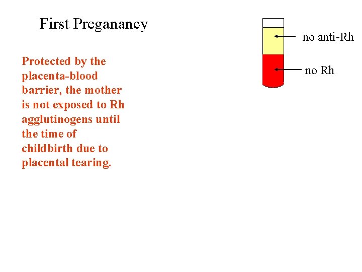
First Preganancy Protected by the placenta-blood barrier, the mother is not exposed to Rh agglutinogens until the time of childbirth due to placental tearing. no anti-Rh no Rh
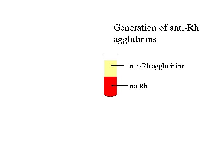
Generation of anti-Rh agglutinins no Rh
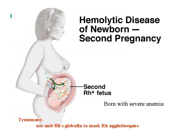
Born with severe anemia Treatment: use anti-Rh globulin to mask Rh agglutinogens
Why does blood clot? • When small blood vessels get broken, blood escapes from the closed circulatory system • Our bodies create a clot which ‘seals’ the damaged blood vessels preventing excessive blood loss and helping to prevent pathogens from entering the body • Prothrombin and fibrinogen are plasma proteins which circulate in the blood • Platelets are cell fragments which also circulate
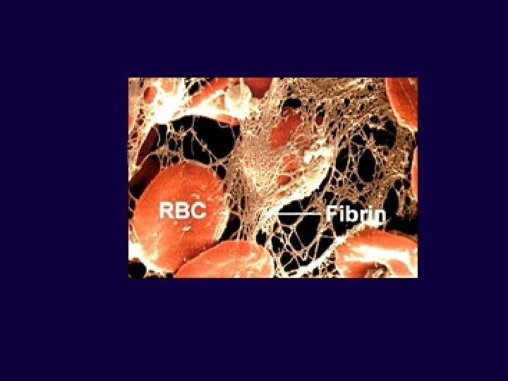
Blood clotting sequence • • Blood vessel is damaged Damaged cells release chemicals which stimulate platelets to adhere to the damaged area • Other platelets begin adhering to those platelets • To strengthen the plug, the damaged tissue and platelets release chemicals called clotting factors which convert prothrombin into thrombin • Thrombin is an active enzyme which catalyses the conversion of soluble fibrinogen into the relatively insoluble fibrin • Fibrin is a fibrous protein which forms a mesh-like network that helps to stabilize the platelet plug • More cellular debris gets trapped in the fibrin mesh and soon a stable clot has formed preventing both further blood loss and entry of pathogens
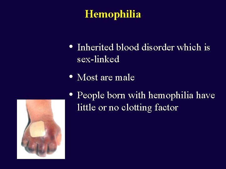
Hemophilia • Inherited blood disorder which is sex-linked • • Most are male People born with hemophilia have little or no clotting factor
Primary Immune Response 1. Macrophage encounters a foreign antigen and engulfs the possible pathogen by phagocytosis 2. Antigens of the invader are displayed on the cell membrane of the macrophage – this is known as antigen presentation 3. Leukocytes known as helper-T cells chemically recognize the antigen being presented and become activated 4. Helper-T cells chemically communicate with the specific B cell type (which has also come in contact with the antigens) that is able to produce the antibody needed
Cell cloning • When a helper-T cell activates a specific B cell, the activated B cell type begins a series of cell division known as cell cloning • Types • Antibody-secreting plasma cells – secrete antibodies immediately and help to fight off the primary infection • Memory cells – do no secrete antibodies during the primary infection, but are long-lived cells which remain circulating in the bloodstream waiting for a subsequent infection
Principles of true immunity • • Challenge and response • Immune system challenged by an antigen during 1 st infection in order to develop an immunity • Macrophages, helper-T cells, B cells Clonal selection • • • Identification of plasma B cells Multiple cell divisions to build up #s of same cell Memory cells • Provide long-term immunity
• Types of immunity Active Immunity • • • Always leads to the production of memory cells Provides long-term immunity Passive Immunity • When an organism acquires antibodies which were produced in another organism • Only the organism which produces the antibodies has the memory cells • • • Mother to fetus through placenta From mother’s colostrum Injection of antibodies in antisera (antivenoms produced for treatment of poisonous snake and spider bites)
How does a vaccine result in immunity? • One cannot be immune to a pathogen before being exposed to it at least once • For many diseases, vaccines have been developed that act as the first exposure to the pathogen • Vaccine is developed by weakening a pathogen and then injecting the pathogen into the body • Methods: • • Selecting a weak strain Heating the pathogen Chemical treatment of pathogen Infection is not prevented, but the secondary immune response is quicker and more intense than the primary response
- Slides: 60