BLOOD PHYSIOLOGY 12 Control of Erythropoiesis Iron Metabolism
 Control of Erythropoiesis, Iron Metabolism and Hemoglobin (3) Red: very important.")
BLOOD PHYSIOLOGY (1+2) Control of Erythropoiesis, Iron Metabolism and Hemoglobin (3) Red: very important. Green: only found in males’ slides. Purple: only found in females’ slides. Gray: notes. Physiology Team 436 – Foundation block lectures 8+9+10 1 Lecture: If work is intended for initial studying. Review: If work is intended for revision.

Objectives � Describe : physical and chemical characteristics of blood � Recognize: the causes of polycythemia. � Define: Erythro-poiesis; leuco-poiesis, thrombopoiesis. � Recognize: hemoglobin structure and its functions. � Discuss: Iron metabolism (absorption, storage and transport). � Describe: Cellular and non-cellular blood components � Describe: different stages of RBC differentiation. � Describe: features of RBC maturation � Describe: regulation of RBC production and erythropoietin hormone secretion in response to hypoxia. � Describe essential elements needed for RBC formation. � Describe: essential elements needed for RBC formation � Describe the process of Vitamin B 12 absorption and its malabsorption. � Describe: the process of Vitamin B 12 absorption and its malabsorption. � Recognize hemoglobin structure and its functions. � Discuss iron metabolism (absorption, storage and transport). � Describe: the fate of old RBC. � Describe: anemia and its causes � Describe the fate of old RBC. � Recognize: blood functions. � Describe anemia and its causes. � Recognize: clinical conditions associated with high level of erythropoietin in the blood. � Recognize causes of polycythemia. � Understand steps of erythropoiesis & its regulation. � 2 Recognize: sites of RBC formation at different developmental age.
 • White Blood. Cells(Leucocytes) • Platelets (Thrombocytes) 1.")
Blood Composition • Red Blood Cells(Erythrocytes) • White Blood. Cells(Leucocytes) • Platelets (Thrombocytes) 1. Cellular components: 2. Plasma (ECF): 3 • NOTE: red blood cells are in millions • Platelets are hundred thousands • white blood cells are in thousands. • 98% water + ions + plasma proteins e. g. (Albumin, globulin, Fibrinogen) • #notice: it’s the same ionic composition as interstitial fluid. (Na, K, PO 4, etc) Note: in males’ slides water is 92%, ions are 7%, and proteins are

Characteristics of Blood Viscosity p. H Temperature Color 4 Osmolarity Regeneratio n • 3 -4 times than Water. • Blood relative viscosity: (4 -5) mainly depends on the numbers of RBC. • Plasma relative viscosity: (1. 6 -2. 4) is mainly involved in plasma protein. • Slightly alkaline, with a p. H of 7. 35 -7. 45. • It has a salty metallic taste and is Sticky. • Hb acts as blood buffer. • about 37 C (slightly higher than normal body temp). • Bright red = O 2 rich; • Dull red = O 2 poor • Plasma osmolality is about 300 mmol/L (Equal to 0. 9% Na. Cl Solution=Isotonic). • Tremendous regenerative capacity.
 5")
Characteristics of Blood Plasma osmotic pressure: Specific gravity: Erythrocyte sedimentatio n rate (ESR) 5 • Is 300 mmol/L or 770 k. Pa • Crystal osmotic pressure: results from Na. Cl and modulates water distribution between inside and outside cells. • Colloid osmotic pressure: results from albumin and regulates water distribution between inside and outside of capillaries. • Total blood: (1. 050 -1. 060) more influenced by RBC. • Plasma: (1. 025 -1. 030) more influenced by plasma protein. • RBC: (1. 090 -1. 092) more influenced by hemoglobin. • It the red blood cell descending distance per hour. • Normal male 0¬ 15 mm/h. • Normal Female 0¬ 20 mm/h.
. 55% is plasma volume. 5 liter")
Blood Volume 45% is packed cells volume (PCV). 55% is plasma volume. 5 liter in adult • • Males => 5 to 5. 5 L Females => 4 to 4. 5 Because females lose every month half a liter of blood due to menstruation 7 The volume of blood cells in a sample of blood after it has been centrifuged (separation of contents). Plasma: Straw colored liquid that serves as a transport medium for blood and platelets. It is called Serum when fibrinogen (coagulating factor) is removed after coagulation. Plasma contains: Electrolytes, clotting factors, antibodies, blood gases, nutrients, and wastes. Ratio of the volume of red blood cells to the total volume of blood. ﺍﻟﺘﺸﺨﻴﺺ ﻓﻲ ﻳﺴﺎﻋﺪﻧﺎ . ﺍﻻﻧﻴﻤﻴﺎ ﺯﻱ ﻛﺜﻴﺮ ﺃﺸﻴﺎﺀ

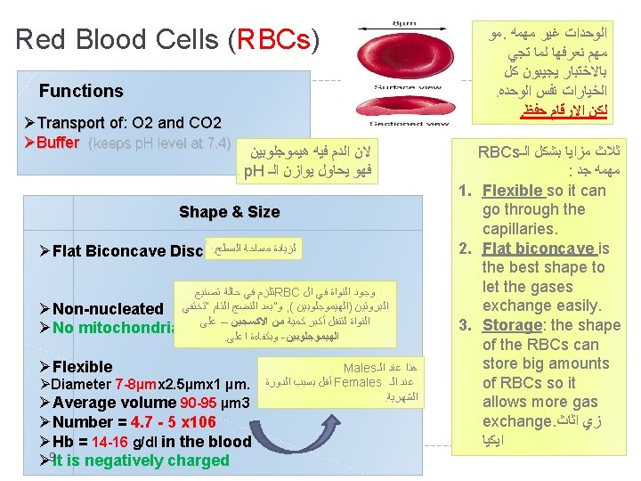
Functions of the Blood 1. Transport � Respiration: O 2, CO 2 � Trophic: nutrients to the tissue � Excretive: waste products (metabolites) from tissues to excretory organs � Regulative: hormones 2. Homoeostasis � Regulation of: Formation of Blood Cells: Haemopoiesis Erythro-poiesis: Erythro-poiesis Formation of RBC (erythrocytes) Leuco-poiesis: Leuco-poiesis Formation of WBC (leucocytes) 1) Body temperature by redistributing blood between skin and internally 2) ECF p. H 3. Protecting against Infections: White Blood Cells, Antibodies 4. Blood clotting: to prevent blood loss 8 Thrombo-poiesis: Thrombo-poiesis Formation of platelets (thrombocytes) * Note: Active cellular marrow is called red marrow; inactive marrow that is infiltrated with fat is called yellow marrow. Cyte: Mature cell Blast: Still not mature cell

Red Blood Cells Cytoskeleton � Skeletal protein network � • Spectrin � • Actin � • Ankyrin � • Tropomyosin � • Proteins 4. 1 and 4. 9 10 � The red cell, as it continuously circulates, must be able to undergo extensive passive deformation and to resist fragmentation. These two essential qualities require a highly deformable yet remarkably stable membrane. Blood cells that lyse(breakdown) without difficulty at low ionic strength are a sign of Hereditary Spherocytosis.

RBCs Enzyme- Carbonic Anhydrase � Carbonic anhydrase is an enzyme that catalyzes the reversible reaction between carbon dioxide (CO 2) and water to form carbonic acid (H 2 CO 3), increasing the rate of this reaction several thousand fold. It plays a major role in CO 2 transport. 11

Production of RBCs are only nucleated in fetus • Early few weeks (1 st 4 months) : embryo Inutero: nucleated RBCs are formed in yolk sac. (nucleus is needed for division) • Middle trimester: mainly in liver & spleen & lymph nodes. • Last months: RBCs are formed in bone marrow of all bones • Bone marrow of flat bones (e. g. scapula, After Birth: skull, . . ) continue to produce RBC into adult life. • In children: all bones produce RBCs until 5 years old. • Shaft of long bones stop producing RBC at puberty while epiphysis continues. • In adults: after 20, membranous bones such as vertebrae, sternum, ribs, and ilia produce RBCs 12

Production of RBC in utero 13

Note: Children need RBC more than adults for their growth. 14
 -Flat bones ( Vertebras (Irregular bones)")
As mentioned in the previous slide: (After birth) -Flat bones ( Vertebras (Irregular bones) continue to Flat bones Sternum, Ribs) and the , Ribs produce RBCs. -Shafts of long bones ( Shafts of long bones Femur, Tibia) stop producing RBCs after puberty. Femur, Tibia 15 Production of RBC

: ﺍﻟﺼﻮﺭﺓ ﻣﺤﺘﻮﻯ ﺷﺮﺡ Multipotential = Pluripotent ﺍﻟﻤﻌﻨﻰ ﻧﻔﺲ Found in bone marrow and cord blood All blood cells are formed from (stem cells) stem cells have two paths: 1 - First Path: “ myeloid“ ➡ that forms RBCs + some types of WBCs + 2 -Second Path: “ lymphoid“ ➡ that forms lymphocytes (another type of WBC) *Lymphocytes are always special ; )* There are different growth factors 16 Note: Stem cells can differentiate into a lot of different cells. Myeloblast gives white blood cells.
 of RBCs Pluri-potential hemato-poietic stem cells: give rise to (create) : all")
Genesis (Production) of RBCs Pluri-potential hemato-poietic stem cells: give rise to (create) : all the other blood cells • • committed cells Committed stem cells for RBC Committed stem cells for WBC Growth of different stem cells are controlled by different growth factors Note Colony-Forming Unit CFU: 17 cells produce colonies of their own type
: 7 Days Committed stem cell: Proerythro-blast (first identifiable cell)")
Stages of RBCs Differentiation (development): 7 Days Committed stem cell: Proerythro-blast (first identifiable cell) Basophil erythro-blast Poly-chromatophil erythro-blast In cases of rapid *fast* RBC production reticlocytes in the circulation Ortho-chromatic erythro-blast Reticulocytes (loses nucleus) Mature erythrocytes Reticulocytes: First cell to appear in Circulation Matures in 24 -48 Hours. Contain remnants of the Golgi apparatus & mitochondria. 18 Note: RBC goes into stages to get its concave structure, Reticulo-cyte is the stage where the cell loses its nucleus and is covered by a net.
 in (Reduction) cell size. Disappearanc e")
Erythro-poiesis RBC development is characterized by Decrease (Reduction) in (Reduction) cell size. Disappearanc e of nucleus. Appearance (Aquisiton) of (Aquisiton) hemoglobin (Hb) ﺍﻟﻬﻴﻤﻮﺟﻠﻮﺑﻴﻦ ﻋﺸﺎﻥ ﺍﻟﻨﻮﺍﻩ ﺗﺨﺘﻔﻲ ﺍﻟﻨﻮﺍﺓ ﻣﺤﻞ ﺣﻞ 19 Memorize steps and shapes in the

Regulation of RBC Production Erythro-poiesis stimulated by: erythro-poietin hormone (which is produced by: the kidney in response to hypoxia (low oxygen in the blood) )ﻭﻫﺬﺍ ﻗﻠﻴﻞ ﺍﻟﺪﻡ ﻓﻲ ﺍﻻﻭﻛﺴﺠﻴﻦ ﻣﺴﺘﻮﻯ ﻳﻜﻮﻥ ﻟﻤﺎ ﻫﺮﻣﻮﻥ ﺗﻔﺮﺯ kidney ﺍﻟـ ﺗﺮﻭﺡ (hypoxia ﻳﺴﻤﻰ ﺍﻟـ ﺗﻮﺍﺯﻥ ﻋﺸﺎﻥ Erythropoietin ﺍﺳﻤﻪ Erythropoiesis Causes of Hypoxia ﺍﻟﺠﺒﺎﻝ ﻓﻮﻕ ﺯﻱ ﻛﺜﻴﺮ ﺍﻭﻛﺴﺠﻴﻦ ﻣﺎﻓﻴﻪ ﺍﻟﺠﻮ Low RBC count (Anaemia) 20 Hemorrhage ﻧﺰﻳﻒ High altitude ﺍﻟﺪﻡ ﻳﻮﺯﻉ ﻣﺎﻳﻘﺪﺭ ﺍﻟﻘﻠﺐ Prolong heart failure Lung disease

Tissue oxygenation and RBC formation 21
 Glycoprotein 90% from renal cortex (kidney) 10% from the liver. Stimulate: the")
Erythro-poietin (Hormone) Glycoprotein 90% from renal cortex (kidney) 10% from the liver. Stimulate: the growth of early stem cells. Stimulate Does not affect: maturation process of RBCs. Does not affect Can be measured in plasma & urine. Can be measured Conditions like: anemia, High altitude, Heart failure, Lung Disease result in: High erythropoietin levels polycythemia Polycythemia = RBCs level is up Erythropoietin => works when RBCs level is down mia ﺑﻤﻘﻄﻊ ﺗﻨﺘﻬﻲ ﻛﻠﻤﻪ ﺃﻲ ﺯﻳﺎﺩﻩ ﻣﻌﻨﺎﻫﺎ The hormone controls the early stages of RBC formation (production of proerythroblasts from stem cells), not the middle or last stages, so erythropoietin makes the cells pass more rapidly through the erythroblastic stages and can increase it to 10 or more times than normal. 22

Role of the kidneys in RBC formation: Healthy kidneys produce a hormone called erythropoietin and prompts the bone marrow to make red blood cells, which then carry oxygen throughout the body. 23 Kidney stimulate the production of Erythropoietin

Essential Elements for Formation and Maturation of RBCs : ﻟﻠﻔﻬﻢ ﻣﻼﺣﻈﺔ Hemo-globin: Heme: iron. Globin: protein Amino acids (Proteins) Iron Formation of Hemoglobin. Formation of globin in hemoglobin Heme Synthesis. Other : Vit B 6, Riboflavin, nicotinic acid, biotin, Vit C, Vit E Vitamins: B 12 and Folic Acid Synthesis of nucleoprotein. Synthesis of DNA Cell division RBC Final maturation Vit C Vit B 6 Iron absorption RBC Synthesis deficiency lead to: Deficiency leads to Anemia and Hemoglobino-pathy 24 Micro-cytic and hypochromic Anemia Megaloblastic (macrocytic) macrocytic Anemia Iron Deficiency lead to: normochromic normocytic Anaemia Hormones: • Androgens, Thyroid, cortisol & growth hormones. • Deficiency lead to Anemia. Essential elements: • Copper, Cobalt, zinc, manganese.

Most of this slide is found only in females’ slides. Vitamin B 12 & Folic acid Important for DNA synthesis and final maturation of RBC. ion (Important for all stages of maturation of RBCs) Dietary source: meat, milk, liver, fat, green vegetables. Deficiency Leads to In shape: Abnormally (in erythropoiesis) large & oval shape RBC Failure of: -nuclear maturation -division 25 In count: Reduced RBC Type of Anemia count & Hb Type of Anemia: Short life span Macrocytic (megaloblastic) anemia
 Anemia MICROcytic hypochromic Anemia MICROcytic It is caused")
Anemia: Deficiency of hemoglobin. MACROcytic (Megaloblastic) Anemia MICROcytic hypochromic Anemia MICROcytic It is caused by the deficiency of vit B 12 and Folic Acid The RBC's are smaller than normal Have an increased zone of central pallor. Note 1 - The hyper-segmented 1 - Neutrophil (WBC) 2 - The RBC are almost as large 2 as the lymphocyte (WBC). 3 -There are fewer RBCs. 326 Increased anisocytosis (variation in size) Increased poikilocytosis (variation in shape) Causes: Iron deficiency and Thalassemia. Most of this slide is found only in females’

Microcytic hypochromic anemia MCV: the average volume of RBCs. smaller size of each RBC 43527 ﺗﻴﻢ ﻣﻦ less hemoglobin in each RBC (MCV<80 fl) (MCH<27 pg) MCH: the average mass of hemoglobin per RBC.

This slide is found only in females’ slides. Mal-absorption of vit. B 12 Pernicious Anemia Causes of deficiency parietal cells ﺍﻟـ ﻓﻴﻪ Stomach ﺍﻟـ Intrinsic factor ﺍﻟـ ﻟﻲ ﺗﻄﻠﻊ ﺍﻟﻠﻲ ﻭﻫﻲ VB 12 absorption needs intrinsic factor secreted by parietal cells of stomach ﺍﻟﻤﻌﺪﺓ ﺟﺪﺍﺭ ﺧﻼﻳﺎ VB 12 + intrinsic factor is absorbed in the terminal Ileum. Intrinsic ﺑﺎﻟـ ﻣﺎﺳﻚ ﻳﻜﻮﻥ VB 12 ﺍﻟـ • ﻻﺯﻡ . ﺗﻤﺘﺼﻪ ﻋﺸﺎﻥ factor • Terminal ileum = ﻓﻲ ﺣﺘﻪ ﺍﺧﺮ Small intestine ﺍﻟـ 28 Inadequate intake Poor absorption due to Intestinal disease Poor absorption = decrease in intrinsic factors
: Iron is needed for the synthesis of hemoglobin, hemoglobin myoglobin cytochrome")
Iron Metabolism (Fe): Iron is needed for the synthesis of hemoglobin, hemoglobin myoglobin cytochrome oxidase, myoglobin cytochrome oxidase peroxidase & catalase. Total Iron in the body = 3 -5 / 4 -5 g Note: Females’ Slides 65% - 75%. . Haemoglobin (3 g) 5% ……. . other hems 1% ……. bound to transferrin (betaglobulin) in blood 15 -30% …… stored iron in the form of ferritin in the liver, spleen and bone marrow. 4% ……. . Muscle Hb (myoglobin) 1% ……. Enzymes (cytochrome) 0. 1% ……… Plasma iron: (transferrin) 29 Note: Males’ Slides

ﺑﺮﺗﻘﺎﻝ ﻋﺼﻴﺮ ﻣﻌﻪ ﻧﺸﺮﺏ ﻳﻨﺼﺤﻮﻥ iron ﻧﺎﺧﺬ ﻟﻤﺎ ﻛﺬﺍ ﻋﺸﺎﻥ ﺑﺎﻻﻣﺘﺼﺎﺹ ﻳﺴﺎﻋﺪ ﻻﻧﻪ ﻟﻴﻪ؟ ( ﺳﻲ )ﻓﻴﺘﺎﻣﻴﻦ Iron Absorption: Decrease ﻋﻨﺪﻫﻢ Stomach diseases ﻋﻨﺪﻫﻢ ﺍﻟﻠﻲ . ﻛﻮﻳﺲ iron ﺍﻟـ ﻳﻤﺘﺼﻮﻥ ﻳﻘﺪﺭﻭﻥ ﻣﺎ in vit. C Iron in food is mostly in oxidized form (Ferric, F+3) Better absorbed in reduced form (Ferrous, F+2 ) Iron in stomach is reduced by gastric acid, Vitamin C ﺍﻻﻣﺘﺼﺎﺹ ﻓﻲ ﻳﺴﺎﻋﺪ What does rate of iron absorption depend on? -The amount of iron stored. When does rate of iron absorption decrease? - when iron stores are saturated (When all the apoferritin is saturated). 30 -Ferritin: intracellular iron-storage protein -Ferritin that is not combined with iron is

Transport and Storage of Iron o Iron transportation: in plasma in the form of Transferrin (apotransferrin + iron). o Normally 30 -40 saturated with Fe: (plasma iron 100 -130 ug/100 ml) o When transferrin is 100% saturated with Fe: ( plasma iron 300 ug/100 ml) Haemosiderin: Iron storage complex only found in cells (not in circulating blood) appears to be 1. a complex of ferritin. 2. denatured ferritin. 3. other material. (not a good supplier of iron when needed) o Iron storage: Ferritin : (apoferritin + iron) (Loose bond) ﻗﺺ ﺃﻮ ﺑﺎﻟﺠﻠﺪ ﺟﺮﺡ ﻃﺮﻳﻖ ﻋﻦ Iron ﺍﻟـ ﻧﻔﻘﺪ Haemosiderin : insoluble complex molecule, (Ferritin + Ferritin) ﻭﺍﻟﺸﻌﺮ ﺍﻷﻈﺎﻓﺮ (Firm bond) Sites : liver, spleen & bone marrow o Daily loss of iron is 0. 6 mg in male & 1. 3 mg/day in females. o Diet provides 10 -20 mg iron per day. 31

Iron excretion Iron losses feces: unabsorbed, dead epithelial cells bile and saliva. Skin: cell, hair, nail, in sweat. Urine Menstruation, pregnancy and child birth 32

Destruction of RBCs RBC life span in circulation = 120 days. Metabolic active cells. Old cells have a fragile cell membrane, the cell will rupture as it passes in narrow sinusoidal capillaries of the spleen, bone marrow & liver. Released Hb is taken up by macrophages in liver, spleen & bone marrow: Hb is broken into its component: ○ Polypeptide amino acids amino acid pool (protein pool = storage). ○ Haem: ○ Iron recycled (reused) iron storage (ferritin form). ○ prophyrin biliverdin bilirubin (secreted by the liver into bile) (excess destruction of RBC can cause Jaundice) 33

Anemia: Definition ○ Decrease number of RBC ○ Decrease Hb Normal ranges of Hb: • Men: Hb 13. 5 to 17. 5 g/d. L • Women: Hb 12. 0 to 15. 5 g/d. L • Infants : Hb 14 – 19 g/d. L Symptoms: Fatigue Tired Dyspnea (shortness of breathing) Pallor Tachycardia heart failure. Greek word (an-haîma) meaning "without blood" 34
 1 - Blood loss")
Causes of Anemia Causes of anemia 3. Haemo-lytic (excessive destruction) 1 - Blood loss Acute ➡ accident (RBC return to normal in 3 to 6 w) Chronic ➡ microcytic hypochromic anemia (ulcer, worms) Acute = ﺣﺎﺩ Chronic = ﻳﻔﻘﺪ ﺯﻱ ﻓﺘﺮﺍﺕ ﻋﻠﻰ ﺍﻟﺪﻡ ﻣﺜﻼ ﺍﻟﺸﻬﺮﻳﻪ ﺍﻟﺪﻭﺭﻩ ﻗﺮﺣﻪ ﻋﻨﺪﻩ ﺍﻟﻠﻲ + , ﺑﻮﺍﺳﻴﺮ , ﺑﺎﻟﻤﻌﺪﻩ ﺑﺎﻻﻣﻌﺎﺀ ﺩﻳﺪﺍﻥ 35 Abnormal cells or Hb 2 - Decrease RBC production 1 - Nutritional causes Iron ➡ microcytic Hypochromic anemia Vit B 12 & Folic acid ➡ megaloblastic anemia Spherocytosis sickle cells 2 - Bone marrow failure: Destruction by cancer, radiation, drugs ➡ Aplastic anaemia. Erythroblastosis fetalis Incompatible blood transfusion Spherocytosis = ﺗﺘﻜﺴﺮ cell membrane ﺑﺎﻟـ ﻣﺸﻜﻠﻪ . ﺑﺴﺮﻋﻪ RBCs ﺍﻟـ Sickle cells = ﻭﺣﺪﻩ ﺷﻔﺮﻩ ﻓﻲ ﺗﻐﻴﺮ . ﻭﺭﺍﺛﻲ , ﻃﻔﺮﻩ ﻓﺤﺪﺛﺖ Incompatible blood transfusion = ﻣﻨﺎﺳﺐ ﻏﻴﺮ ﺩﻡ ﻟﻠﺸﺨﺺ ﻧﻘﻞ ﻟﻤﺎ
: is the volume or percentage of RBC in the blood. Hgb :")
Hct (Hematocrit): is the volume or percentage of RBC in the blood. Hgb : Hemoglobin 36 ﻃﺒﻴﻌﻲ RBCs ﺍﻝ ﺣﺠﻢ ﺍﻟﺒﻼﺯﻣﺎ ﺣﺠﻢ ﺯﻳﺎﺩﺓ ﻭﻟﻜﻦ . Hct ﺗﻘﻠﻴﻞ ﺍﻟﻰ ﺃﺪﺕ
 in the RBCs”")
Polycythemia “Increased number of RBC or high concentration of Hb (hemoglobin) in the RBCs” Primary (Polycythemia Rubra Vera - PRV): uncontrolled RBC production. ” Secondary to hypoxia: high altitude (physiological), chronic respiratory or cardiac disease 37 Types : • • True Absolute • Hypoxia = ﺍﻻﻭﻛﺴﺠﻴﻦ ﻧﻘﺺ • Primary = ) ﺑﺎﻟﻤﻨﺎﻋﻪ • . ﻣﺤﻤﺮ ﺍﻟﺠﻠﺪ ﻳﺼﻴﺮ ( ﺍﻛﺜﺮ ﺍﻟﻨﺴﺎﺀ ﻋﻨﺪ • Secondary = ﻳﺤﺪﺙ ﺑﻤﻌﻨﻰ polycythemia ﻳﺤﺪﺙ hypoxia ﻳﺼﻴﺮ ﻟﻤﺎ ﻳﻌﻨﻲ Polycythemia ﻓﻴﺤﺪﺙ ﺍﻻﻭﻛﺴﺠﻴﻦ ﻳﻘﻞ ﻫﻨﺎ ﺯﻱ , ﺍﺧﺮ ﻟﺴﺒﺐ ﻧﺘﻴﺠﺔ

Polycythemia Cont. Polycythemia is either caused by: 1 - Increase in RBC, therefore, increase in hemoglobin. 2 - Increase in hemoglobin content of RBC WITHOUT increase in RBC count. 3 - Decrease in plasma therefore hemoglobin is more dominant and apparent. End result always: an increase in hemoglobin. 38 Relative: Haemoconcentration » loss of body fluid in vomiting, diarrhea and sweating. Note: Polycythemia is always due to an ABNORMAL increase or decrease.
")
Most of this slide is found only in females’ slides. Structure of Hemoglobin (Hb) Structure ﺍﻟـ ﻣﻌﺮﻓﻪ ﻣﻬﻢ Hb molecules is made of 4 chains, each formed of heme & polypeptide chain (globin) heme Polypeptide chain (globin) globin Hemoglobin chain Hemoglobin structure: Protoporphyrin ring + iron (F 2+) Hemoglobin holds/contains: (4 MOLECULES of hydrogen) OR (8 ATOMS of hydrogen) 39
 2")
Types of Hb abnorm al normal Hb A (α Hb A 2β 2) 2 alpha, 2 beta chains. (adult Hb) Most common (98%) Abnormality in the polypeptide chain delta Hb A 2 (α Hb A 2 2δ 2) (2%) gamma Hb F (α Hb F 2γ 2) (Hb of intrauterine life). (80 -90%) 40 abnormal Hb (hemoglobin-pathy) e. g thalassemias, sickle cell (Hb. S). Hb F = high nifty of O 2 ﻳﺘﺤﻮﻝ ﻳﻜﺒﺮ ﻭﻟﻤﺎ , ﺑﺎﻟﺒﻴﺒﻲ ﻳﻜﻮﻥ + Hb A ﺍﻟﻰ

Function of Hemoglobin Carbon monoxide : ﻳﻤﺴﻚ ﺍﻟﻜﺮﺑﻮﻥ ﺍﻛﺴﻴﺪ ﺍﻭﻝ ﻳﺻﻞ ﻳﺨﻠﻴﻪ ﻭﻻ ﺑﺎﻟﻬﻴﻤﻮﺟﻠﻮﺑﻴﻦ binds ﻻﻧﻪ O 2 ﺍﻟـ irreversibly ﺍﻟﻨﺎﺭ ﺷﺐ ﺯﻱ ﺍﺧﺘﻨﺎﻕ ﻓﻴﺼﻴﺮ ﻣﻐﻠﻘﻪ ﺑﻐﺮﻑ ﺍﻟﺪﻓﺎﻳﺎﺕ ﺍﻭ ﺑﺎﻟﺨﻴﺎﻡ . ﺗﻬﻮﻳﻪ ﺑﺪﻭﻥ Hb reversibly binds to O 2 to form oxyhemoglobin, affected by p. H, temperature, H+ Carriage of O 2 Function of HB In smokers: Hb binds to CO 2= carboxyhemaglobin 41 Carriage of CO 2 buffer

This slide is found only in males’ slides. Quantity of Hemoglobin in Blood o RBCs have the maximum ability to concentrate hemoglobin in the cell fluid up to about 34 grams in each 100 milliliters of cells. o • Each gram of hemoglobin can combine with 1. 34 ml of oxygen if Hb is 100% saturated with O 2 Carrying Capacity of Blood: o It is the amount of O 2 carried by 100 ml of blood. o Calculated by: Hb conc x 1. 34. o Example: 16 x 1. 34 = 21. 4 ml 42

Different Cell Morphology 43 Megaloblastic anemia = Macrocytic

Classification of RBCs by Size & Color Hypochromia Paler “Decrease Hb” Normochromia Normal colored Hyperchromia Darker “Increase Hb” 44 Size Microcytes Small Normocytes Normal Macrocytes Large
 (= 80 TO 90 fl OR 83")
RBC Indices � Mean corpuscular Volume (MCV) (= 80 TO 90 fl OR 83 Cubic um) Mean corpuscular Hemoglobin (MCH) (= 27 – 32 picogram) Mean corpuscular Hemoglobin Concentration (MCHC) (MCHC= 30 – 36 gm/dl) * Fl : fimto liter *gm/dl : gram per Deci liter 45

46

Control of Erythropoiesis 47

Percentages & Numbers Blood volume : 5 liter in adult 55%plasma 45% is packed cells volume (PCV) 1% buffy coat Plasma (ECF): 98% water Diameter 7 -8µmx 2. 5µmx 1 µm. Average volume 90 -95 µm 3 Number 4. 7 - 5 x 10⁶ Hb 14 -16 g/dl in the blood Total Iron in the body 3 -4 -5 g Normally: 30 -40% saturated with Fe: plasma iron 100 -130 ug/100 ml When transferrin is 100% saturated with Fe: plasma iron 300 ug / 100 ml Daily loss of iron is 0. 6 mg in male & 1. 3 mg/day in females. Normal ranges of Hb: Men: 13. 5 to 17. 5 g/d. L Women: 12. 0 to 15. 5 g/d. L Infants : 14 – 19 g/d. L 48

Thank you! . ﻋﻤﻼ ﺃﺤﺴﻦ ﻣﻦ ﺃﺠﺮ ﻳﻀﻴﻊ ﻻ ﺍﻟﻠﻪ ﺃﻦ ﺗﻌﻠﻢ ﺃﻨﺖ ﻭ ﺍﻋﻤﻞ ، ﺩﻣﻌﺔ ﻟﺘﻤﺴﺢ ﺍﻋﻤﻞ ، ﺑﺴﻤﺔ ﻟﺘﺮﺳﻢ ﺍﻋﻤﻞ The Physiology 436 Team: Lina Alwakeel Fahad Al Fayez Ibrahim Al Deeri Rana Barassain Hassan Al Shammari Heba Alnasser Abdullah Al Otaibi Munira Aldofayan Abdullah Al Subhi Sara Alshamrani Al Subaei Sundus Alhawamda Omar Al Babteen Foad Fathi Ruba Ali Faisal Al Fawaz Rehab Alanazi Muhammad Al Norah Alshabib Aayed Nouf Alaqeeli Muhammad Al Mutlaq Nasser Abu Dujeen Buthaina Almajed Waleed Al Asqah Alaa Alaqeel 49 Team Leaders: Qaiss Almuhaideb Lulwah Alshiha Contact us: Physiology 436@gmail. com @Physiology 436
- Slides: 49