Blood Part 2 Classification of WBCs WBCs are

Blood – Part 2

Classification of WBCs � WBCs are classified into two major groups, depending on whether or not they contain visible granules in their cytoplasm. 1. Granulocytes – Granule containing WBCs. 2. Agranulocytes – Lack visible cytoplasmic granules.
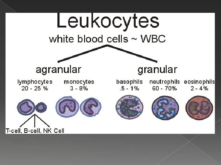

Granulocytes � Granulocytes – Granule containing WBCs. › Have lobed nuclei �Typically consist of several rounded nuclear areas connected by thin strands of nuclear material. › The granules in their cytoplasm stain specifically with Wright’s stain. › Granulocytes include the: 1. Neutrophils 2. Eosinophils 3. Basophils

Neutrophils � Multilobed nucleus. � Very fine granules that respond to both acid and basic stains. � The cytoplasm as a whole stains pink. � Avid phagocytes at sites of acute infection.

Eosinophils � Blue-red nucleus. › Resembles an old fashioned telephone receiver. Large brick-red cytoplasmic granules. � Their number increases rapidly during allergies and infections by parasitic worms (ex: tapeworms and flatworms) �

Basophils � Rarest of the WBCs. � Contain large histaminecontaining granules that stain dark blue. › Histamine – Inflammatory chemical that makes blood vessels leaky and attracts other WBCs to the inflammatory site.

Agranulocytes � Agranulocytes – lack visible cytoplasmic granules. › Their nuclei is closer to the norm – that is spherical, oval, or kidney shaped. › The agranulocytes include: 1. Lymphocytes 2. Monocytes

Lymphocytes � Have a large, dark purple nucleus that occupies most of the cell volume. � Slightly larger than RBCs. � Tend to take up residence in lymphatic tissues, where they play an important role in the immune response.

Monocytes � Largest of the WBCs. � Except for their more abundant cytoplasm and indented nucleus, they resemble large lymphocytes. � When they migrate into the tissues, they change into macrophages with huge appetites. � Macrophages are very important in fighting chronic infections.

Platelets � Platelets – One of the irregular cell fragments of blood. › Not cells in the strict sense; They are fragments of bizarre multinucleate cells called megakaryocytes. �Megakaryocytes rupture releasing thousands of anucleate “pieces” that quickly seal themselves off from the surrounding fluids.

Platelets � Appear as darkly staining, irregularly shaped bodies scattered among the other blood cells. � Needed for the clotting process.

Hematopoiesis � Hematopoiesis – Blood cell formation. › Occurs in red bone marrow. �This tissue is found chiefly in the: 1. 2. 3. 4. Flat bones of the skull and pelvis Ribs Sternum Proximal epiphyses of the humerus and femur › After the cells mature, they are discharged into the blood vessels surrounding the area.

Hemocytoblast � All the formed elements arise from a common type of stem cell, the hemocytoblast. › Hemocytoblasts resides in the bone marrow. › Their development differs and once a cell is committed to a specific blood pathway it cannot change.

The Aging of RBCs Because they are anucleate, RBCs are unable to synthesize proteins, grow, or divide. � As they age, RBCs become more rigid and begin to fragment, or fall apart in 100 -120 days. � Their remains are eliminated by phagocytes in the liver, spleen, and other body tissues. � Lost cells are replaced by the division of hemocytoblasts. �

Developing RBCs � The developing RBCs divide many times and then begin synthesizing huge amounts of Hb. › When enough Hb has been accumulated, the nucleus and most organelles are ejected and the cell collapses inward. � The entire developmental process from hemocytoblast to mature RBC takes 3 -5 days.

Erythropoietin � The rate of erythrocyte production is controlled by a hormone called erythropoietin. › Normally a small amount of erythropoietin circulates in the blood at all times, and RBC are formed at a fairly constant rate. › The kidneys play the major role in producing this hormone. �If blood levels of O 2 begin to decline, the kidneys step up their release of erythropoietin. �An excessive amount of O 2 in the bloodstream, depresses erythropoietin release.

Formation of Leukocytes and Platelets � The formation of leukocytes is stimulated by two hormones: 1. Colony Stimulating Factors (CSFs) 2. Interleukins � The production of platelets is accelerated by the hormone thrombopoietin.
- Slides: 18