
Blood Human Anatomy Physiology 1 The functions of
Blood Human Anatomy & Physiology 1
The functions of blood are: • to transport nutrients, oxygen, wastes, & hormones • to help maintain the stability of interstitial fluid (the fluid between cells) • to distribute heat • to transport substances between body cells & the external environment (helping to maintain homeostasis) Human Anatomy & Physiology 2
Blood: Blood Cells & Fluid The solid elements of blood (aka “formed” elements) make up about 45% of blood volume; they are the • red blood cells (RBCs) called erythrocytes • white blood cells (WBCs) called leukocytes • platelets called thrombocytes The blood volume of an average sized male is 5 liters. • males have more blood than females (1. 500 gallons to 0. 875 gallons) Human Anatomy & Physiology 3
Blood: Blood Cells & Fluid 1. Plasma (a clear, straw-colored liquid) makes up about 55% of blood volume. 2. Plasma consists of clotting factors, antibodies, water, amino acids, carbohydrates, lipids, vitamins, hormones, electrolytes, & cellular wastes Human Anatomy & Physiology 4

Plasma vs Serum is the liquid part of blood after clotting; therefore it is devoid of clotting factors (no fibrinogen) Plasma is the liquid, cellfree part of blood that contains clotting factors (fibrinogen) Serum = plasma - fibrinogen Human Anatomy & Physiology 5
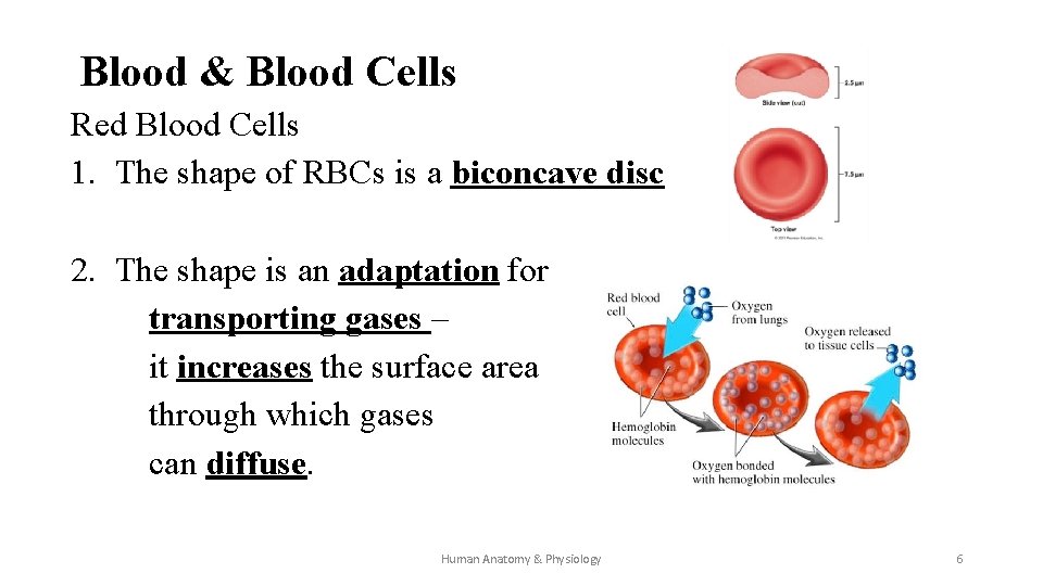
Blood & Blood Cells Red Blood Cells 1. The shape of RBCs is a biconcave disc 2. The shape is an adaptation for transporting gases – it increases the surface area through which gases can diffuse. Human Anatomy & Physiology 6
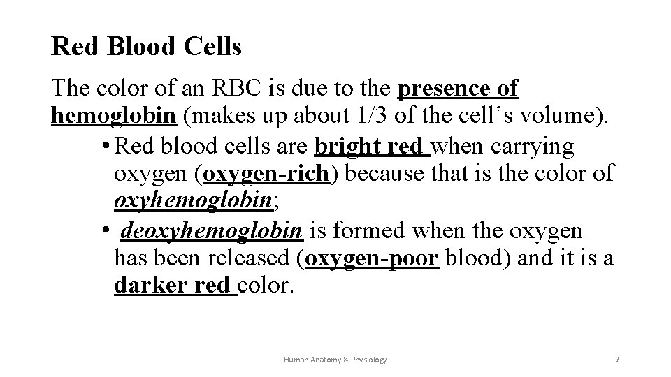
Red Blood Cells The color of an RBC is due to the presence of hemoglobin (makes up about 1/3 of the cell’s volume). • Red blood cells are bright red when carrying oxygen (oxygen-rich) because that is the color of oxyhemoglobin; • deoxyhemoglobin is formed when the oxygen has been released (oxygen-poor blood) and it is a darker red color. Human Anatomy & Physiology 7
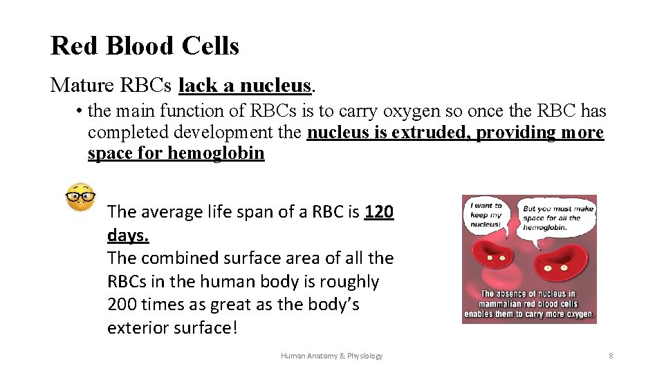
Red Blood Cells Mature RBCs lack a nucleus. • the main function of RBCs is to carry oxygen so once the RBC has completed development the nucleus is extruded, providing more space for hemoglobin The average life span of a RBC is 120 days. The combined surface area of all the RBCs in the human body is roughly 200 times as great as the body’s exterior surface! Human Anatomy & Physiology 8
Red Blood Cells RBCs are produced in different places at different life stages. • in the embryo & early fetal stages, RBCs are produced in the yolk sac • in late fetal stages, RBCs are produced in the liver & spleen • after birth, RBCs are produced in red bone marrow Human Anatomy & Physiology 9
Red Blood Cell Production Dietary factors that influence the production of RBCs are • folic acid & vitamin B 12 – essential for DNA synthesis; new blood cells are constantly being produced to replace worn out & damaged cells – DNA is necessary for the cell production • iron – essential for the production of hemoglobin Human Anatomy & Physiology 10
Red Blood Cell Counts & Production The typical red blood cell count is • 4, 600, 000 -6, 200, 000 cells per mm 3 for males and • 4, 500, 000 -5, 100, 000 cells per mm 3 for females. RBC production is controlled by negative feedback. When prolonged exposure to low oxygen levels are experienced, the kidneys & liver release the hormone erythropoietin which stimulates hematopoiesis (production of RBCs) 11
Blood & Blood Production 1) Oxygen deficiency is detected 2) Kidneys & liver release erythropoietin which travels to target cells in red bone marrow 3) Erythropoiesis begins & within a few days new RBCs appear in circulating blood 4) Oxygen levels increase; when “normal” set point is reached, erythropoietin release decreases & the rate of RBC production returns to normal Human Anatomy & Physiology 12
Red Blood Cells: Sickle –Cell Anemia In individuals affected with sickle-cell anemia, a single DNA base change causes an incorrect amino acid to be added to the globin protein chain. The defective hemoglobin crystallizes in a low oxygen environment, causing the RBCs to bend into a sickle shape. • The sickle-shaped cells cause circulation blockage in small blood vessels causing joint pain and damaging organs Human Anatomy & Physiology 13
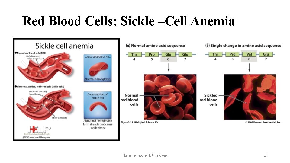
Red Blood Cells: Sickle –Cell Anemia Human Anatomy & Physiology 14
Destruction of RBCs As RBCs age they become less elastic, less flexible, & more fragile and are damaged as they pass through capillaries. Damaged RBCs are phagocytized (destroyed) by macrophages located in the spleen and liver. Human Anatomy & Physiology 15
Destruction of RBCs • The hemoglobin breaks down into heme (the ironcontaining portion) and globin (the protein portion). • The heme is further decomposed into biliverdin (a green pigment) which is eventually broken down into bilirubin (an orange pigment). Both biliverdin & bilirubin are excreted in bile. • The iron from hemoglobin may be transported to the red bone marrow tissue responsible for hematopoiesis to be reused in the production of RBCs Human Anatomy & Physiology 16
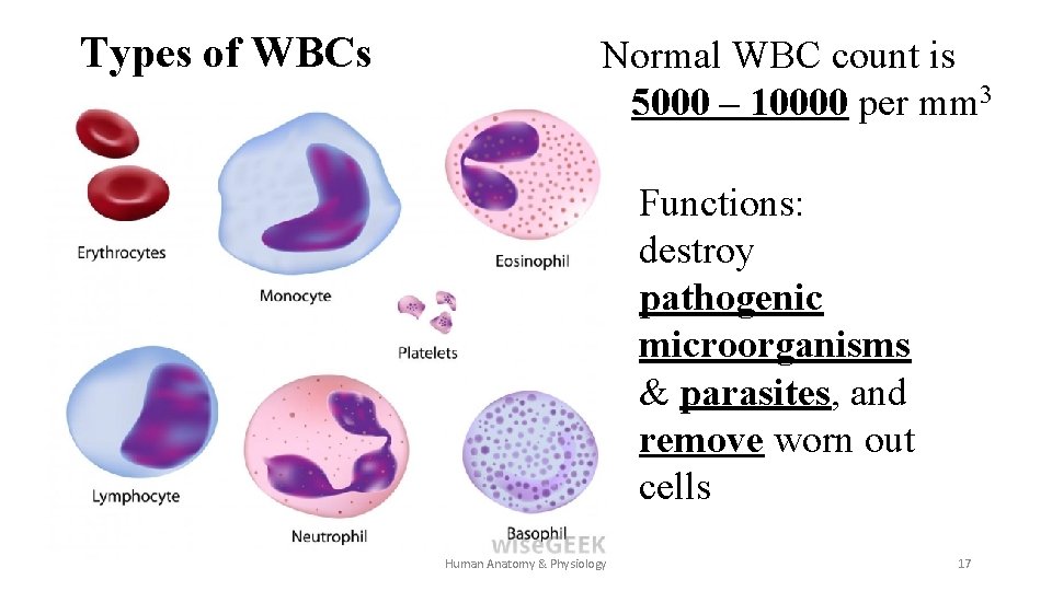
Types of WBCs Normal WBC count is 5000 – 10000 per mm 3 Functions: destroy pathogenic microorganisms & parasites, and remove worn out cells Human Anatomy & Physiology 17
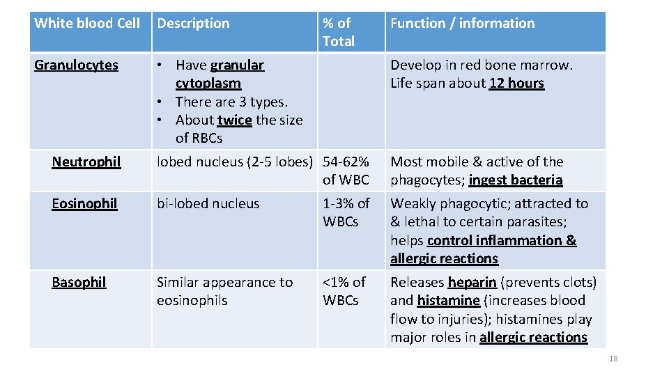
White blood Cell Description Granulocytes • Have granular cytoplasm • There are 3 types. • About twice the size of RBCs lobed nucleus (2 -5 lobes) 54 -62% of WBC bi-lobed nucleus 1 -3% of WBCs Develop in red bone marrow. Life span about 12 hours Similar appearance to eosinophils Releases heparin (prevents clots) and histamine (increases blood flow to injuries); histamines play major roles in allergic reactions Neutrophil Eosinophil Basophil % of Total <1% of WBCs Function / information Most mobile & active of the phagocytes; ingest bacteria Weakly phagocytic; attracted to & lethal to certain parasites; helps control inflammation & allergic reactions 18
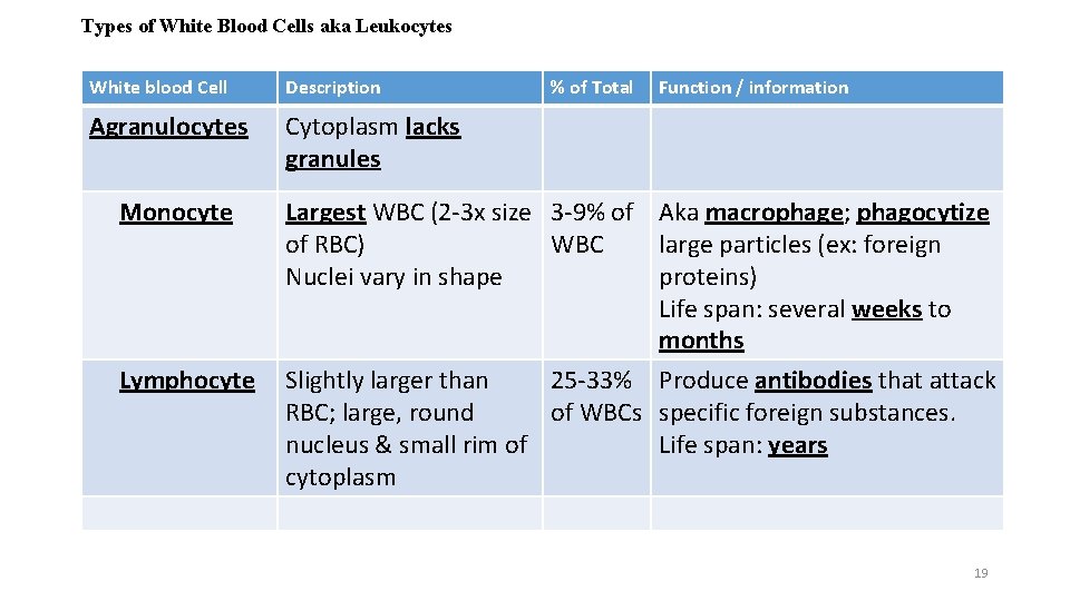
Types of White Blood Cells aka Leukocytes White blood Cell Description Agranulocytes Cytoplasm lacks granules Monocyte Lymphocyte % of Total Function / information Largest WBC (2 -3 x size 3 -9% of Aka macrophage; phagocytize of RBC) WBC large particles (ex: foreign Nuclei vary in shape proteins) Life span: several weeks to months Slightly larger than 25 -33% Produce antibodies that attack RBC; large, round of WBCs specific foreign substances. nucleus & small rim of Life span: years cytoplasm 19
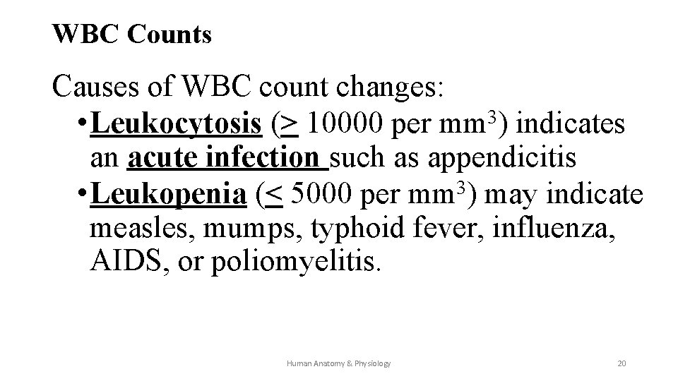
WBC Counts Causes of WBC count changes: 3 • Leukocytosis (> 10000 per mm ) indicates an acute infection such as appendicitis 3 • Leukopenia (< 5000 per mm ) may indicate measles, mumps, typhoid fever, influenza, AIDS, or poliomyelitis. Human Anatomy & Physiology 20
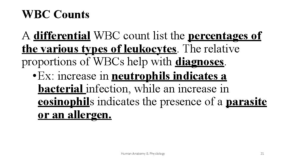
WBC Counts A differential WBC count list the percentages of the various types of leukocytes. The relative proportions of WBCs help with diagnoses. • Ex: increase in neutrophils indicates a bacterial infection, while an increase in eosinophils indicates the presence of a parasite or an allergen. Human Anatomy & Physiology 21
Platelets • Platelets (aka thrombocytes) arise from large cells in the marrow called megakaryocytes. • The megakaryocytes shatter into small fragments (platelets) that contain cytoplasm but no nucleus. 22
Platelets • Megakaryocytes & platelets increase in numbers in response to the hormone thrombopoietin. • Platelets move like an amoeba, have a life span of about 10 days, and play a vital role in blood clotting. 23
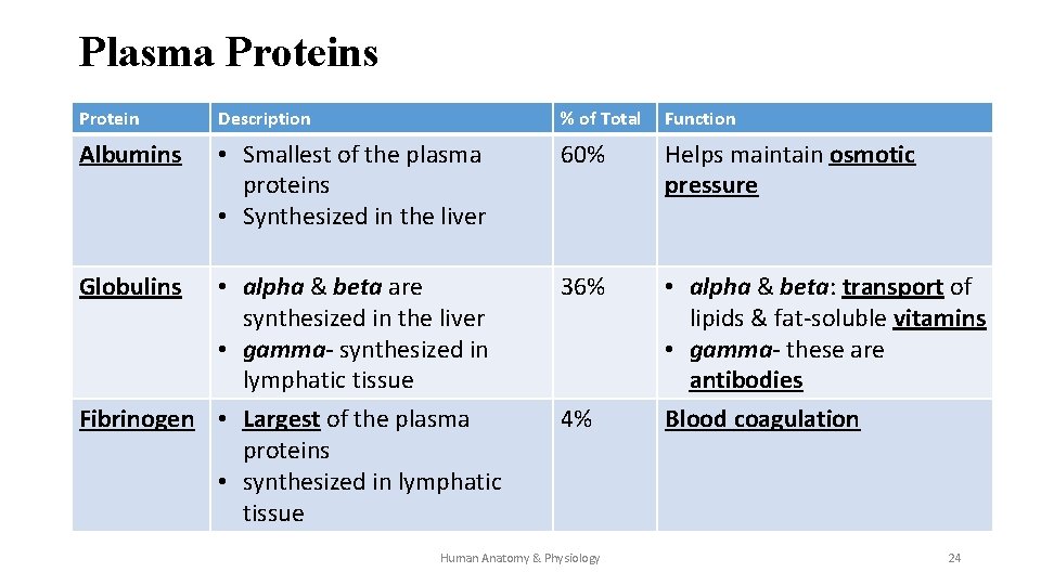
Plasma Proteins Protein Description % of Total Function Albumins • Smallest of the plasma proteins • Synthesized in the liver 60% Helps maintain osmotic pressure 36% • alpha & beta: transport of lipids & fat-soluble vitamins • gamma- these are antibodies Blood coagulation Globulins • alpha & beta are synthesized in the liver • gamma- synthesized in lymphatic tissue Fibrinogen • Largest of the plasma proteins • synthesized in lymphatic tissue 4% Human Anatomy & Physiology 24
Blood Plasma: Nutrients & Gases • Oxygen, nitrogen, carbon dioxide are the gases found on blood plasma. • The nutrients found in blood plasma are amino acids, simple sugars, nucleotides, & lipids. • Non-protein nitrogenous substances found in plasma are amino acids, urea, & uric acid (these are products of protein & nucleic acid digestion) ØLevels of these substances in the plasma can be used as a measure of kidney & liver function. Human Anatomy & Physiology 25
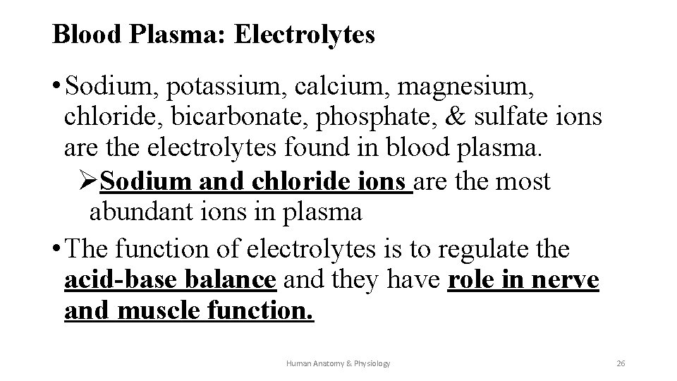
Blood Plasma: Electrolytes • Sodium, potassium, calcium, magnesium, chloride, bicarbonate, phosphate, & sulfate ions are the electrolytes found in blood plasma. ØSodium and chloride ions are the most abundant ions in plasma • The function of electrolytes is to regulate the acid-base balance and they have role in nerve and muscle function. Human Anatomy & Physiology 26
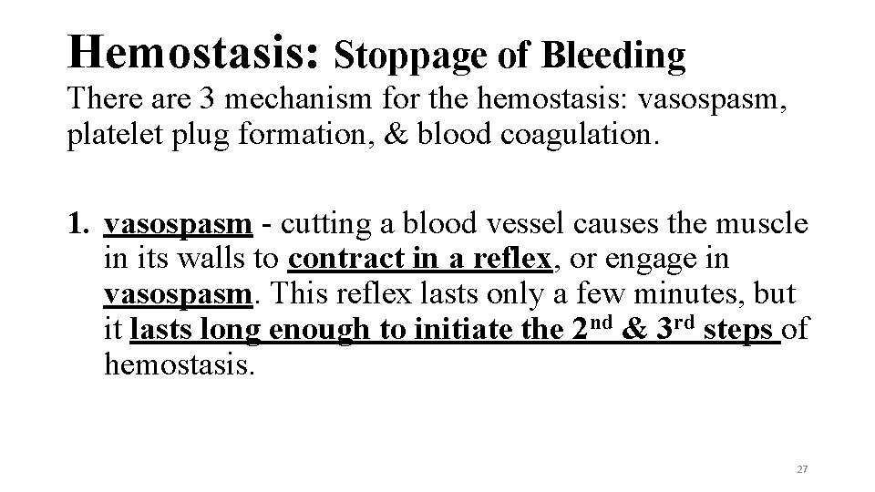
Hemostasis: Stoppage of Bleeding There are 3 mechanism for the hemostasis: vasospasm, platelet plug formation, & blood coagulation. 1. vasospasm - cutting a blood vessel causes the muscle in its walls to contract in a reflex, or engage in vasospasm. This reflex lasts only a few minutes, but it lasts long enough to initiate the 2 nd & 3 rd steps of hemostasis. 27
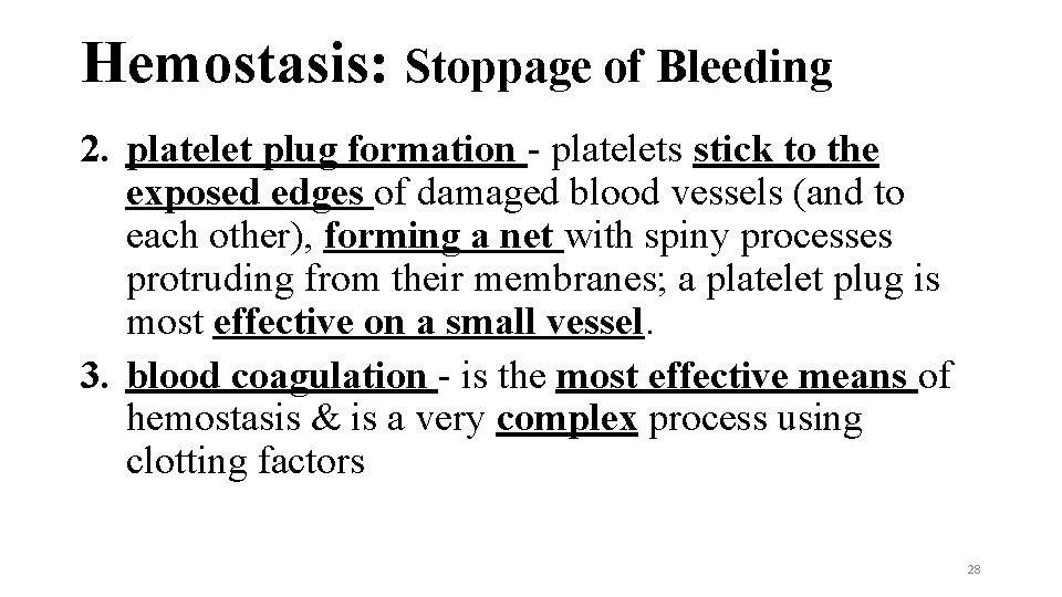
Hemostasis: Stoppage of Bleeding 2. platelet plug formation - platelets stick to the exposed edges of damaged blood vessels (and to each other), forming a net with spiny processes protruding from their membranes; a platelet plug is most effective on a small vessel. 3. blood coagulation - is the most effective means of hemostasis & is a very complex process using clotting factors 28
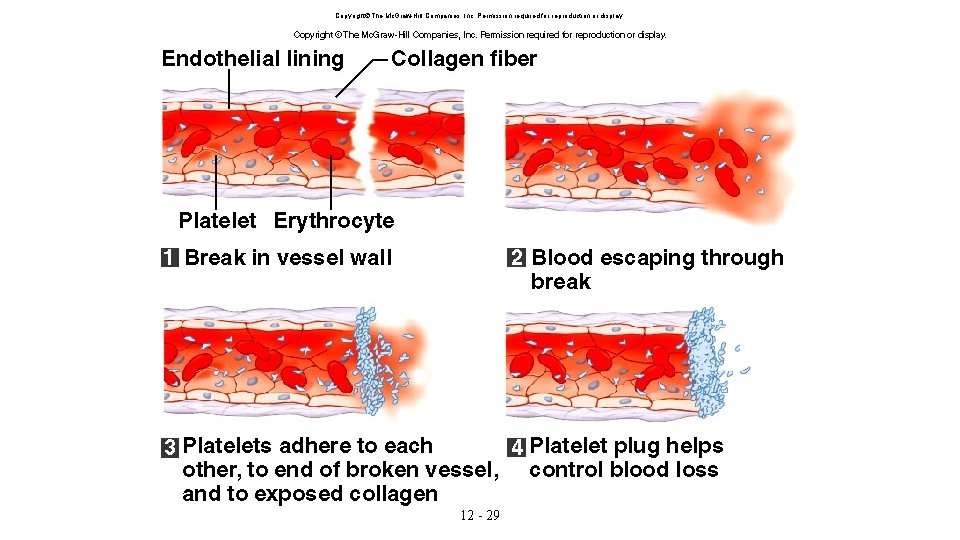
Copyright The Mc. Graw-Hill Companies, Inc. Permission required for reproduction or display. 12 - 29
Hemostasis – Blood Clot Formation Major events in the blood-clotting mechanism: 1. in the presence of calcium, prothrombin converts to thrombin 2. thrombin catalyzes the conversion of fibrinogen to long strands of fibrin 3. fibrin sticks to exposed surfaces of blood vessels forming a mesh to trap other blood elements (WBCs, RBCs, & platelets). 4. the mass formed is a blood clot. Human Anatomy & Physiology 30
Blood Clots Normal blood flow keeps the concentration of thrombin too low to cause clotting. • When blood flow is diminished or when blood pools, the concentration of thrombin increases and can lead to a blood clot or a thrombus. Thrombi vs emboli • A thrombus is a blood clot abnormally formed in a blood vessel that is not moving. • An embolus is a fragment of a clot that breaks off & is carried away by blood flow. Human Anatomy & Physiology 31
Thrombus and embolus Thrombi or emboli can be formed under any of the following circumstances: • atrial fibrillation (the most common type of cardiac arrhythmia – rate or rhythm of heartbeat) • incompetent venous valves • immobility • the formation of plaques in atherosclerosis Human Anatomy & Physiology 32
Blood Groups- Genetics Basic ABO blood group has 3 alleles (versions of the gene): IA , IB, and i alleles. IA and IB are co-dominant and i is recessive. • Type A blood can be homozygous (IA IA) or heterozygous (IA i) • Type B blood can be homozygous (IB IB) or heterozygous (IB i) • Type AB blood is IA IB • Type O blood is ii Rh blood group has 2 alleles: Rh+ and Rh • Rh+ type blood can be homozygous (Rh+ Rh+) or heterozygous (Rh+ Rh-) • Rh- type blood is Rh- Rh 33
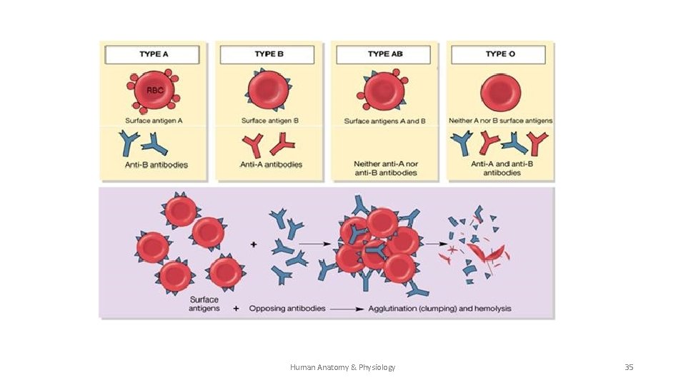
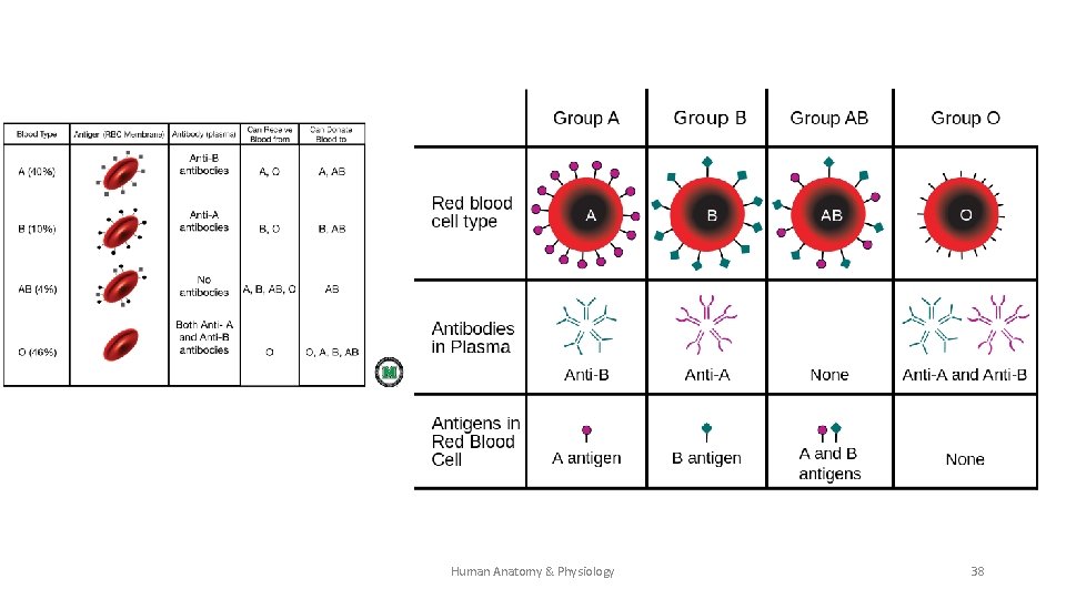
Blood Groups & Transfusions Agglutinogens & Agglutinins • Antigens (Agglutinogens) are molecules on the surface of RBCs that determine the ABO blood type. • Antibodies (agglutinins) are molecules found in the plasma that react with antigens, and if incompatible, will “clump” together (or agglutinate). ØNote: ABO antibodies form in an infants blood within 2 to 8 months following birth. These antibodies are specific for blood type. Whenever “A” antigen is absent from the body anti-A antibodies form; when “B” antigen is absent, anti-B antibodies form. Human Anatomy & Physiology 34
Human Anatomy & Physiology 35
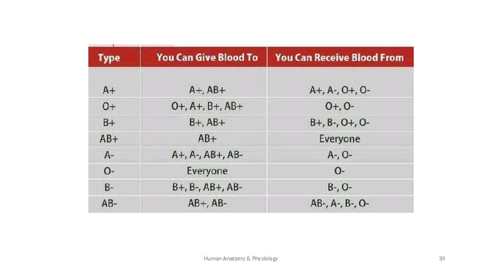
VI. 12. 5 Blood Groups & Transfusions B. ABO Blood Group 1. The presence, or lack, of specific antigens (agglutinogens) determine blood type: • A antigen only – type A blood • B antigen only – type B blood • both A & B antigens - type AB blood • Neither A nor B antigen – type O blood 2. If incompatible antigens and antibodies are mixed “clumping” (clinical term is agglutination) and transfusion reactions can occur (may have serious & life threatening consequences – see greenbox on p. 548) Human Anatomy & Physiology 36
VI. 12. 5 Blood Groups & Transfusions C. Rh Blood Group 1. There are several Rh antigens that determine if a blood type is Rh+ or Rh-. The most important is antigen D. 2. Anti-Rh antibodies do not form spontaneously. Anti-Rh antibodies form only in Rh- individuals and ONLY in response to stimulation (through exposure to Rh antigen). Human Anatomy & Physiology 37
Human Anatomy & Physiology 38
Human Anatomy & Physiology 39
Blood Groups & Transfusions 3. Rh Blood Group Erythoblastosis fetalis occurs when a woman who has already developed anti-Rh antibodies (called hemolysins and are due to a previous exposure to Rh antigens) becomes pregnant with an Rh+ fetus. The woman’s antibodies can cross the placental membrane & destroy the fetal red blood cells. This condition is rare today because physicians carefully track Rh status of pregnant females. A Rh- female who might carry a Rh+ fetus is given a Rho. GAM shot. Antibodies in the injection bind to & shield any Rh+ fetal cells. Human Anatomy & Physiology P. Wilson 40
- Slides: 40