BLOOD Honors Anatomy Chapter 17 BLOOD is a

BLOOD Honors Anatomy Chapter 17

BLOOD � is a type of CT made up of scattered cells & a liquid matrix � well oxygenated- scarlet � poorly oxygenated – dark red
 RBCs � WBCs � Platelets (plts) � 2.")
WHAT’S IN BLOOD? 1. Cells (45%) RBCs � WBCs � Platelets (plts) � 2. Plasma (55%) � water, a. a. , proteins, carbohydrates, lipids, vitamins, hormones, electrolytes, cellular waste

HEMATOCRIT � vol of blood cells in a sample of blood � blood centrifuged then % cells figured � normal levels: �Newborns: 55 -68% � 10 yr olds: 36 -40% �Women: 38 -46% �Men: 42 -54%

FUNCTIONS OF BLOOD � transports � substances maintains homeostasis in the body �regulates body temperature �maintains normal p. H in body tissues �maintains adequate fluid vol. �protects against blood loss (hemostasis) �prevents infection (abys, WBCs)

HEMO = BLOOD � hemophobia: fear of blood � hemostasis: bleeding is under control � hematocyte: blood cell � hematemesis: vomiting blood � hematuria: bloody urine � hematopoiesis: formation of blood cells

PLASMA � ~90% water � Functions: �transport nutrients, gases, vitamins, hormones �maintain fluid & electrolyte balance �maintains normal p. H

PLASMA PROTEINS 1. Albumins made in liver/ 60% of plasma proteins � maintain osmotic pressure & blood vol. � 2. Globulins α & β, from liver � transport lipids & fat-soluble vitamins � 3. Fibrinogen from liver, largest of plasma proteins � in blood clotting fibrin �

RBCS � erythrocytes, �formed � shape: hematocytes, corpuscles in bone marrow biconcave disc �allows for optimal surface area for diffusion of O 2 & CO 2 � 5 million/mm 3 � no nucleus �so � live no cell division about 120 days (no nucleus, mitochondria) �then phagocytosed in liver & spleen
 � 2. hemoglobin:")
RBCS FUNCTIONS 1. transport O 2 thru out body (lungs cells) � 2. hemoglobin: (hgb) large protein that O 2 attaches to inside RBC transports CO 2 (~20%) thru out body (cells lungs)

HEMOGLOBIN

HEMOGLOBIN � oxyhemoglobin: �plenty of oxygen being carried in RBCs, blood is bright red � deoxyhemoglobin: �not carrying much oxygen, blood is burgundy -red
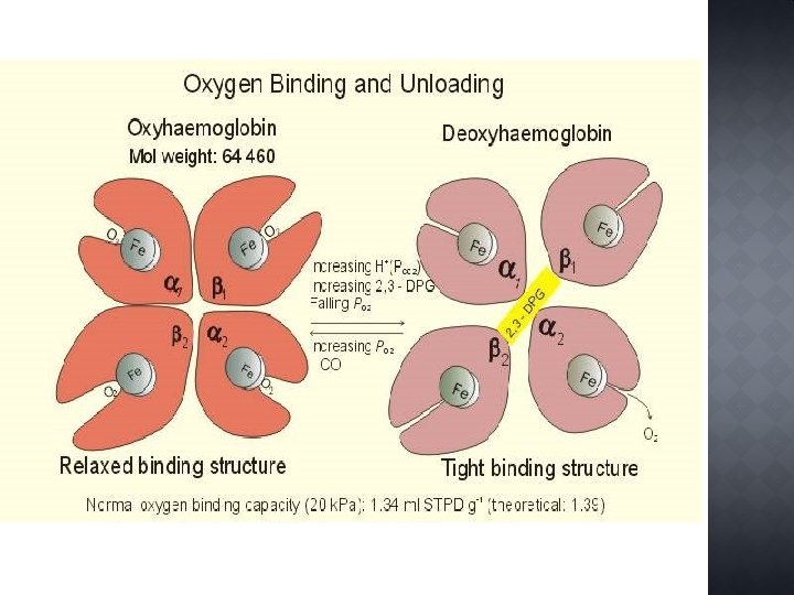

IRON � critical element needed to make hgb & normal RBCs � most of body’s Fe is in RBCs �in heme portion

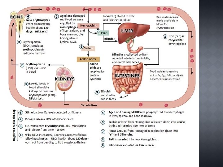
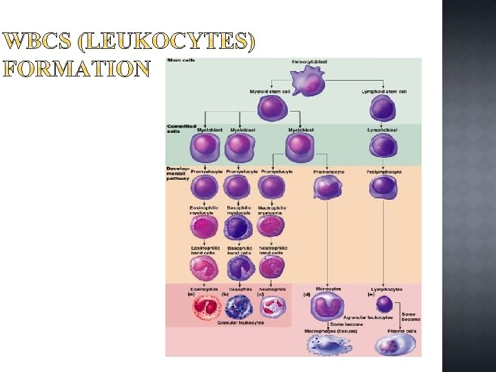
� formation of blood cells � in red bone marrow �RBCs arise from hematopoietic stem cells

� ~15 days from stem cell to reticulocyte � enter blood stream transporting oxygen � ~2 more days mature RBC � 1 – 2% of all erythrocytes �count gives indication of rate of blood cell formation

� RBCs lifespan 100 – 120 days �cannot make new proteins or organelles (no nucleus) �become rigid, fragile block smaller vessels �spleen filters out damaged RBCs �engulfed by macrophages

� “lacking blood” � condition in which O 2 carrying capacity is too low to support normal metabolism � it‘s a sign of some disorder not a disease � 3 basic causes: 1. Blood loss 2. RBC production low 3. RBC destruction more than normal

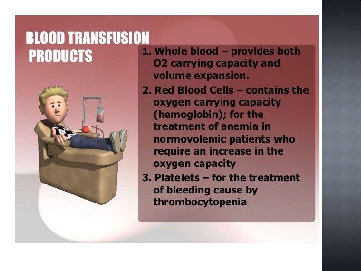
� Hemorrhagic Anemia Acute 1. rapid, heavy � tx‘d with blood transfusions � Chronic 2. slight, persistent blood loss � example: GI bleed, menorrhagia � tx‘d by treating underying cause �

Fe-deficiency Anemia 1. often 2° to hemorrhagic anemia � or from lo Fe consumption in diet � RBCs small: microcytic, pale � tx‘d: Fe supplements or increase in diet � Pernicious Anemia 2. � � � autoimmune disease elderly’s stomach mucosa cells that release intrinsic factor damaged less absorption of Vit B 12 in intestines RBCs macrocytic, grow but cannot divide tx‘d: B 12 shots

�Microcytic RBCs �Macrocytic RBCs

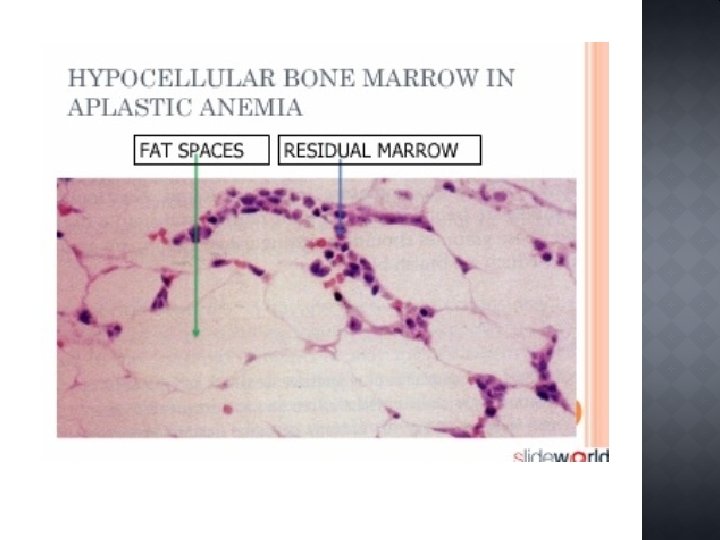
� most cases, cause unknown � Hallmark: marrow destruction �all blood cells depleted: see clotting defects, immunity impaired � tx‘d: stopgap: transfusions �stem cells/ bone marrow transplant

Thalassemias 1. Mediterreanean descent, many subtypes � 1 of globin chains absent or faulty � mild severe cases � tx‘d: monthly transfusions � Sickle Cell 2. Β chains have 1 abnormal amino acid � RBCs sickle in conditions of lo O 2 block small blood vessels lo O 2 delivery causes pain, SOB � tx‘d: transfusions/ O 2 � new: inhale nitric oxide dilates vessels �
 � leukocytes � general function: defend the body against pathogens")
WHITE BLOOD CELLS (WBCS) � leukocytes � general function: defend the body against pathogens

WHITE BLOOD CELLS Type Name Function Granulocytes Neutrophils aka PMNs polymorphoneutrophils very active in phagocyting bacteria & are present in large #s in pus of wounds, most common of all types, normal= 60% of WBCs Eosinophils attack parasites, control allergic reactions 2% of WBC count (granular cytoplasm) Picture
")
WHITE BLOOD CELLS type Name Function Granulocytes continued Basophils produces heparin (prevents blood clots) & histamines (inflammatory reaction) 1% of WBC Agranulaocytes (lacking granular cytoplasm) Monocytes precursors of macrophages; 6% of WBC Lymphocytes main cell of immune system 30% of WBC Picture
 � thrombocytes � cell fragments formed from megakaryocyte, live ~4 days �")
PLATELETS (PLTS) � thrombocytes � cell fragments formed from megakaryocyte, live ~4 days � help initiate formation of blood clots �release clotting factors
: anythin that induces")
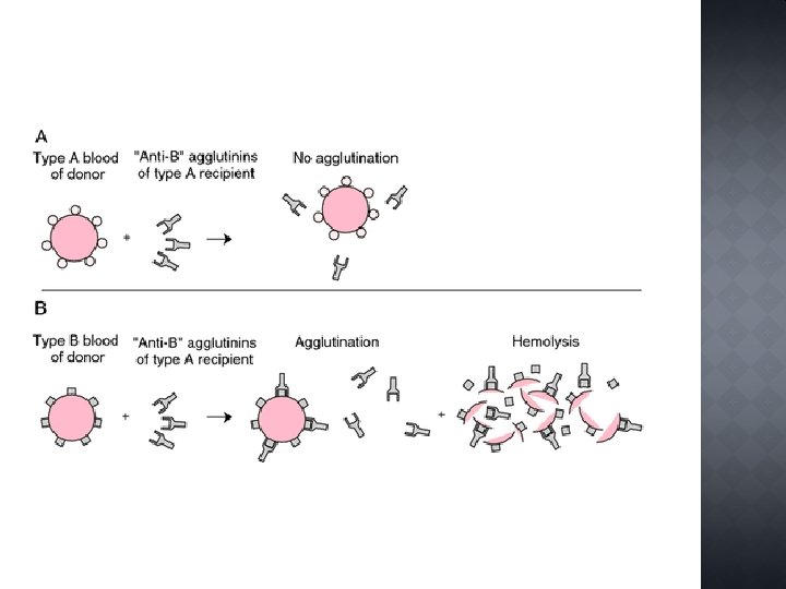
� glycoproteins on plasma membranes of RBCs act as antigens (agns): anythin that induces immune response � RBCs from different blood type will agglutinate (clump) � ABO & Rh blood groups cause vigorous transfusion reactions if not properly matched
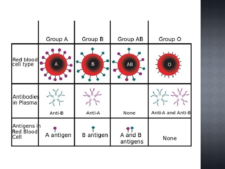

� ~85% of Americans Rh+ � antibodies not spontaneously in human blood
- Slides: 37