BLOOD GROUPS Red very important Green only found

BLOOD GROUPS Red: very important. Green: only found in males’ slides. Purple: only found in females’ slides. Gray: notes. Physiology Team 436 – Foundation block lecture 11 1 Lecture: If work is intended for initial studying Review: If work is intended for revision.

Objectives �Describe ABO blood groups types. �Recognize Agglutinin in plasma. �Describe genetic inheritance of Blood groups. �Recognize transfusion reactions. �Describe Rhesus blood groups. �Describe causes of hemolytic disease of the newborn. 2

Blood Group System � Blood Groups are determined by: Antigens on the surface of RBCs. Remember: Antigens can stimulate the immune system to produce antibodies. � Examples of antigens: v [glycoproteins, complex oligosaccharides that differ in their terminal sugar. ] � About 20 blood group systems are known: v [E. g. ABO System, Rh-System, MNS System, Kell System, Lewis v Karl Landsteiner is an Austrian scientist that: System, Duffy, Lutheran, KIDD] • � The two most common blood groups: v ABO Blood group System. v Rh (Rhesus)Blood group System. 3 • • Discovered the ABO Blood group system in 1901 Discovered Rh factor in 1930 along with Alexander S. Wiener Noble prize in Physiology/Medicine in 1930

ABO & Rh Blood group systems ABO System Rh System v Depends on : the presence of one or both or neither of the two blood antigens (A) and (B) on the surface of the RBC. v Four main ABO groups: A, B, AB, O v A and B blood groups are dominant over the O blood group. v A and B group genes are co-dominant. v Depends on: the presence or absence of the Rhesus antigen (D) on the surface of RBC. (Co-dominance: form of dominance in which the alleles of a gene pair in a heterozygote are fully expressed thereby resulting in offspring with a phenotype that is neither dominant nor recessive) v Presence of D (individual is Rh+ve) [85%] v Absence of D (individual is Rh–ve) [15%] v Rhesus antigens: Dd, Cc, Ee v (Clinically most important is D) v Locus of alleles responsible for Rh Factor is on the long arm of chromosome 1. v Each person has two copies of genes coding for their ABO blood group (one maternal and one paternal in origin) v Locus of alleles responsible for the ABO system is on the long arm of chromosome 9. 4 Locus: a particular position or place where v They were named Rhesus antigens or Rh antigens because something occurs. the same antigens are present in the Rhesus monkey.
 (A&B)")
Definitions Agglutinogens Agglutinin Agglutination • It is Blood group Antigens (on RBCs membrane) (A&B) • It is The respective antibody to antigen • Or It is Antibodies in serum (In Plasma) • Agglutinins are immunoglobulins (Ig. M & Ig. G): Proteins which appear in the plasma or body fluids in response to administration of antigens. • It is a Reaction (clumping) between Agglutinogens on RBC membrane and the respective Anti. Body • In transfusion reaction If a person with blood group A was transfused with blood of group B which contains anti-A in plasma • The anti-A in plasma of recipient with blood group B will agglutinate the transfused cell (A). • The clumped cells plug (block) small blood vessels. • Sometimes causes hemolysis. Landsteiner Law (1900): v If an agglutinogen (antigen) is present on the RBC of an individual, the corresponding agglutinin (antibody) must be absent in the plasma of that individual and viceversa. This law is only applicable to ABO blood grouping system. 5

Genetic Determination of Agglutinogens Genotype for each blood group: A AA , AO B BB , BO AB O OO Uses of genotypes: • Sorting disputes in paternal dispute • Frequency of ABO has ethnic variation 6 These are genotypes )ﻃﺮﺍﺯ ( ﺟﻴﻨﻲ

Agglutinin /Antibodies �Examples of Agglutinins: Anti-A & Anti-B antibodies. Anti-D antibodies. • NOT naturally-occurring antibodies. • Acquired by: • Transfusion of Rh-ve individual with Rh+ve blood. • Rh-ve pregnancy with Rh+ve faetus. • Are considered as Ig. G class. Example: if my blood type is B+, I will have antigen B and • Can cross placenta. • Naturally occurring antibodies. • Not present at birth, appear 2 -8/12 weeks (2 to 8 months) after birth. • Triggered by A & B antigens in food and bacteria. • Are considered as Ig. M class. (large molecule) • Cannot cross placenta. antigen D, and I will have anti A. 7

Agglutinin Titer of Anti- A&B Agglutinins at Different Ages The Rh immune response Formation of anti-Rh agglutinin: Rh +ve blood • 2 to 8 months after birth, an infant begins to produce agglutinins. • A maximum titer is usually reached at 8 to 10 years of age, and this gradually declines throughout the remaining years of life. 8 Transfused for the first time Rh –ve person Anti-Rh agglutinins develop slowly (2 -4 months). Once produced they persist for years and can produce serious reactions during 2 nd transfusion. (so, it can only be done once). Titer: the concentration of an antibody, as determined by finding the highest dilution at which it is still able to cause agglutination of the antigen.
 A Agglutinin (Antibody) Anti-B")
Comparing between Agglutinogens & Agglutinin Blood Group A Agglutinogen (Antigen) A Agglutinin (Antibody) Anti-B B B Anti-A AB A &B - - Anti A+Anti B O Example: If a baby has blood group (antigen) A, and is infected with a bacteria with antigen A, nothing will happen. If the baby is infected with bacteria with antigen B, so he will produce antibody Anti B. 9

Agglutination • If there is agglutination in the blood drop when Anti-A was used that means the patient's blood group is A. • Also: Anti-B → agglutination → blood group B. • If there is agglutination in the blood drop when we used both Anti. A and Anti-B that means the patient's blood group is AB. • If there isn’t agglutination in the drop patient blood when we used both Anti-A and Anti-B that mean patient's blood group is O. When the antigen and antibody are the same type, it can be dangerous and may lead to anemia. 10

Agglutination 11 Girls slide : circles from doctor

Importance of blood groups In blood transfusion. In preventing hemolytic disease (Rh incompatibility between mother and fetus) In paternity disputes (to determine the father) In medicolegal cases ( )ﺍﻟﺠﺮﺍﺋﻢ In knowing susceptibility to disease: 1. 2. 3. 4. 5. • • Group O: duodenal cancer. Group A: carcinoma of stomach, pancreas & salivary glands. * Color just to make it clear for the next slides 12
 � Blood test before transfusion :")
1. Blood Transfusions ( Donors and Recipients ) � Blood test before transfusion : A drop pf patient RBC Blood group type of patient (recipient) Anti-A Anti-D Anti-B (next slide) • Cross-matching : donor RBC + recipient’s (patient) serum (plasma) • People with blood group O are called “universal donors” people with blood group AB are called “universal recipients”. 13

Blood group type of patient Blood being tested Type AB (contains agglutinogens A and B; agglutinates with both serum) Type A (contains agglutinogen A; agglutinates with anti-A) Type B (contains agglutinogen B; agglutinates with anti-B) Type O (contains no agglutinogens; does not agglutinate with either serum) 14 serum Anti-A Anti-B

2. Complications of Blood Transfusions 1 - Immune reaction: incompatible blood transfusions leading to immediate or delayed reaction, fever, hemolysis, allergic reaction. (appears within 10 to 15 minutes of transfusion) 2 - Transmission of diseases: malaria, syphilis, viral hepatitis, and AIDs virus. 3 - Iron overload: due to multi-transfusion in case of sickle cell anemia and thalassemia. 4 - Acute kidney failure: (reaction to mismatched transfusions ) 15
 of Mismatched Transfusions § 2 Types: � Immediate: • • Agglutination Hemolysis")
Hazards (Risks) of Mismatched Transfusions § 2 Types: � Immediate: • • Agglutination Hemolysis Fever Allergic reaction Acute renal shutdown Renal vasoconstriction Circulatory shock Tubular blockage 16 � Delayed: • • Sensitization Thrombophlebitis

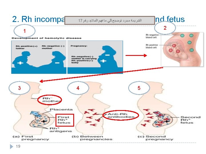
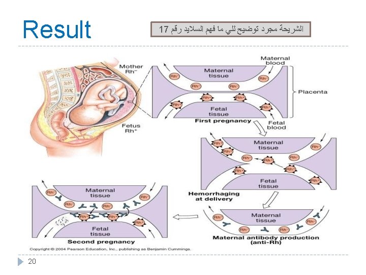
3. Rh incompatibility between mother and fetus � Mother is Rh –ve and the first baby is Rh +ve (has D antigen) : (father is +ve which is more dominant therefore : baby is +ve) � At delivery: Fetal Rh +ve RBC cross to maternal blood. • The mother will develop anti-D after delivery. If it was the opposite: mom is positive • First child escapes & is safe. and baby is negative, nothing will • happen. If the mother was transfused before with Rh +ve blood before, the first child will be affected. 17 � Second fetus (baby): Rh +ve If Rh +ve: Antibody-D crosses placenta from mom to baby and destroys fetal Rh +ve RBCs. � Outcome: • Hemolytic disease of the newborn. •
 1. Hemolytic anemia: If severe: is treated")
Hemolytic disease of new born (Erythroblastosis fetalis) 1. Hemolytic anemia: If severe: is treated with exchange transfusion: Replace baby’s blood with Rh –ve RBC (several times). 2. Hydrops fetalis: (death in utero) 3. Kernicterus : is a brain damage in newborn with sever jaundice (jaundice : caused by high level of bilirubin in baby’s blood )) Prevalence of disease: 1 st pregnancy: 0% 2 nd pregnancy: 3% 3 rd pregnancy: 10% Prevention: After 1 st childbirth : Injecting the mother with antibody. D immediately after 1 st childbirth. Antenatal (during pregnancy) : prophylaxis. 21
 Blood groups cant be used to prove paternity,")
3. Paternity (to determine the father) Blood groups cant be used to prove paternity, but can be used to disprove it. Red : what we Mother Question 1: are sure about Nora: blood type A and Ahmad: blood type B Have a baby: blood type O. Can Ahmad be the father? Possible Answer 1: Phenotype genotypes Ahmad can be the father, but not 100% A AA or AO sure B BB or BO O OO Question 2: Answer 2: The father can NOT be type O. Here we disproved paternity. Mother: blood type A Daughter: blood type B Can the father be blood type O? 22 from question Blue: we solved Father A O B O Red : what we are sure about from the question Bab y OO Mother A O or A O Fathe O r Baby B O/B

Thank you! . ﻋﻤﻼ ﺃﺤﺴﻦ ﻣﻦ ﺃﺠﺮ ﻳﻀﻴﻊ ﻻ ﺍﻟﻠﻪ ﺃﻦ ﺗﻌﻠﻢ ﺃﻨﺖ ﻭ ﺍﻋﻤﻞ ، ﺩﻣﻌﺔ ﻟﺘﻤﺴﺢ ﺍﻋﻤﻞ ، ﺑﺴﻤﺔ ﻟﺘﺮﺳﻢ ﺍﻋﻤﻞ The Physiology 436 Team: Lina Alwakeel Fahad Al Fayez Ibrahim Al Deeri Rana Barassain Hassan Al Shammari Heba Alnasser Abdullah Al Otaibi Munira Aldofayan Abdullah Al Subhi Al Subaei Sara Alshamrani Omar Al Babteen Sundus Alhawamda Foad Fathi Ruba Ali Faisal Al Fawaz Muhammad Al Rehab Alanazi Aayed Norah Alshabib Muhammad Al Mutlaq Nasser Abu Dujeen Nouf Alaqeeli Waleed Al Asqah Buthaina Almajed Alaa Alaqeel 23 Team Leaders: Qaiss Almuhaideb Lulwah Alshiha Contact us: Physiology 436@gmail. com @Physiology 436
- Slides: 23