
Blood Gas Analysis MOTIVATION FOR LEARNING ABOUT BLOOD
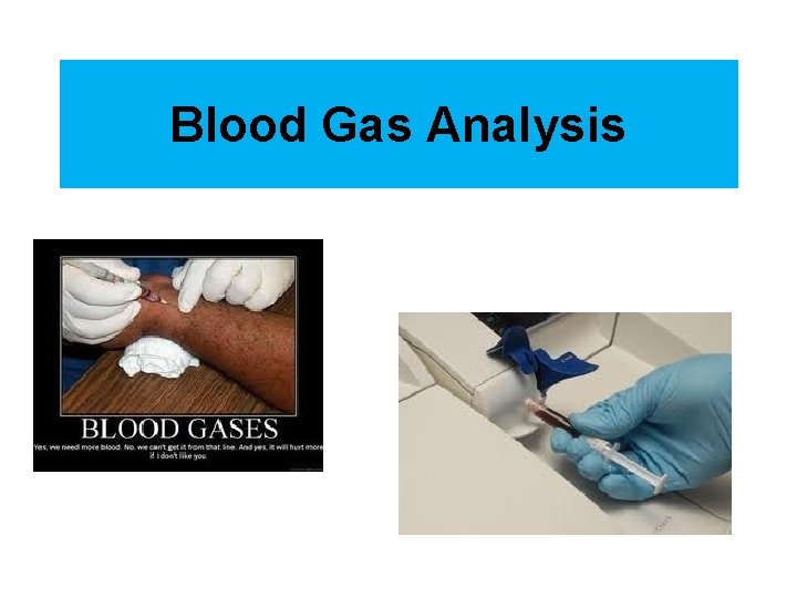
Blood Gas Analysis

MOTIVATION FOR LEARNING ABOUT BLOOD GAS INTERPRETATION
MOTIVATION In a survey conducted at a university teaching hospital, 70% of the participating physicians claimed that they were well versed in the diagnosis of acid-base disorders and that they needed no assistance in the interpretation of arterial blood gases (ABGs). These same physicians were then given a series of ABG measurements to interpret, and they correctly interpreted only 40% of the test samples. Hingston DM. A computerized interpretation of arterial p. H and blood gas data: do physicians need it? Respir Care 1982; 27: 809 -815. From: THE ICU BOOK - 2 nd Ed. (1998)
MOTIVATION A survey at another teaching hospital revealed that incorrect acid-base interpretations led to errors in patient management in one-third of the ABG samples analyzed. Broughton JO, Kennedy TC. Interpretation of arterial blood gases by computer. Chest 1984; 85: 148 -149. From: THE ICU BOOK - 2 nd Ed. (1998)
MOTIVATION These surveys reveal serious deficiencies in an area that tends to be ignored. This can cause trouble in the ICU, where 9 of every 10 patients may have an acid-base disorder. Gilfix BM, Bique M, Magder S. A physical chemical approach to the analysis of acid-base balance in the clinical setting. J Crit Care 1993; 8: 187 -197. From: THE ICU BOOK - 2 nd Ed. (1998)
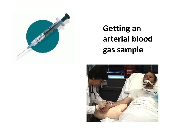
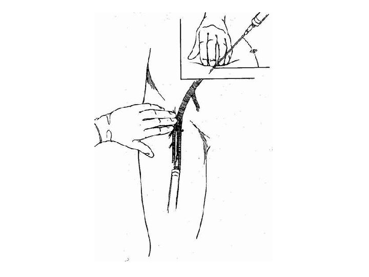
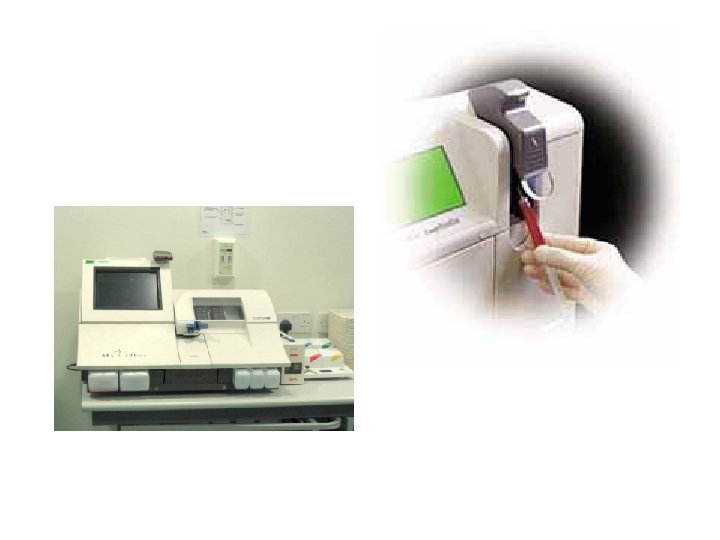
Getting an arterial blood gas sample
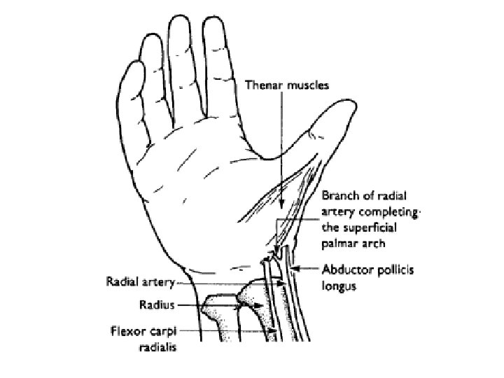
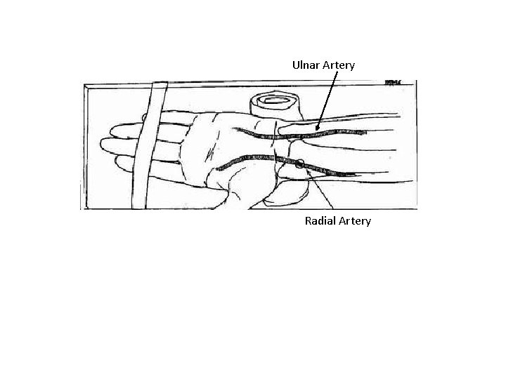
Ulnar Artery Radial Artery
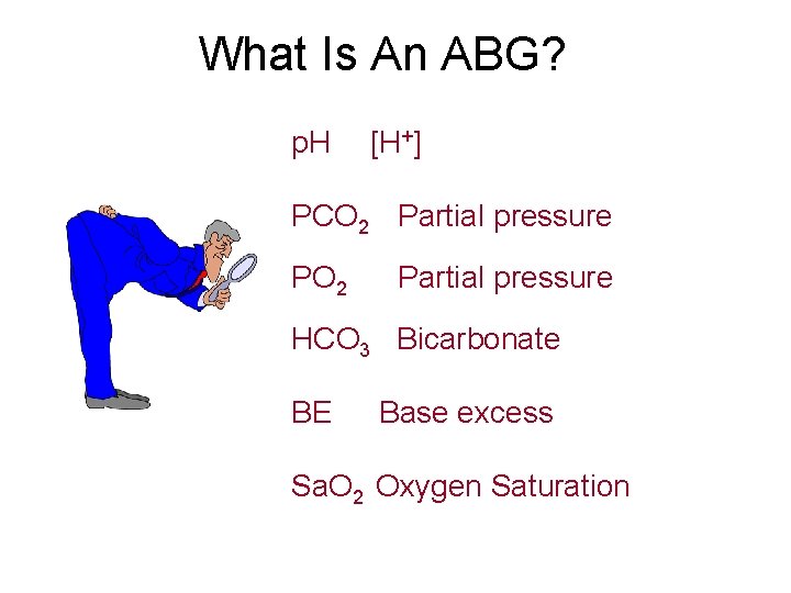
What Is An ABG? p. H [H+] PCO 2 Partial pressure PO 2 Partial pressure HCO 3 Bicarbonate BE Base excess Sa. O 2 Oxygen Saturation
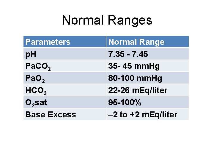
Normal Ranges Parameters p. H Pa. CO 2 Pa. O 2 HCO 3 O 2 sat Base Excess Normal Range 7. 35 - 7. 45 35 - 45 mm. Hg 80 -100 mm. Hg 22 -26 m. Eq/liter 95 -100% – 2 to +2 m. Eq/liter
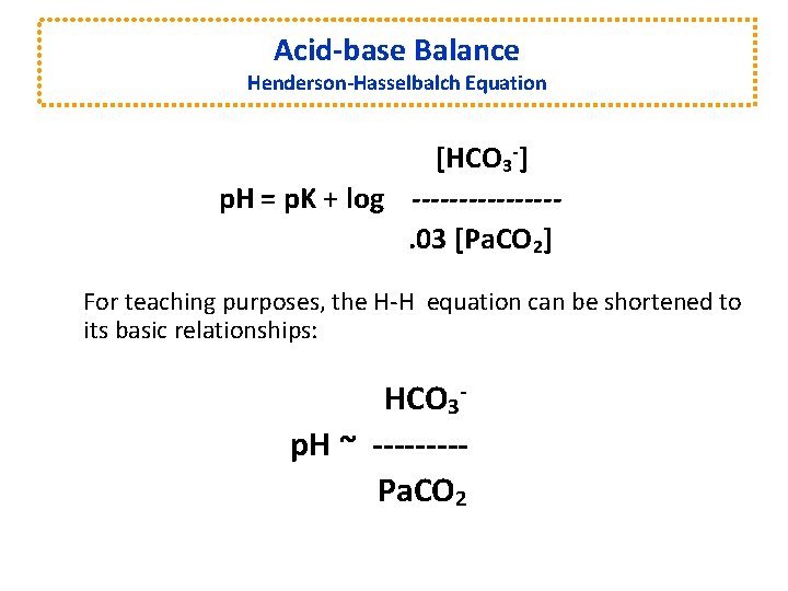
Acid-base Balance Henderson-Hasselbalch Equation [HCO 3 -] p. H = p. K + log --------. 03 [Pa. CO 2] For teaching purposes, the H-H equation can be shortened to its basic relationships: HCO 3 p. H ~ ----Pa. CO 2
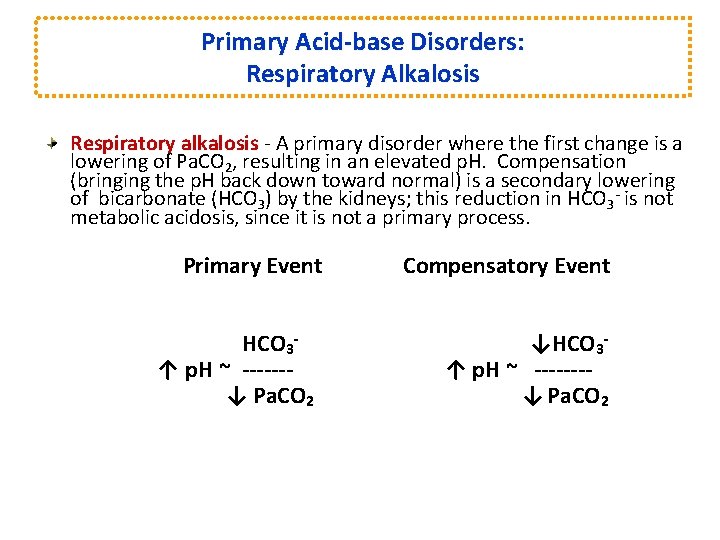
Primary Acid-base Disorders: Respiratory Alkalosis Respiratory alkalosis - A primary disorder where the first change is a lowering of Pa. CO 2, resulting in an elevated p. H. Compensation (bringing the p. H back down toward normal) is a secondary lowering of bicarbonate (HCO 3) by the kidneys; this reduction in HCO 3 - is not metabolic acidosis, since it is not a primary process. Primary Event HCO 3↑ p. H ~ ------↓ Pa. CO 2 Compensatory Event ↓HCO 3↑ p. H ~ -------↓ Pa. CO 2
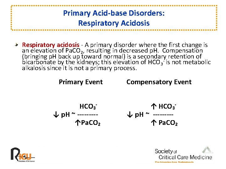
Primary Acid-base Disorders: Respiratory Acidosis Respiratory acidosis - A primary disorder where the first change is an elevation of Pa. CO 2, resulting in decreased p. H. Compensation (bringing p. H back up toward normal) is a secondary retention of bicarbonate by the kidneys; this elevation of HCO 3 - is not metabolic alkalosis since it is not a primary process. Primary Event HCO 3↓ p. H ~ ----↑Pa. CO 2 Compensatory Event ↑ HCO 3↓ p. H ~ ----↑ Pa. CO 2
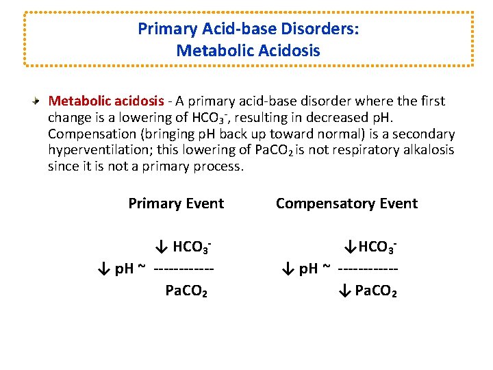
Primary Acid-base Disorders: Metabolic Acidosis Metabolic acidosis - A primary acid-base disorder where the first change is a lowering of HCO 3 -, resulting in decreased p. H. Compensation (bringing p. H back up toward normal) is a secondary hyperventilation; this lowering of Pa. CO 2 is not respiratory alkalosis since it is not a primary process. Primary Event ↓ HCO 3↓ p. H ~ ------Pa. CO 2 Compensatory Event ↓HCO 3↓ p. H ~ ------↓ Pa. CO 2
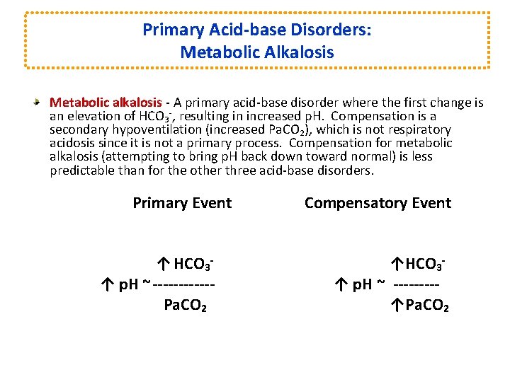
Primary Acid-base Disorders: Metabolic Alkalosis Metabolic alkalosis - A primary acid-base disorder where the first change is an elevation of HCO 3 -, resulting in increased p. H. Compensation is a secondary hypoventilation (increased Pa. CO 2), which is not respiratory acidosis since it is not a primary process. Compensation for metabolic alkalosis (attempting to bring p. H back down toward normal) is less predictable than for the other three acid-base disorders. Primary Event ↑ HCO 3↑ p. H ~------Pa. CO 2 Compensatory Event ↑HCO 3↑ p. H ~ ----↑Pa. CO 2
Steps to an Arterial Blood Gas Interpretation • Acid-base evaluation requires a focus on three of the reported components: Ø p. H Ø Pa. CO 2 Ø HCO 3
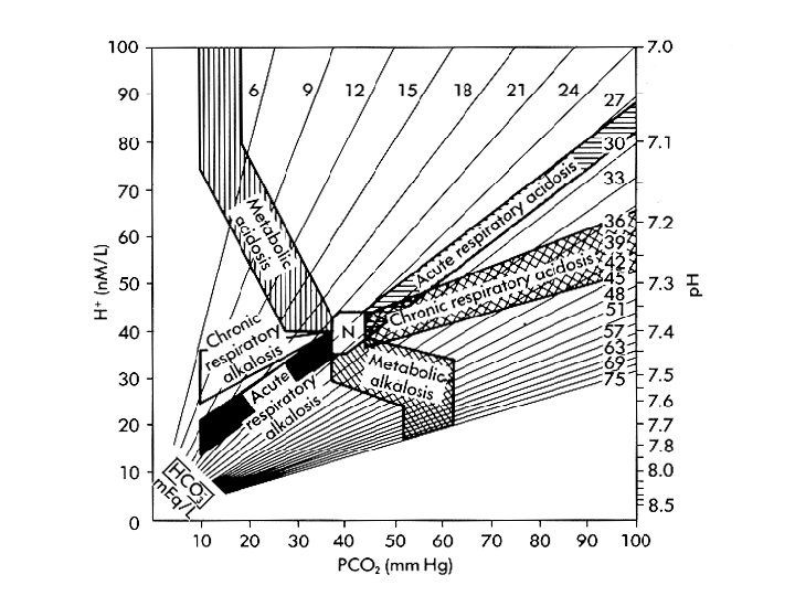
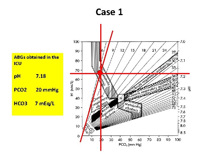
Case 1 ABGs obtained in the ICU p. H 7. 18 PCO 2 20 mm. Hg HCO 3 7 m. Eq/L
Case 2 Arterial blood gas sample is taken, revealing the following: p. H PCO 2 : : 7. 30 65 mm Hg
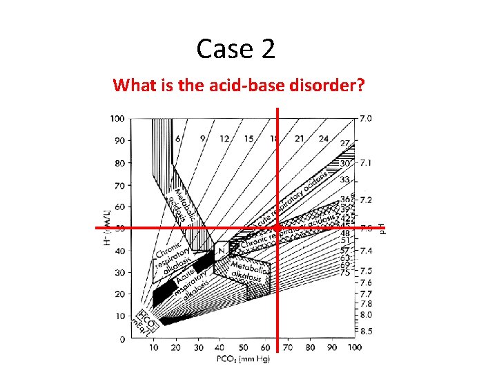
Case 2 What is the acid-base disorder?
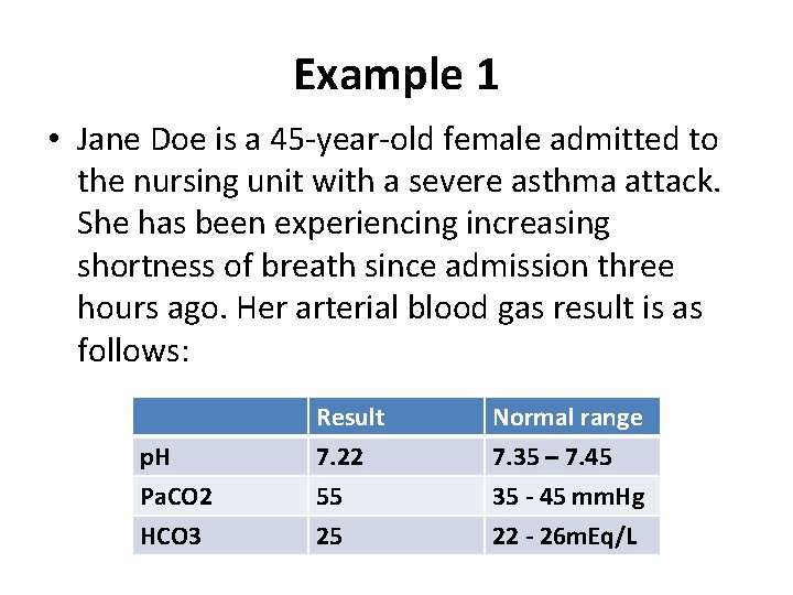
Example 1 • Jane Doe is a 45 -year-old female admitted to the nursing unit with a severe asthma attack. She has been experiencing increasing shortness of breath since admission three hours ago. Her arterial blood gas result is as follows: p. H Pa. CO 2 HCO 3 Result 7. 22 55 25 Normal range 7. 35 – 7. 45 35 - 45 mm. Hg 22 - 26 m. Eq/L
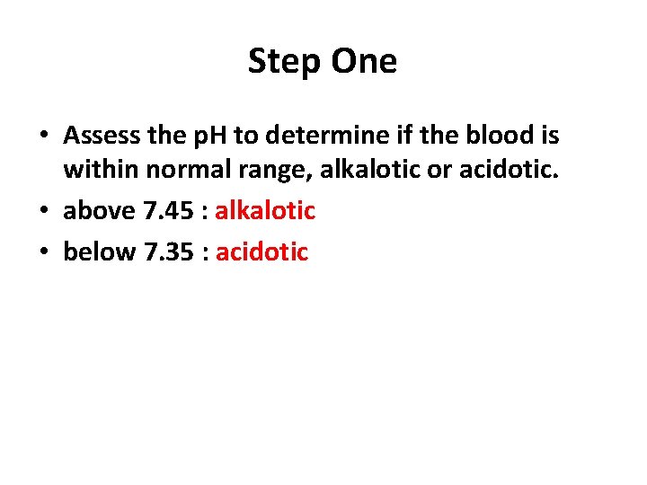
Step One • Assess the p. H to determine if the blood is within normal range, alkalotic or acidotic. • above 7. 45 : alkalotic • below 7. 35 : acidotic
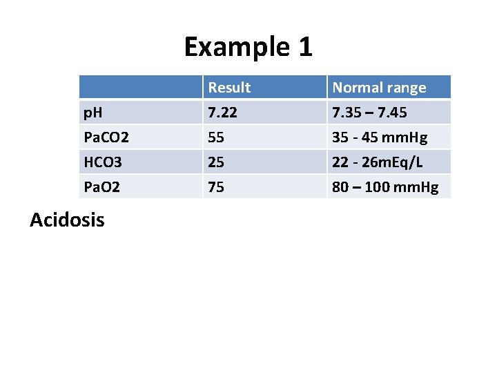
Example 1 p. H Pa. CO 2 HCO 3 Result 7. 22 55 25 Normal range 7. 35 – 7. 45 35 - 45 mm. Hg 22 - 26 m. Eq/L Pa. O 2 75 80 – 100 mm. Hg Acidosis
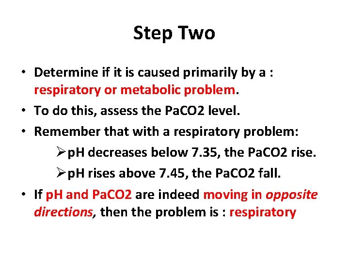
Step Two • Determine if it is caused primarily by a : respiratory or metabolic problem. • To do this, assess the Pa. CO 2 level. • Remember that with a respiratory problem: Øp. H decreases below 7. 35, the Pa. CO 2 rise. Øp. H rises above 7. 45, the Pa. CO 2 fall. • If p. H and Pa. CO 2 are indeed moving in opposite directions, then the problem is : respiratory
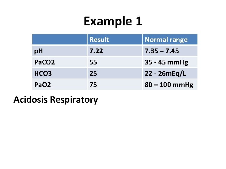
Example 1 p. H Pa. CO 2 HCO 3 Result 7. 22 55 25 Normal range 7. 35 – 7. 45 35 - 45 mm. Hg 22 - 26 m. Eq/L Pa. O 2 75 80 – 100 mm. Hg Acidosis Respiratory
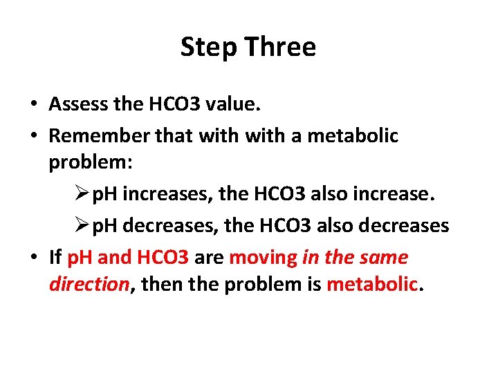
Step Three • Assess the HCO 3 value. • Remember that with a metabolic problem: Øp. H increases, the HCO 3 also increase. Øp. H decreases, the HCO 3 also decreases • If p. H and HCO 3 are moving in the same direction, then the problem is metabolic.
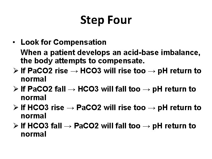
Step Four • Look for Compensation When a patient develops an acid-base imbalance, the body attempts to compensate. Ø If Pa. CO 2 rise → HCO 3 will rise too → p. H return to normal Ø If Pa. CO 2 fall → HCO 3 will fall too → p. H return to normal Ø If HCO 3 rise → Pa. CO 2 will rise too → p. H return to normal Ø If HCO 3 fall → Pa. CO 2 will fall too → p. H return to normal
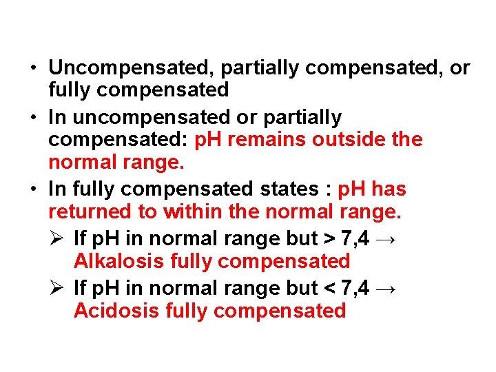
• Uncompensated, partially compensated, or fully compensated • In uncompensated or partially compensated: p. H remains outside the normal range. • In fully compensated states : p. H has returned to within the normal range. Ø If p. H in normal range but > 7, 4 → Alkalosis fully compensated Ø If p. H in normal range but < 7, 4 → Acidosis fully compensated
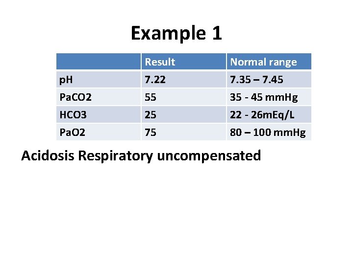
Example 1 p. H Pa. CO 2 HCO 3 Result 7. 22 55 25 Normal range 7. 35 – 7. 45 35 - 45 mm. Hg 22 - 26 m. Eq/L Pa. O 2 75 80 – 100 mm. Hg Acidosis Respiratory uncompensated

Step Five • Assess the Pa. O 2. • A value below 80 mm Hg can indicate hypoxemia, depending on the age of the patient. • If Pa. O 2 < 80 mm. Hg : Hypoxemia • If Pa. O 2 > 100 mm. Hg : Hyperoxemia
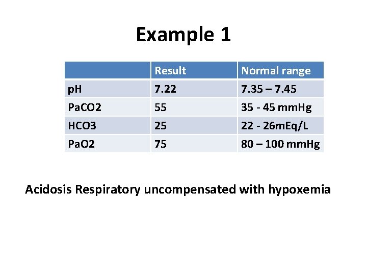
Example 1 p. H Pa. CO 2 HCO 3 Result 7. 22 55 25 Normal range 7. 35 – 7. 45 35 - 45 mm. Hg 22 - 26 m. Eq/L Pa. O 2 75 80 – 100 mm. Hg Acidosis Respiratory uncompensated with hypoxemia
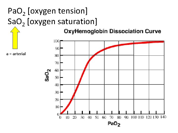
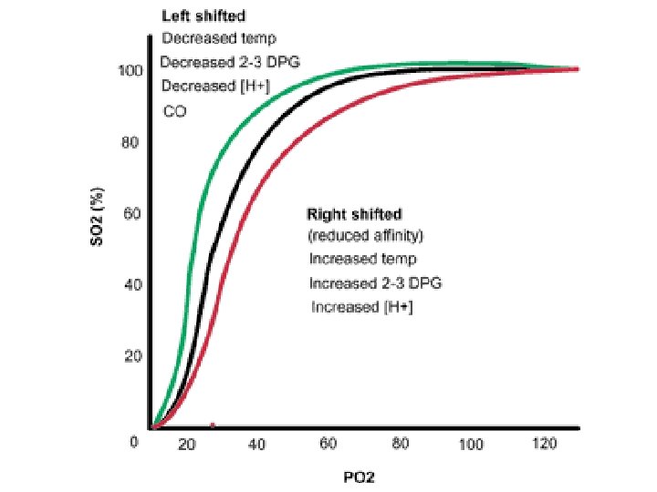
Pa. O 2 [oxygen tension] Sa. O 2 [oxygen saturation] a = arterial
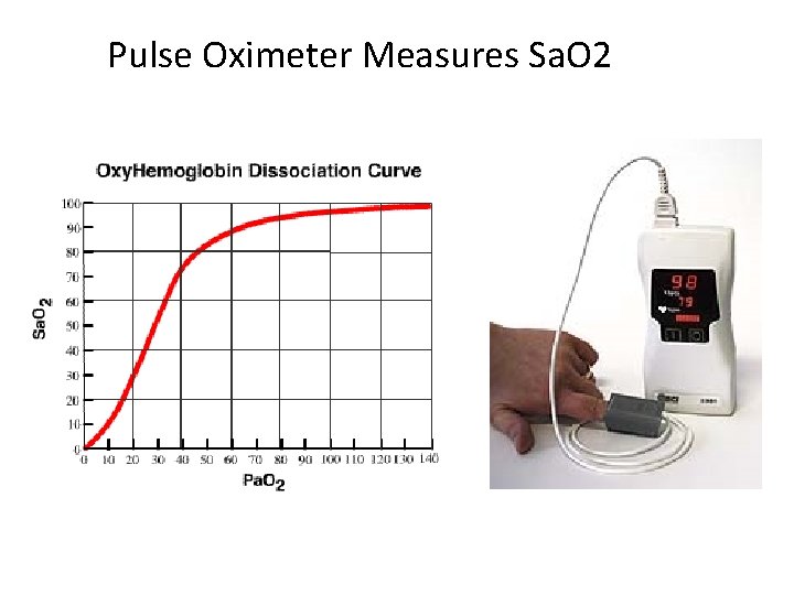
Pulse Oximeter Measures Sa. O 2
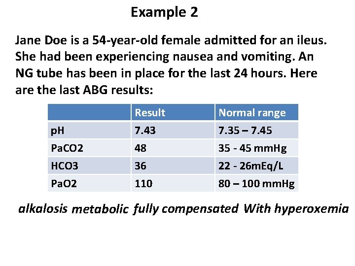
Example 2 Jane Doe is a 54 -year-old female admitted for an ileus. She had been experiencing nausea and vomiting. An NG tube has been in place for the last 24 hours. Here are the last ABG results: p. H Pa. CO 2 HCO 3 Result 7. 43 48 36 Normal range 7. 35 – 7. 45 35 - 45 mm. Hg 22 - 26 m. Eq/L Pa. O 2 110 80 – 100 mm. Hg alkalosis metabolic fully compensated With hyperoxemia
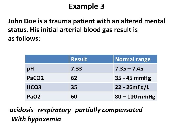
Example 3 John Doe is a trauma patient with an altered mental status. His initial arterial blood gas result is as follows: p. H Pa. CO 2 HCO 3 Result 7. 33 62 35 Normal range 7. 35 – 7. 45 35 - 45 mm. Hg 22 - 26 m. Eq/L Pa. O 2 60 80 – 100 mm. Hg acidosis respiratory partially compensated With hypoxemia
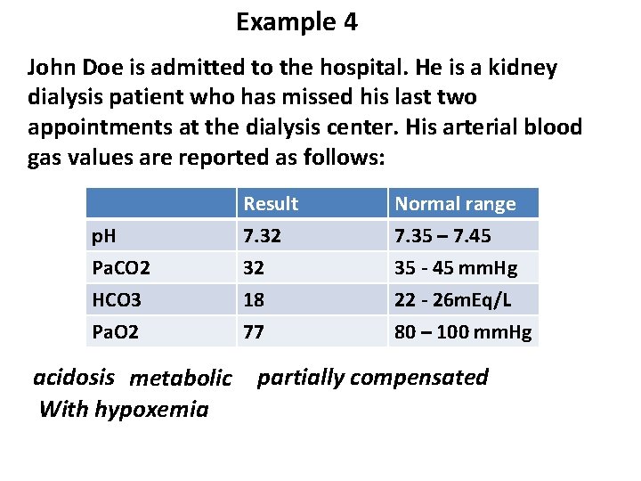
Example 4 John Doe is admitted to the hospital. He is a kidney dialysis patient who has missed his last two appointments at the dialysis center. His arterial blood gas values are reported as follows: p. H Pa. CO 2 HCO 3 Result 7. 32 32 18 Normal range 7. 35 – 7. 45 35 - 45 mm. Hg 22 - 26 m. Eq/L Pa. O 2 77 80 – 100 mm. Hg acidosis metabolic With hypoxemia partially compensated
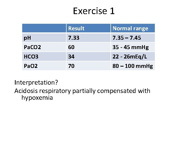
Exercise 1 p. H Pa. CO 2 HCO 3 Result 7. 33 60 34 Normal range 7. 35 – 7. 45 35 - 45 mm. Hg 22 - 26 m. Eq/L Pa. O 2 70 80 – 100 mm. Hg Interpretation? Acidosis respiratory partially compensated with hypoxemia
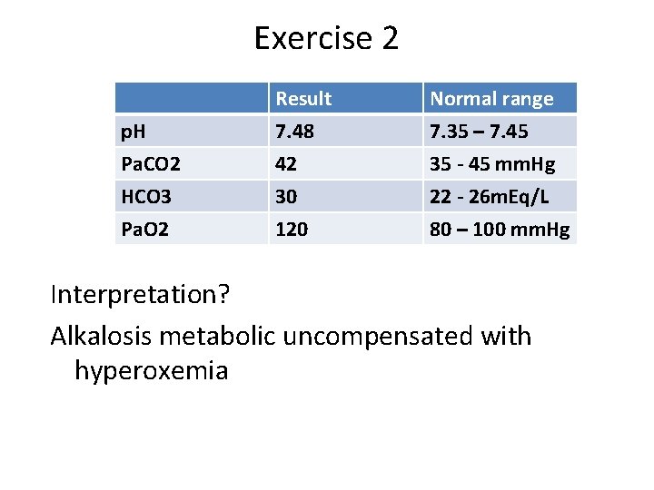
Exercise 2 p. H Pa. CO 2 HCO 3 Result 7. 48 42 30 Normal range 7. 35 – 7. 45 35 - 45 mm. Hg 22 - 26 m. Eq/L Pa. O 2 120 80 – 100 mm. Hg Interpretation? Alkalosis metabolic uncompensated with hyperoxemia
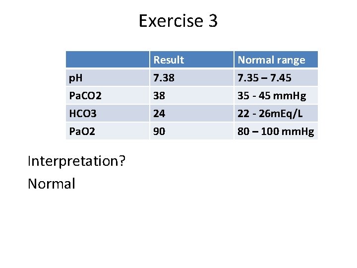
Exercise 3 p. H Pa. CO 2 HCO 3 Result 7. 38 38 24 Normal range 7. 35 – 7. 45 35 - 45 mm. Hg 22 - 26 m. Eq/L Pa. O 2 90 80 – 100 mm. Hg Interpretation? Normal
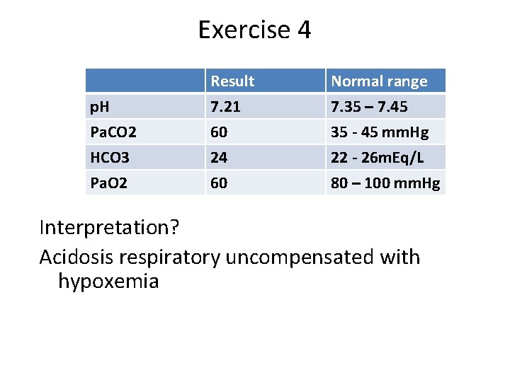
Exercise 4 p. H Pa. CO 2 HCO 3 Result 7. 21 60 24 Normal range 7. 35 – 7. 45 35 - 45 mm. Hg 22 - 26 m. Eq/L Pa. O 2 60 80 – 100 mm. Hg Interpretation? Acidosis respiratory uncompensated with hypoxemia
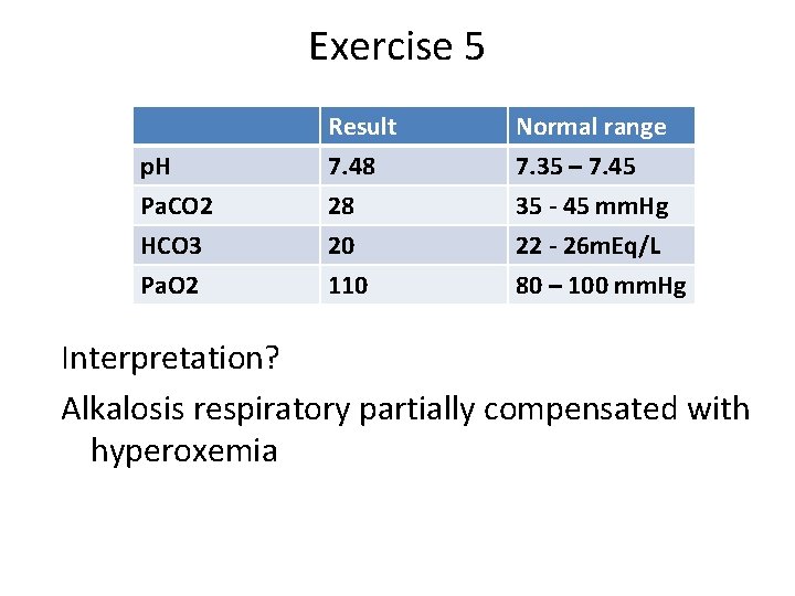
Exercise 5 p. H Pa. CO 2 HCO 3 Result 7. 48 28 20 Normal range 7. 35 – 7. 45 35 - 45 mm. Hg 22 - 26 m. Eq/L Pa. O 2 110 80 – 100 mm. Hg Interpretation? Alkalosis respiratory partially compensated with hyperoxemia
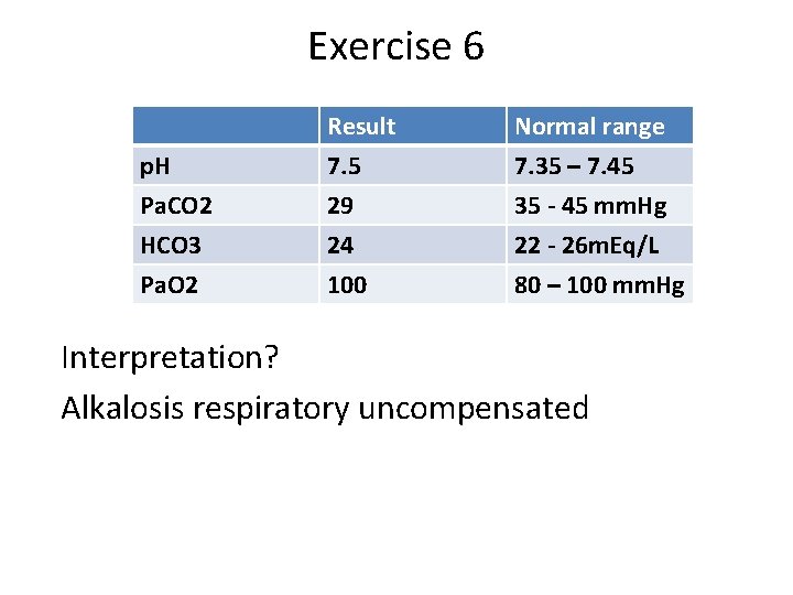
Exercise 6 p. H Pa. CO 2 HCO 3 Result 7. 5 29 24 Normal range 7. 35 – 7. 45 35 - 45 mm. Hg 22 - 26 m. Eq/L Pa. O 2 100 80 – 100 mm. Hg Interpretation? Alkalosis respiratory uncompensated
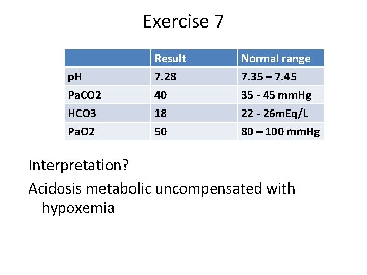
Exercise 7 p. H Pa. CO 2 HCO 3 Result 7. 28 40 18 Normal range 7. 35 – 7. 45 35 - 45 mm. Hg 22 - 26 m. Eq/L Pa. O 2 50 80 – 100 mm. Hg Interpretation? Acidosis metabolic uncompensated with hypoxemia
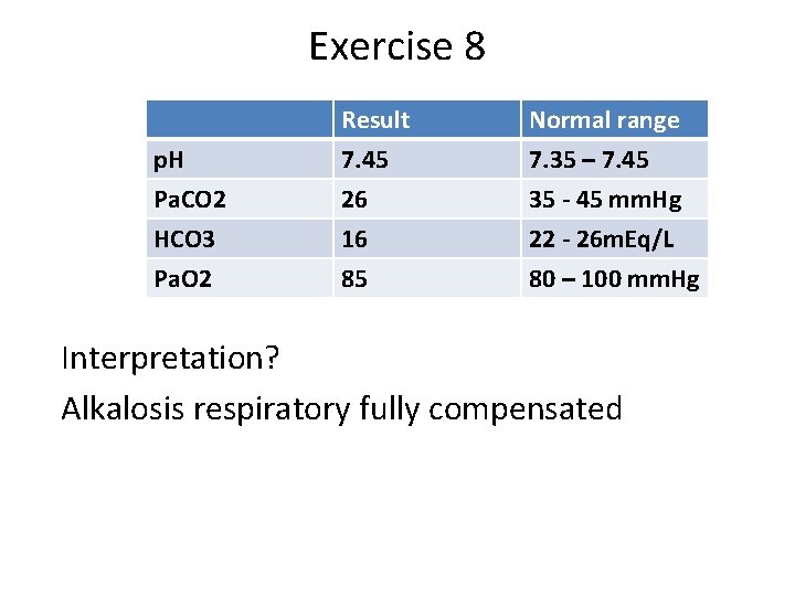
Exercise 8 p. H Pa. CO 2 HCO 3 Result 7. 45 26 16 Normal range 7. 35 – 7. 45 35 - 45 mm. Hg 22 - 26 m. Eq/L Pa. O 2 85 80 – 100 mm. Hg Interpretation? Alkalosis respiratory fully compensated
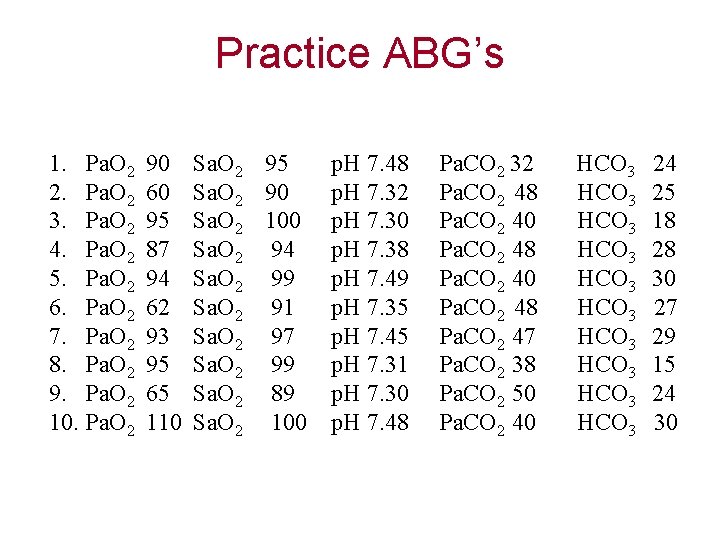
Practice ABG’s 1. Pa. O 2 2. Pa. O 2 3. Pa. O 2 4. Pa. O 2 5. Pa. O 2 6. Pa. O 2 7. Pa. O 2 8. Pa. O 2 9. Pa. O 2 10. Pa. O 2 90 60 95 87 94 62 93 95 65 110 Sa. O 2 95 Sa. O 2 90 Sa. O 2 100 Sa. O 2 94 Sa. O 2 99 Sa. O 2 91 Sa. O 2 97 Sa. O 2 99 Sa. O 2 89 Sa. O 2 100 p. H 7. 48 p. H 7. 32 p. H 7. 30 p. H 7. 38 p. H 7. 49 p. H 7. 35 p. H 7. 45 p. H 7. 31 p. H 7. 30 p. H 7. 48 Pa. CO 2 32 Pa. CO 2 48 Pa. CO 2 40 Pa. CO 2 48 Pa. CO 2 47 Pa. CO 2 38 Pa. CO 2 50 Pa. CO 2 40 HCO 3 HCO 3 HCO 3 24 25 18 28 30 27 29 15 24 30
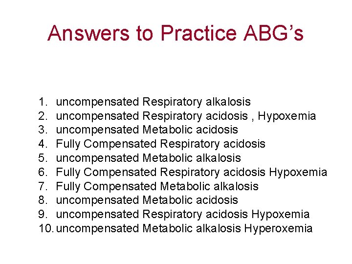
Answers to Practice ABG’s 1. uncompensated Respiratory alkalosis 2. uncompensated Respiratory acidosis , Hypoxemia 3. uncompensated Metabolic acidosis 4. Fully Compensated Respiratory acidosis 5. uncompensated Metabolic alkalosis 6. Fully Compensated Respiratory acidosis Hypoxemia 7. Fully Compensated Metabolic alkalosis 8. uncompensated Metabolic acidosis 9. uncompensated Respiratory acidosis Hypoxemia 10. uncompensated Metabolic alkalosis Hyperoxemia
Mixed respiratory and metabolic acid-base disorder
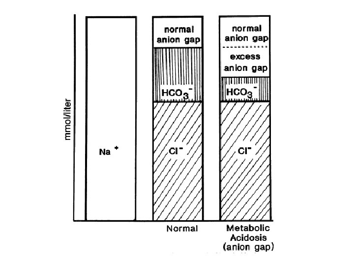
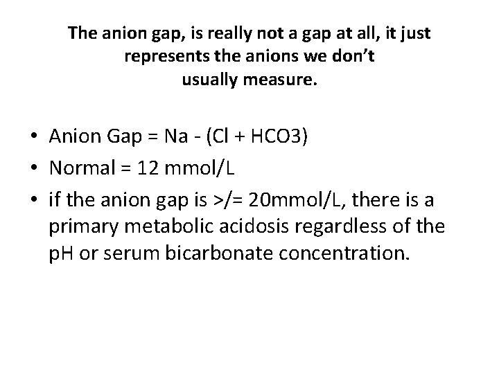
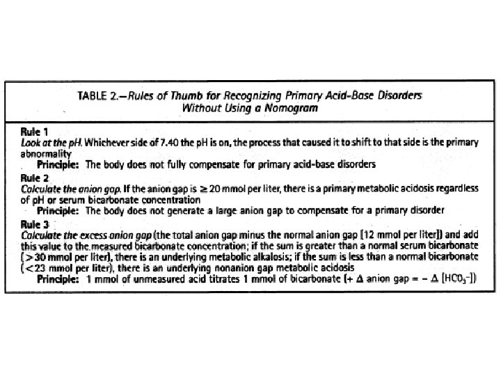
The anion gap, is really not a gap at all, it just represents the anions we don’t usually measure. • Anion Gap = Na - (Cl + HCO 3) • Normal = 12 mmol/L • if the anion gap is >/= 20 mmol/L, there is a primary metabolic acidosis regardless of the p. H or serum bicarbonate concentration.
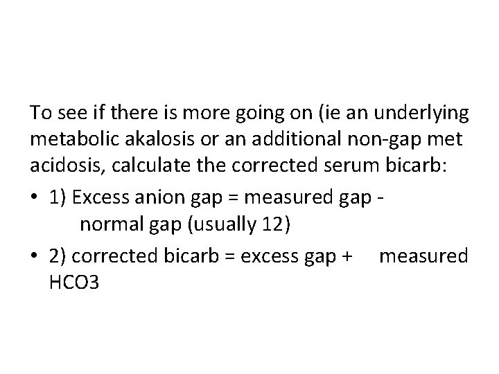
To see if there is more going on (ie an underlying metabolic akalosis or an additional non-gap met acidosis, calculate the corrected serum bicarb: • 1) Excess anion gap = measured gap normal gap (usually 12) • 2) corrected bicarb = excess gap + measured HCO 3
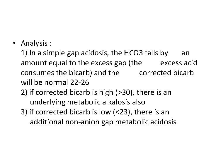
• Analysis : 1) In a simple gap acidosis, the HCO 3 falls by an amount equal to the excess gap (the excess acid consumes the bicarb) and the corrected bicarb will be normal 22 -26 2) if corrected bicarb is high (>30), there is an underlying metabolic alkalosis also 3) if corrected bicarb is low (<23), there is an additional non-anion gap metabolic acidosis
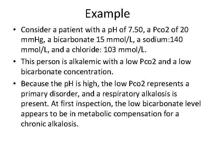
Example • Consider a patient with a p. H of 7. 50, a Pco 2 of 20 mm. Hg, a bicarbonate 15 mmol/L, a sodium: 140 mmol/L, and a chloride: 103 mmol/L. • This person is alkalemic with a low Pco 2 and a low bicarbonate concentration. • Because the p. H is high, the low Pco 2 represents a primary disorder, and a respiratory alkalosis is present. At first inspection, the low bicarbonate level appears to be in metabolic compensation for a chronic alkalosis.
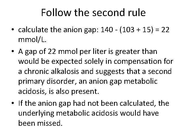
Follow the second rule • calculate the anion gap: 140 - (103 + 15) = 22 mmol/L. • A gap of 22 mmol per liter is greater than would be expected solely in compensation for a chronic alkalosis and suggests that a second primary disorder, an anion gap metabolic acidosis, is also present. • If the anion gap had not been calculated, the underlying metabolic acidosis would have been missed.
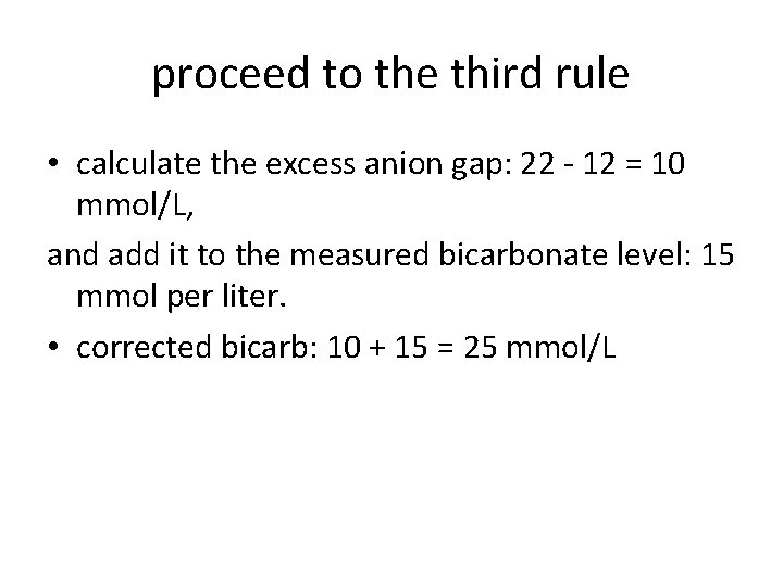
proceed to the third rule • calculate the excess anion gap: 22 - 12 = 10 mmol/L, and add it to the measured bicarbonate level: 15 mmol per liter. • corrected bicarb: 10 + 15 = 25 mmol/L
• Result: normal, indicating that no further primary abnormalities are present. • This patient had ingested a large quantity of aspirin and displayed the centrally mediated respiratory alkalosis and the anion gap metabolic acidosis associated with salicylate overdose.
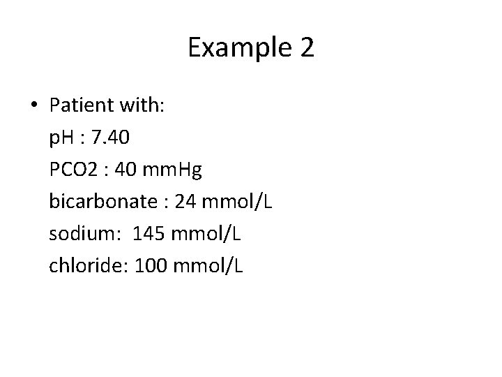
Example 2 • Patient with: p. H : 7. 40 PCO 2 : 40 mm. Hg bicarbonate : 24 mmol/L sodium: 145 mmol/L chloride: 100 mmol/L
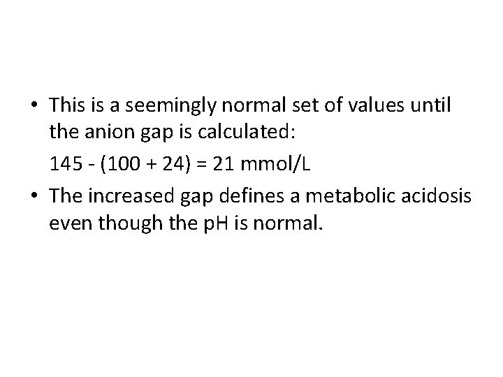
• This is a seemingly normal set of values until the anion gap is calculated: 145 - (100 + 24) = 21 mmol/L • The increased gap defines a metabolic acidosis even though the p. H is normal.
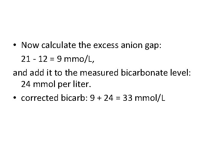
• Now calculate the excess anion gap: 21 - 12 = 9 mmo/L, and add it to the measured bicarbonate level: 24 mmol per liter. • corrected bicarb: 9 + 24 = 33 mmol/L
• The sum, 33 mmol/L, is higher than a normal bicarbonate concentration, indicating a metabolic alkalosis is also present. • Result: Metabolic Acidosis and Metabolic Alkalosis • These laboratory values are from a patient with chronic renal failure (causing the metabolic acidosis) who began vomiting (hence the metabolic alkalosis) as his uremia worsened.
• The acute alkalosis of vomiting off set the chronic acidosis of renal failure, resulting in normal p. H. Without a systematic approach to acid-base disorders, including calculation of the anion gap and of the excess anion gap, these mixed acid-base disorders could easily have been overlooked.
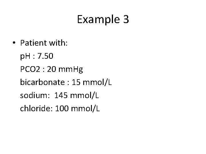
Example 3 • Patient with: p. H : 7. 50 PCO 2 : 20 mm. Hg bicarbonate : 15 mmol/L sodium: 145 mmol/L chloride: 100 mmol/L
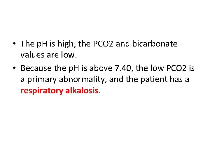
• The p. H is high, the PCO 2 and bicarbonate values are low. • Because the p. H is above 7. 40, the low PCO 2 is a primary abnormality, and the patient has a respiratory alkalosis.
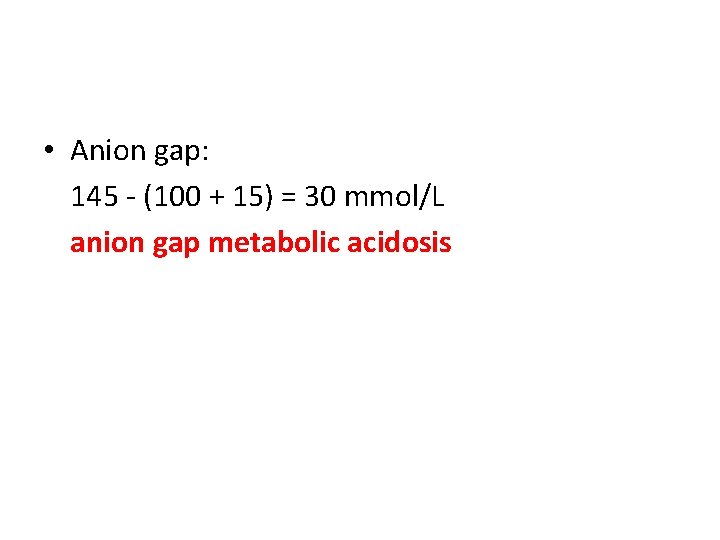
• Anion gap: 145 - (100 + 15) = 30 mmol/L anion gap metabolic acidosis
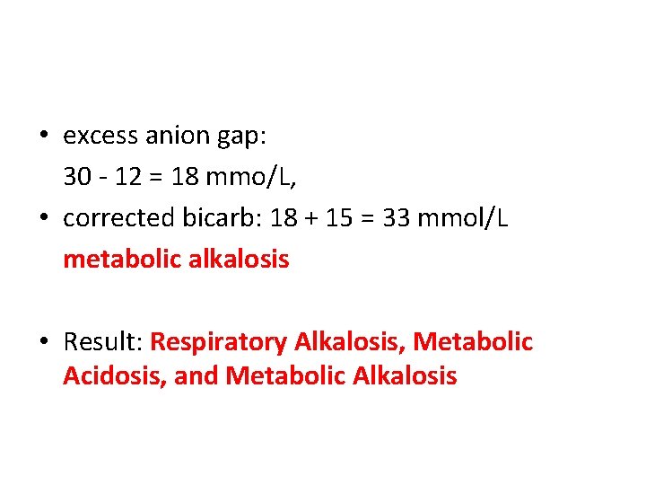
• excess anion gap: 30 - 12 = 18 mmo/L, • corrected bicarb: 18 + 15 = 33 mmol/L metabolic alkalosis • Result: Respiratory Alkalosis, Metabolic Acidosis, and Metabolic Alkalosis
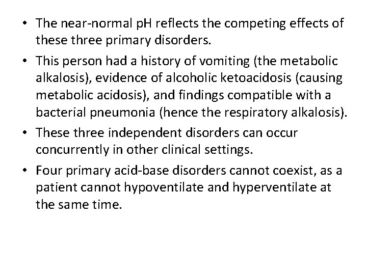
• The near-normal p. H reflects the competing effects of these three primary disorders. • This person had a history of vomiting (the metabolic alkalosis), evidence of alcoholic ketoacidosis (causing metabolic acidosis), and findings compatible with a bacterial pneumonia (hence the respiratory alkalosis). • These three independent disorders can occur concurrently in other clinical settings. • Four primary acid-base disorders cannot coexist, as a patient cannot hypoventilate and hyperventilate at the same time.
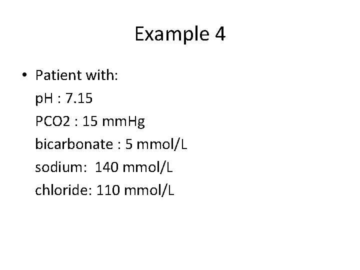
Example 4 • Patient with: p. H : 7. 15 PCO 2 : 15 mm. Hg bicarbonate : 5 mmol/L sodium: 140 mmol/L chloride: 110 mmol/L

• Anion gap: 140 - (110 + 5) = 25 mmol/L anion gap metabolic acidosis
• excess anion gap: 25 - 12 = 13 mmo/L, • corrected bicarb: 13 + 5 = 18 mmol/L nonanion gap metabolic acidosis • Result: Anion Gap and Nonanion Gap Metabolic Acidoses
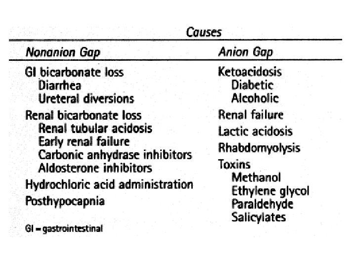
• suggesting that additional gastrointestinal or renal loss of bicarbonate has occurred and that both an anion gap and a nonanion gap metabolic acidosis are present. • Diabetic ketoacidosis was responsible for the anion gap acidosis in this patient. The nonanion gap (hyperchloremic) acidosis is that phenomenon observed in the recovery phase of diabetic ketoacidosis due to failure to regenerate bicarbonate from ketoacids lost in the urine. • If the sum of the excess anion gap and the measured bicarbonate had not been calculated, the underlying nongap acidosis would not have been appreciated.

Thank You
- Slides: 75