Blood circulation Section 1 Summary blood circulation Def
 Section 1 Summary blood circulation (血液循环): Def : blood continuously circulatory")
Blood circulation (血液循环) Section 1 Summary blood circulation (血液循环): Def : blood continuously circulatory flow in cardiovascular system (是指血液在心血管系统内周而复始地循环流动)
: muscle-pump , dynamic organ. (肌肉泵, 动力器官) artery (动脉): the vessel which can")
heart (心脏): muscle-pump , dynamic organ. (肌肉泵, 动力器官) artery (动脉): the vessel which can guide blood to leave heart and flow into tissues. (引导血液离开心脏流向各组织器官的管道). vein (静脉): the vessel which can guide blood to return heart. (引导血液回心的管道) capillary (毛细血管) : the place of substance exchange. (进行物质交换的场所)
")
Systemic circulation and pulmonary circulation (体循环与 肺循环)
 2. 1 Autorhythmicity (自动节律性) Def:")
Section 2 The structures and functions of myocardium (心肌的结构与机能特性) 2. 1 Autorhythmicity (自动节律性) Def: the ability of an excitable cell to rhythmically initiate its own action potentials (组织细胞能自动发生节律性兴奋的特性) 衡量指标: the frequency of automatic excitation (自动兴奋的频率)
 Structure basis : intercalated disc (闰盘) 心肌的机能合体性 myocardial “ all")
2. 2 functional synplasm(机能合胞体) Structure basis : intercalated disc (闰盘) 心肌的机能合体性 myocardial “ all or none ”phenomenon (心肌收缩的“全或无”现象)
 The types of myocardium cell (心肌细胞的类型): 1) normal")
2. 3 Specific conduction system (特殊传导系统) The types of myocardium cell (心肌细胞的类型): 1) normal myocardium (普通的心肌细胞 ): working cardiac cell (non-autorhythmic cell): include atrial muscle and ventricular muscle, have excitability , conductivity, and contractibility, no autorhythmicity. 作细胞 (非自律细胞), 包括心房肌、心室肌, 具兴奋性, 传导性, 收缩性, 无自律性
 Specific myocardium (特殊分化的心肌细胞 ): rhythmic cell and non-autorhythmic cell, include P cell and")
2) Specific myocardium (特殊分化的心肌细胞 ): rhythmic cell and non-autorhythmic cell, include P cell and Purkinje cell, have excitability, autorhythmicity and conductivity, no contractibility, also have capacity to produce and conduct rhythmic excitation, 自律细胞和非自律细胞, 包括P细胞和浦肯野氏细胞, 有 兴奋性, 自律性和传导性, 基本无收缩性, 具有产生和传导 节律性兴奋的能力
 artrio-ventricular node , A-V node (房室结) 0. 05")
sino-artrial node , S-A node (窦房结) artrio-ventricular node , A-V node (房室结) 0. 05 m/s atrioventricular bundle and bilateral bundle (房室束及左右束支) 2 -5 m/s Purkinje fiber(浦肯野氏纤维) 2 -5 m/s Fig. 7 -1 atrioventricular delay (房室延搁) and significance :
: S-A node , right atrium wall (窦房结, 右心房内壁) Latent pacemaker(潜在起搏点): a kind of")
Pacemaker(正常起搏点): S-A node , right atrium wall (窦房结, 右心房内壁) Latent pacemaker(潜在起搏点): a kind of autorhythmic tissue which don’t express autorhythmicity in normal circumstances. (正常情况下不表现本身自律性的自律组织。)
 : the latent pacemaker which can control cardiac excitation and palmus in")
Ectopic pacemaker(异位起搏点) : the latent pacemaker which can control cardiac excitation and palmus in abnormal circumstances. (异常情况下控制心脏兴奋和跳动的潜在起搏点. ) Artificial pacemaker (人 起博器)
")
2. 4 The AP of myocardial cell ------ bioeletricity phenomenon of myocardial cell (心肌细胞的AP---心肌细胞的生物电现象)
 2. different myocardial cell have different transmembrane")
1. different from the skeletal muscle. (不同于骨骼肌) 2. different myocardial cell have different transmembrane potential extent, persistent time, wave form and mechanism. (不同的心肌细胞,其跨膜电位的幅度、 持续 时间、波形、产生机制亦不相同)
 1. Absolute refractory period & effective")
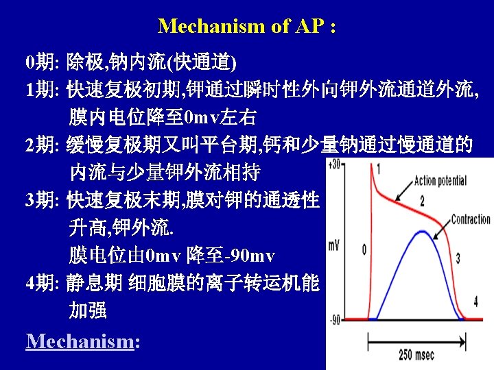
2. 5 The refractory period of heart (心脏的不应期) 1. Absolute refractory period & effective refractory period, ARP&ERP (绝对不应期与有效不应期): 0 — -55 mv, -60 mv , can’t generate AP. 2. Relative refractory period, RRP (相对不应期): -60 — -80 mv, > threshold intensity (阈强度), can cause AP. 3. Superanormal period, SNP(超常期): -80 — -90 mv, < threshold intensity , can cause AP Fig. 7 -16
: premature systole or extrasystole & compensatory pause")
Significance of long effective refractory period (有效不应期长的意义): premature systole or extrasystole & compensatory pause (期前收缩与代偿间歇) Fig. 7 -17
 Basic waveform (基本波形): P , QRS, T Significance: P:")
2. 6 Electrocardiogram, ECG (心电图) Basic waveform (基本波形): P , QRS, T Significance: P: T: Fig. 7 -18 QRS: P-R period 7 -20
 3. 1 Def. cardiac cycle")
Section 3 The dynamics change of cardiac cycle (心动周期的力学变化) 3. 1 Def. cardiac cycle (心动周期) : one period of systole and diastole (心脏由收缩到舒张的整个过程)
 3. 2. 1 Period of atrial systole")
3. 2 Ejection process of heart (心脏的射血过程) 3. 2. 1 Period of atrial systole (心房收缩期): end-diastolic volume (心舒末期容积) 3. 2. 2 Period of ventricular systole (心室收缩期):
 period of isovolumic contraction (等容收缩期): semilunar valve is still closed , atrioventricular valve")
1) period of isovolumic contraction (等容收缩期): semilunar valve is still closed , atrioventricular valve is closed (半月瓣仍关, 房室瓣关)
 period of rapid ejection (快速射血期): semilunar valve is opened, ejecting (半月瓣开, 射血) 3)")
2) period of rapid ejection (快速射血期): semilunar valve is opened, ejecting (半月瓣开, 射血) 3) period of slow ejection (减慢射血期): end-systolic volume (心缩末期容积 )
: 1) period of isovolumic relaxation (等容舒张期):")
3. 2. 3 Period of ventricular relaxation (心室舒张期): 1) period of isovolumic relaxation (等容舒张期): semilunar valve is closed , atrioventricular valve is closed (半月瓣关, 房室瓣仍关)
 period of rapid filling (快速充盈期): atrioventricular valve is opened. (房室瓣开) 3) period of")
2) period of rapid filling (快速充盈期): atrioventricular valve is opened. (房室瓣开) 3) period of reduced filling(减慢充盈期 ): Fig 7 -22
 First heart sound (第一心音) : the tone is low")
3. 3 heart sound (心音) First heart sound (第一心音) : the tone is low and long, mostly caused by closing of atrioventricular valve, it is symbol of cardiac contraction. (音调低而长, 主要由于房室瓣关闭引起, 标志心缩开始)
 : the tone is high and short, mostly caused by")
Second heart sound (第二心音) : the tone is high and short, mostly caused by closing of semilunar valve, it is symbol of cardiac relaxation. (音调高而短, 主要由于半月瓣关闭引起, 标志 心舒开始)
 Cardiac output (心输出量) : the volume")
Section 4 Cardiac output and its regulation (心输出量及其调节) Cardiac output (心输出量) : the volume of blood pumped by each ventricle each minute; equals stroke volume times heart rate. (一侧心室每分钟射出的血量)
")
Cardiac Output = Heart Rate×Stroke Volume CO=HR×SV =75 × 70 =5250(ml)
 CO (Cardiac output) quiet (安静时): 5 L/min movement (运动时): 180 -200")
Cardiac reserve (心力储备) CO (Cardiac output) quiet (安静时): 5 L/min movement (运动时): 180 -200 150 -170 25 -30 L/min
: maximal volume of blood pumped by each ventricle each minute. (一侧心室每分钟射出的最大血量)")
Maximal output (最大输出量): maximal volume of blood pumped by each ventricle each minute. (一侧心室每分钟射出的最大血量) Cardiac reserve(心力贮备): the ability that cardiac output is increased along with the need of body metabolism. (心输出量随机体代谢需要而增加的能力)
 Nerve regulation (神经调节): sympathetic nerve (交感神经)")
4. 1 The regulation of heat rate (心率的调节) Nerve regulation (神经调节): sympathetic nerve (交感神经) vagus nerve (+) (迷走神经) (-) Fig. 7 -2 Hormone regulation(激素调节): adrenal hormone 肾上腺素(+)
 4. 2. 1 heterometric autoregulation (异长调节) cardiac")
4. 2 Regulation of cardiac output (心搏量的调节) 4. 2. 1 heterometric autoregulation (异长调节) cardiac regulation (心脏自身的调节) cardiac law (心脏定律) Fig 7 -29
 Nerve regulation (神经调节) sympathetic nerve (交感神经) (+) vagus")
4. 2. 2 homometric regulation (等长调节) Nerve regulation (神经调节) sympathetic nerve (交感神经) (+) vagus nerve (迷走神经) (-)
 adrenal hormone (肾上腺素) (+) , thyroid hormone (甲状腺素) (+)")
Hormone regulation (激素调节) adrenal hormone (肾上腺素) (+) , thyroid hormone (甲状腺素) (+)
 5. 1 Structures and function characters of")
Section 5 physiology of blood vessel (血管生理) 5. 1 Structures and function characters of various vessels (各类血管的结构与机能特点)
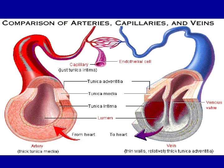
 1). Aorta (大动脉) windvessel (弹性贮器血管) Function:buffering high pressure and")
5. 1. 1 artery (动脉) 1). Aorta (大动脉) windvessel (弹性贮器血管) Function:buffering high pressure and being related to blood flow continually (缓冲高压与血流的持续 流动有关) Elasticity and age (弹性与年龄):
. Distribution vessel (分配血管) Function: transfusing blood to organs and tissues (输送血液至器官组织)")
2). Distribution vessel (分配血管) Function: transfusing blood to organs and tissues (输送血液至器官组织)
. Arteriole (微动脉) resistance vessel before capillary (Cap前阻力血管) Structure characters : vessel wall is")
3). Arteriole (微动脉) resistance vessel before capillary (Cap前阻力血管) Structure characters : vessel wall is mainly smooth muscle. (管壁以平滑肌为主. )

Function: 1 regulating peripheral resistance/BP; 调节外周阻力/ BP calamus is small, velocity of flow is high; (管径小, 流速快)

Function: 2 controlling local blood flow , blood flow is in direct proportion to r 4 , and related to continuous flow of blood. (控制局部血灌流量, 血流量与r 4成正比 与血流持续有关. )
: aorta (主大A) small artery (小A) arteriole(微A)")
Blood pressure fall (血压降落): aorta (主大A) small artery (小A) arteriole(微A)
 capacitance vessel (容量血管) Function: contain 60 -70% blood (容纳")
5. 1. 2 vein (静脉) capacitance vessel (容量血管) Function: contain 60 -70% blood (容纳 60 -70%的血)
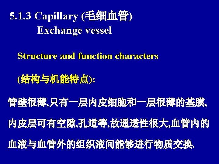
 Function:The place that substances exchange (物质交换的场所)")
Exchange vessel (交换血管) Function:The place that substances exchange (物质交换的场所)
 BP (血压): The blood side pressure")
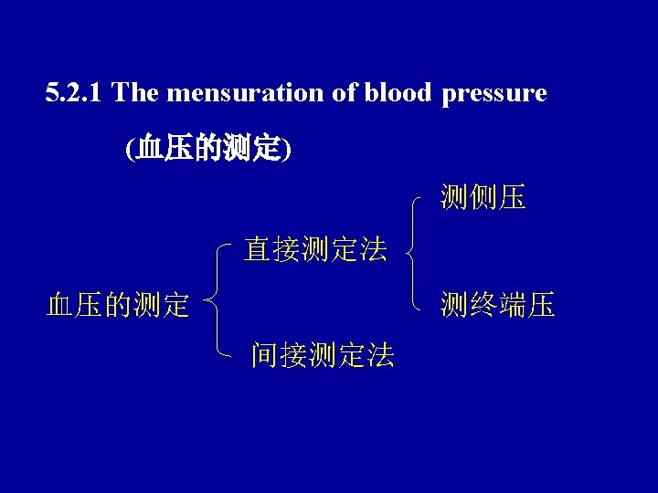
5. 2 Blood pressure and its mensuration (血压与血压的测定) BP (血压): The blood side pressure of unit area blood vessel wall (血液对单位面积血 管壁的侧压力) Unit(单位): k. Pa/mm. Hg
 Basic factors: ① enough blood")
5. 2. 2 The forming of blood pressure (血压的形成) Basic factors: ① enough blood volume in cardiovascular center (心血管内有足够的血量) ② ventricular ejection (心室射血 ) ③ peripheral resistance (外周阻力)
 Systolic pressure(收缩压) when ventricle is contracting, the")
5. 2. 3 Arterial blood pressure (动脉血压) Systolic pressure(收缩压) when ventricle is contracting, the maximal arterial pressure is called the systolic pressure. (心室收缩时动脉压升到的最高值。) Normal (正常值): 100 -120 mm. Hg / 13. 3 -16 k. Pa
 when ventricle is relaxing, the minimal arterial pressure is called diastolic")
Diastolic pressure (舒张压) when ventricle is relaxing, the minimal arterial pressure is called diastolic pressure. (心室舒张时动脉压降到的最低值) Normal (正常值): 60 -80 mm. Hg / 8 -10. 6 k. Pa Blood pressure, age, sex and others (血压与年龄, 性别及其它) :
 1) VP (V血压) 1. Peripheral")
5. 2. 4 Venous pressure & venous return (静脉血压和静脉回流) 1) VP (V血压) 1. Peripheral VP (外周V压) VP of different organs (各器官V的血压) 15 -20 mm. Hg
 VP of main line in right atrium and thoracic cavity")
2. Central VP (中心V压) VP of main line in right atrium and thoracic cavity (右心房和胸腔内大V的血压) 0 -12 mm. Hg V return power = Peripheral VP - Central VP V回流动力= 外周V压 - 中心V压
 1. Systole (心脏收缩) 心肌收缩力量 正变 (左、右心衰) 2. Gravity")
Effect factors of venous return (影响V回流的因素) 1. Systole (心脏收缩) 心肌收缩力量 正变 (左、右心衰) 2. Gravity and pose (重力与体位) 血液重力引起静水压 lie: VP are approximately same in different part. 各处V压大致相同 stand: VP are different in different parts , 各处V压大不相同
 The degree of V engorge is affected by transmura pressure.")
Gravity and pose (重力与体位) The degree of V engorge is affected by transmura pressure. (V充盈程度受跨壁压的影响大) VP of lower parts are expanded because of the transmura pressure increased, the blood volume are also increased. (身体低垂部分的V压因跨壁压增大而扩张, 容纳 血量增多)
 Inspiration (吸气) (+) 4. Extrusion")
3. Respiratory movement / respiratory pump (呼吸运动 / 呼吸泵) Inspiration (吸气) (+) 4. Extrusion action of skeletal muscle /muscle pump (骨骼肌的挤压作用 /肌泵) Muscle contraction (+) (肌肉收缩)
 Def : the blood circulation in microangium between arteriole and")
5. 3 Microcirculation (微循环) Def : the blood circulation in microangium between arteriole and venule. (微动脉和微静脉之间微血管中的血液循环)
 arteriole(微动脉); metarteriole(后微动脉); precapillary sphincter (毛细血管前括约肌); true")
5. 3. 1 The component of microcirculation (微循环的组成) arteriole(微动脉); metarteriole(后微动脉); precapillary sphincter (毛细血管前括约肌); true capillary (真毛细血管); thoroughfare channel (通血毛细血管); arteriovenous anastomoses (动-静脉吻合支); venule (微静脉) Fig 7 -38
: precapillary resistance vessel ; master valve (毛细血管前阻力血管; 总闸门) 2. Capillary (毛细血管): Structure")
1. Arteriole(微动脉): precapillary resistance vessel ; master valve (毛细血管前阻力血管; 总闸门) 2. Capillary (毛细血管): Structure characters : big area , slow velocity, high permeability (面积大, 流速慢, 通透性大) Fig. 7 -39 Function: exchange of substances 3. Venule(微静脉): capacitance vessel (容量血管)
 1. diffusion(扩散): •")
5. 3. 2 The methods and mechanisms of substance exchange (物质交换的方式和机制) 1. diffusion(扩散): • Concentration (浓度), • Distance(距离), • Area(面积), • Temperature(温度), • molecular weight(分子量), • membranous permeability(膜的通透性等)
 Starling hypothesis(Starling假说) Power (动力): effective filtration pressure = (Cap")
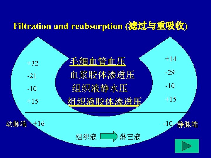
2. Filtration and reabsorption (滤过与重吸收) Starling hypothesis(Starling假说) Power (动力): effective filtration pressure = (Cap pressure + colloid osmotic pressure of tissue fluid)- (colloid osmotic pressure of blood plasma + lentic pressure of tissue fluid) 有效滤过压 =(Cap压+ 组织液胶体渗透压)— (血浆胶体渗透压+组织液静水压)
: Effective filtration pressure of Cap A terminal is positive , the fluid is")
Process(过程): Effective filtration pressure of Cap A terminal is positive , the fluid is filtrated and forms tissue fluid; effective filtration pressure of Cap V terminal is negative , tissue fluid returns Cap A端的有效滤过压为正值,液体滤出而 成组织液;Cap V端的有效滤过压为负值,组 织液回流入Cap。
 Two – dimensional action (双向作用) Exchange of macromolecular protein (大分子蛋白质的交换)")
4. Pinocytosis (胞饮作用) Two – dimensional action (双向作用) Exchange of macromolecular protein (大分子蛋白质的交换)
 5. 4. 1 Component (组成): lymphatic capillary, lymphatic vessel,")
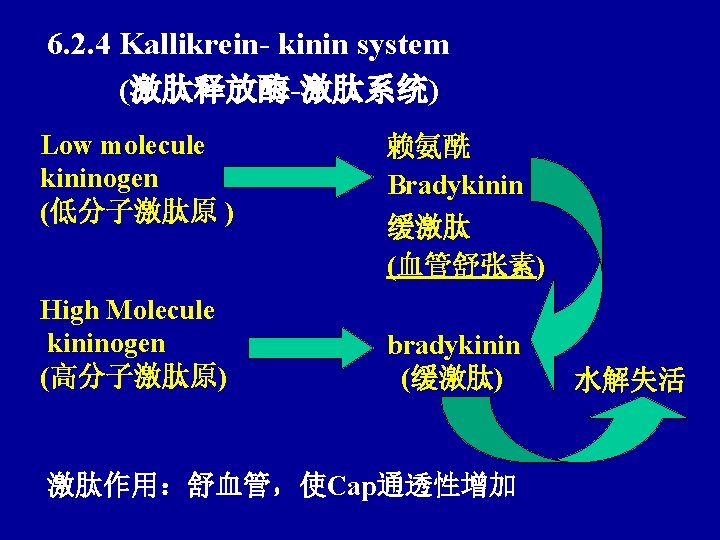
5. 4 Lymph circulation (淋巴循环) 5. 4. 1 Component (组成): lymphatic capillary, lymphatic vessel, lymph node. (毛细淋巴管, 淋巴结. ) permeability : lymphatic capillary > capillary (毛细淋巴管通透性 >毛细血管)
 1. The")
5. 4. 2 Effect factors of lymph fluid forming and return (影响淋巴液生成与回流的因素) 1. The pressure difference between tissue fluid and lymphatic capillary (组织液与毛细淋巴管内的压力差) 2. Skeletal muscle movement (骨骼肌运动)
 1. Regulating the balance")
5. 4. 3 The physiological significance of lymphatic return (淋巴回流的生理意义) 1. Regulating the balance of blood volume and tissue fluid volume. (调节血量与组织液量的平衡) 2. Recovering protein of tissue fluid. (对组织液中的蛋白质的回收作用)
 4. Defense barrier action of")
3. The transportation of Chyle vessel to fat (乳糜管对脂肪的运输) 4. Defense barrier action of lymph node (淋巴结的防御屏障作用) macrophage (巨噬细胞) lymphocyte (淋巴细胞)
 6. 1 Nervous regulation (神经调节) 6. 1.")
Section 6 Regulation of cardiovascular activity (心血管活动的调节) 6. 1 Nervous regulation (神经调节) 6. 1. 1 Cardiovascular innervation (心血管的N支配) 1). Cardiac innervation (心脏的N支配)
: T 1 -5 (NE+β 1 R) different")
Cardiac sympathetic nerve and its regulation (心交感N及其作用): T 1 -5 (NE+β 1 R) different parts of heart (NE+β 1 R) 心脏各部分 Function:正性变时变力变传导
: Dorsal nucleus and ambiguous nucleus of medulla")
Cardiac vagus nerve and its regulation (心迷走N及其作用): Dorsal nucleus and ambiguous nucleus of medulla (ACh+MR) different (延髓背核、疑核) (ACh+MR)心脏各部分 parts of heart Function:负性变时变力变传导 2). vessel innervation (血管的N支配)
: The nerve fiber which can cause contraction of smooth muscle of")
Vasoconstrictor fiber (缩血管神经纤维): The nerve fiber which can cause contraction of smooth muscle of blood vessel (能引起血管平滑肌收缩的神经纤维) Sympathetic vasoconstrictor nerve (交感缩血管N) :
 Contraction")
T 1 --L 2. 3 NE+smooth muscle of blood vessel αR (NE+血管 平滑肌αR) Contraction (收缩)

Function: keeping tonic contraction of blood vessel smooth muscle, peripheral resistance is increased, the organic refuse is reduced. (维持血管壁平滑肌的紧张性收缩, 外周阻力升 高, 该器官血液灌流量下降. )
: ① blood vessel of skin > blood vessel of skeletal muscle")
Distribution density (分布密度): ① blood vessel of skin > blood vessel of skeletal muscle and viscus > blood vessel of cardiac muscle and cerebrovascular system (皮肤血管 > 骨骼肌及内脏血管 >心肌血管及脑 血管) ②A>V ③ the thinner of the calibre, the higher of the distribution density. (口径愈细,分布密度愈高)
: ① sympathetic vasodilator nerve fiber ACh…. . . MR (交感舒血管Nf) ②")
Vasodilator fiber (舒血管神经纤维): ① sympathetic vasodilator nerve fiber ACh…. . . MR (交感舒血管Nf) ② parasympathetic vasodilator nerve fiber (副交感舒血管Nf) ACh…. . . MR
 Concentrative site of neuron which related with cardiovascular")
6. 1. 2 Cardiovascular center (心血管中枢) Concentrative site of neuron which related with cardiovascular reflex (与心血管反射有关的神经元集中的部位。)
 Integration (整合): Compose a uniform and interactional physiological")
3. Hypothalamus and cerebral cortex (下丘脑和大脑皮层) Integration (整合): Compose a uniform and interactional physiological process from various physiological effects. (是指把各种不同的生理反应组成一个统一的相 互配合的生理过程. )
 : Root in hypothalamus and cerebral cortex (源于下丘脑和大脑皮层) Defence reaction(防御反应):")
Sympathetic vasodilator N (交感舒血管N) : Root in hypothalamus and cerebral cortex (源于下丘脑和大脑皮层) Defence reaction(防御反应): Hypothalamus (下丘脑)
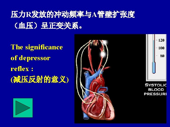
 1. Carotid sinus 、aortic arch baroreceptor reflex (颈A窦、主A弓压力感受性反射)")
6. 1. 3 Cardiovascular reflex (心血管反射) 1. Carotid sinus 、aortic arch baroreceptor reflex (颈A窦、主A弓压力感受性反射) An autonomically mediated reflex response that influences the heart and blood vessels to oppose a change in mean arterial blood pressure. (血压变化经压力感受器等反射弧活动而维持 血压于稳态的反射。)
 carotid sinus (颈A窦) Pressure R (压力R) aortic arch (主A弓) Sinus nerve")
Blood pressure (血压) carotid sinus (颈A窦) Pressure R (压力R) aortic arch (主A弓) Sinus nerve (窦N) Aortic nerve (主A神经) Spinal bulb (延脑) Cardiac sympathetic tone 心交感紧张(-) sympathetic vasoconstrictor tone 交感缩血管紧张(-) cardiac vagal ton 心迷走紧张(+) Cardiovisceral vessel (心血管活动) Blood pressure (血压)
 Receptor (感受器): carotid body")
2. Chemoreceptor reflex of Carotid body and aortic body (颈A体、主A体化学感受性反射) Receptor (感受器): carotid body , aortic body (颈A体, 主A体) chemoreceptor (化学感受器) Afferent nerve (传入N): glossopharyngeal. N (舌咽N) vagus N (迷走N)
![Process: P CO 2 [H]+ P O 2 carotid body (颈A体) chemoreceptor (化学感受器) aortic](http://slidetodoc.com/presentation_image_h2/6ac0f30c212e25a7f36342a38ee8d8ed/image-83.jpg "Process: P CO 2 [H]+ P O 2 carotid body (颈A体) chemoreceptor (化学感受器) aortic")
Process: P CO 2 [H]+ P O 2 carotid body (颈A体) chemoreceptor (化学感受器) aortic body (主A体) cardiovascular center medulla 心血管中枢(+) 心输出量 (延髓) respiration center 呼吸中枢(+) 外周阻力 舌咽N 迷走N 血压

Significance : When the organism in hypoxia、 asphyxia and low A pressure, it can regulate cardiovascular activity obviously to supply blood 、 oxygen for heart and brain. (意义: 在低氧、窒息、A压过低、酸中毒时才对 心血管动起明显调节作用以保证脑心供血供氧. )
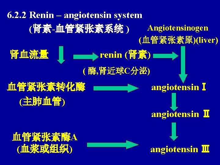
 6. 2. 1 Adrenine and norepinephrine ( 肾上腺素与去甲肾上腺素) Adrenine")
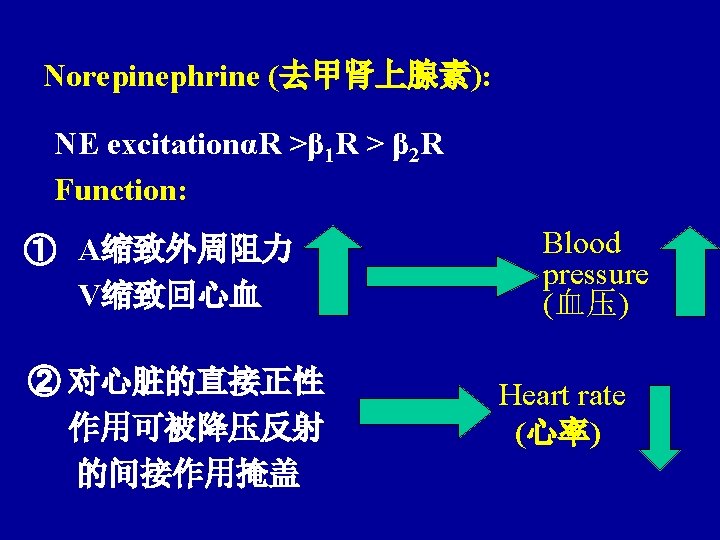
6. 2 Humoral regulation (体液调节) 6. 2. 1 Adrenine and norepinephrine ( 肾上腺素与去甲肾上腺素) Adrenine (肾上腺素) exciting αR, βR Function: ① Heart: 正性变时变力变传导 (β 1 R) BP

②Blood vessel: Excitation of blood vesselαR of skin gastrointestinal and kidney lead to vessel contraction ; while excitation of blood vessel β 2 R of skeletal muscle 、 liver and heart lead to vessel relaxation. The change of peripheral resistance is not obvious, blood is redistributed. 皮肤、肾、胃肠等处血管α R兴奋致血管收缩;而 骨骼肌、肝、心等处血管β 2 R兴奋致血管舒张,外周阻 力变化不大 血液重分配。 (肌、心优先供血)
 Compared with AngⅡ(比较AngⅡ: ) ① Vasoconstrictor function is weaker (缩血管作用较弱)")
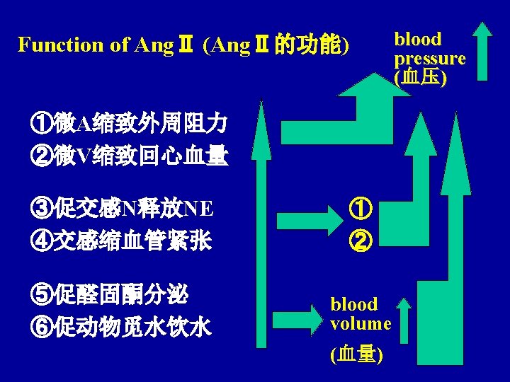
Function of AngⅢ (AngⅢ的功能) Compared with AngⅡ(比较AngⅡ: ) ① Vasoconstrictor function is weaker (缩血管作用较弱) ② The function that accelerate aldosterone synthesis and release is stronger (促醛固酮合成释放作用较强)
")
6. 2. 3 ADH 下丘脑视上核、 室旁核合成ADH neurohypophysis reserve and release into blood (神经垂体贮存并 释放入血) Vasoconstriction 外周阻力 (血管收缩 ) Blood 肾保水 volume BP
 心房受牵 血量增多 头低足高 浸入水中 ANF function")
6. 2. 5 Atrial natriuretic factor ANF (心钠素) 心房受牵 血量增多 头低足高 浸入水中 ANF function Diuresis (利尿) relaxing vessel (舒血管) ADH renin-Ang-aldosterone (肾素-Ang-醛固酮)
” 2. back“introduction” 3. over 4. back to menu")
1. enter“Chapter 8 Respiration (呼吸 )” 2. back“introduction” 3. over 4. back to menu
- Slides: 94