Blood Chapter 12 What are the 3 functions

Blood: Chapter 12

What are the 3 functions of blood? Transport nutrients, oxygen, wastes, and hormones 2. Helps maintain the stability of fluid 3. Distributes heat 1.
** White blood cells (WBC)**")
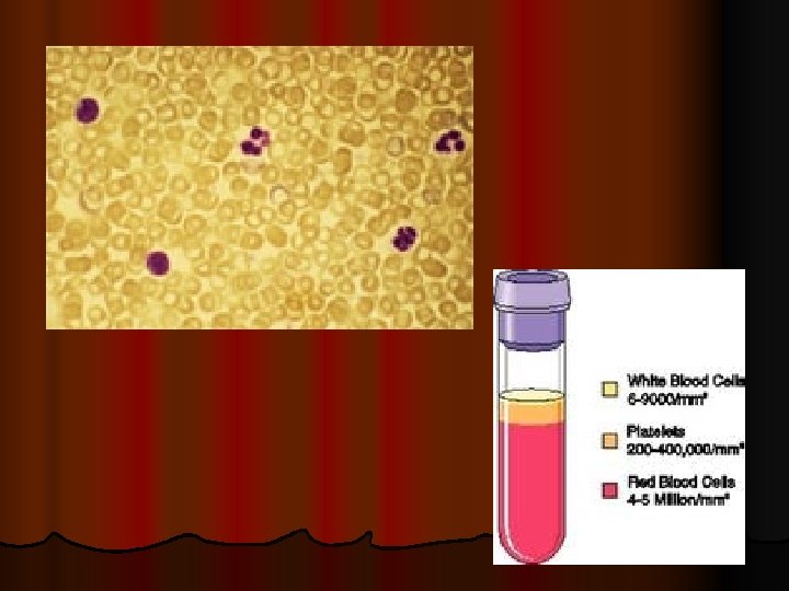
4 components: 1. 2. 3. 4. Red blood cells (RBC)** White blood cells (WBC)** Platelets** Plasma **together are called the “formed elements”
 Hematocrit: (about 45%)")
Average blood volume for an adult: 5. 3 quarts (5 L) Hematocrit: (about 45%) -cells by volume (the rest is plasma) -mostly RBC

Plasma is made up of: l l l l l Water Amino acids Proteins Carbohydrates Lipids Vitamins Hormones Electrolytes Cellular wastes
? l Erythrocytes l They have")
What is another name for red blood cells (RBC)? l Erythrocytes l They have biconcave discs Why do they have this shape? 1. Increases surface area through which gases can diffuse 2. Puts the cells membrane closer to the hemoglobin within the RBC

How much of a RBC is made up of hemoglobin? About 1/3 What is this responsible for? the color of blood When it’s combined with oxygen, it is called… oxyhemoglobin (bright red) When it’s NOT combined with oxygen, it is called… deoxyhemoglobin (dark red)

Why does someone become cyanotic? l May become cyanotic when they become hypoxic (low oxygen levels). l Low temperature can also cause cyanosis, because it decreases blood flow What does this mean? Their skin will appear to have a bluish tint because of the high concentration of deoxyhemoglobin

What is the RBCC? l Red Blood Cell Count What is the normal range for males and females? Males: 4. 6 -6. 2 million cells/mm 3 Females: 4. 2 -5. 4 million cells/mm 3

How does RBC affect oxygen-carrying capacity? l They are directly proportional l Changes in RBC may affect their health

What is hemopoiesis? l Red Blood Cell Formation Where does it first occur? Before birth: yolk sac, liver, spleen After birth: in red bone marrow
 What")
What is the average life span of a RBC? 120 days (4 months) What does erythropoietin do for the blood? Erythropoietin is a hormone that controls the rate of RBC formation. The kidneys (& liver) release this in response to oxygen deficiency to make more RBC)

What causes anemia? l Too few RBC or too little hemoglobin l Reduces the oxygen-carrying capacity of the blood Symptoms: may appear pale, lethargic -pregnant women are especially prone to this because of the hematocrit decrease

What destroys damaged RBC? macrophages 2 components of hemoglobin: 1. heme: iron-containing portion A. Iron B. Biliverdin (a greenish pigment) 2. globin: a protein

How are bilirubin and biliverdin related? l Biliverdin is eventually converted into bilirubin (an orange pigment) l Both are excreted in bile as bile pigments

What is jaundice? l An accumulation of bilirubin l Turns skin and eyes yellowish l May be result of immature liver cells l Ultraviolet (UV) light breaks down bilirubin (this is why they use the “bili lights” with newborns
 l Aka Leukocytes What is their main function? To protect")
White Blood Cells (WBC) l Aka Leukocytes What is their main function? To protect against disease What do WBCs develop from? Hemocytoblasts in response from hormones

What are the 5 types of WBC? A. Granulocytes -have granular cytoplasm -about 2 x size of RBC -short life span: about 12 hours -develop in red bone marrow 1. neutrophils: 54 -62% of WBC 2. eosinophils: 1 -3% of WBC 3. basophils: less than 1% of WBC

B. Agranulocytes -lack granules 4. monocytes: 3 -9% of WBC -largest blood cell -2 -3 x diameter of RBC 5. lymphocytes: 25 -33% of WBC -formed in lymphatic system organs and red bone marrow -only slightly larger than RBC

How do WBCs function to protect against disease? l Some kill/phagocytize bacterial cells l Produce proteins (antibodies) that destroy foreign particles Neutrophils/monocytes: contain lysosomes; engulf bacteria Eosinophils: attracted to and kill certain parasites; help control inflammation/allergic reactions Basophils: some contain heparin (prevents blood clot) and/or histamine (increase blood flow to injured tissues) -aids in allergic reactions Lymphocytes: important for immunity

What is WBCC? Normal range? White Blood Cell Count Normal: 5, 000 -10, 000 cells -this number changes in response to abnormal conditions

l Increase: may indicate infection -excess of 10, 000 cells/mm 3 = leukocytosis -indicates acute infection (appendicitis) Decrease: -below 5, 000 cells/mm 3 = leukopenia -typhoid fever, influenza, measles, mumps, chicken pox, AIDS, polio

DIFF l Differential white blood cell count l Lists % of types of WBC

Blood Platelets l Aka thrombocytes l Where do they arise from? -megakaryocytes -platelets fragment off (so they are not actually complete cells) -these develop from hemocytoblasts in response to thrombopoietin

What hormone is responsible for directing hemocytoblasts to make more megakaryocytes? l thrombopoietin

Normal Platelet count: l 130, 000 -360, 000 cells/mm 3 They also: -lack a nucleus -are less than ½ size of RBC

Functions: l 1. help close breaks in blood vessels l 2. initiate blood clots **together these control blood loss

12. 3 Plasma What percentage of plasma is water? 92% Functions: 1. transporting nutrients, gases, vitamins 2. help regulate fluid 3. maintain a favorable p. H
 in plasma Are these used")
Plasma proteins l Most abundant of dissolved substances (solutes) in plasma Are these used for energy sources? No

3 main groups: 1. Albumins: smallest of plasma proteins -accounts for 60% -synthesized in the liver What do they help regulate? Osmotic pressure -regulate water movement between blood and tissues, therefore help control blood volume and therefore blood pressure

2. Globulins -accounts for 36% 3 types: alpha, beta, gamma Where are they produced? alpha & beta = liver gamma = lymphatic tissues What is their primary function? Transport of lipids and fat-soluble vitamins

3. Fibrinogens -accounts for 4% What is their primary function? Blood coagulation Are they the smallest or largest plasma protein? largest *see summary chart on pg. 539

Gases and Nutrients 2 most important blood gases: oxygen and carbon dioxide also nitrogen (but no physiological function)

What are the plasma nutrients? Amino acids -plasma carries to liver, where they are used to manufacture proteins 2. Glucose -plasma transports these from small intestine to liver, where it is stored as glycogen or fat 1.
, phospholipids, and cholesterol -combine with proteins to form lipoprotein")
3. Lipids -includes fats (triglycerides), phospholipids, and cholesterol -combine with proteins to form lipoprotein complexes -consists of phospholipid, cholesterol, and protein around a triglyceride
: high concentration of triglycerides Low-density lipoproteins (LDL): high concentration of")
Very low-density lipoproteins (VLDL): high concentration of triglycerides Low-density lipoproteins (LDL): high concentration of cholesterol -major cholesterol-carrying lipoproteins (bad cholesterol!) High-density lipoproteins (HDL): high concentration of protein -lower concentration of lipids (Good cholesterol!)

What are three nonprotein nitrogenous substances? Amino acids 2. Urea 3. Uric acid 1. Give examples of electrolytes found in blood plasma: Na, K, Ca, Mg, Cl, Bicarbonate, P, Sulfate ions

14. 4 What is hemostasis? The stoppage of bleeding What 3 actions may help limit/prevent blood loss following an injury to a blood vessel? 1. blood vessel spasm 2. platelet plug formation 3. blood coagulation

1. Blood Vessel Spasm l Vasospasm: lasts a few minutes; effects for 30 minutes What has formed by the end of the vasospasm? Platelet plug What hormone do platelets release that causes further vasoconstriction? serotonin
 1. any rough")
2. Platelet plug formation What do platelets adhere to? (3 things) 1. any rough surface and to the collagen 2. to collagen underlying the lining 3. to each other, forming a platelet plug What type of blood vessel breaks can a platelet plug control? Small breaks; larger break would require a blood clot

3. Blood coagulation What is the most effective hemostatic mechanism? Coagulation What does this cause? Formation of a blood clot What are another name for clotting factors? biochemicals

Describe the major characteristic of each: 1. 2. 3. Fibrin: converted from fibrinogen Prothrombin: liver constantly produces this Thrombin: Converted from prothrombin

What is serum? Plasma-clotting factors -clear, yellow liquid that remains after the clot forms Coagulation is an example of a positive feedback system. Explain this. The original action stimulates more of the same type of action (clotting) -can only function like this for a short time

Thrombus vs. embolus l Thrombus: a blood clot abnormally formed in a vessel l Embolus: a dislodged blood clot (or fragment) that breaks loose and is carried away by the blood flow

l Atheroschlerosis: accumulation of fatty deposits in the arteries l Coronary thrombosis: a blood clot in a vessel that supplies the heart l Cerebral thrombosis: a blood clot forming in a vessel that supplies the brain l Pulmonary embolism: a blood clot that travels a vessel leading to the lungs l Infaction: when a blood clot kills tissues the vessels serve (Myocardial infarction= heart attack)

14. 5 What is agglutination? The clumping of red blood cells following a transfusion reaction
: red blood cell")
What is the difference between antigens and antibodies? Antigens (aka agglutinogens): red blood cell surface molecules Antibodies (aka agglutinins): proteins carried in plasma

What are the symptoms of agglutination? Anxiety, breathing difficulty, facial flushing, headache, severe pain

What is the ABO blood group? A, B, and O What 2 things is it based upon? -based on the presence of two major antigens: antigen A and antigen B

How many antigen combinations do red blood cells have on their surfaces? 4 What are they? 1. only A 2. only B 3. both A & B 4. neither A or B

What type of blood goes along with the previous combinations? Only A: Type A Only B: Type B Both A & B: Type AB Neither A or B: Type O

Blood type Antibodies present Antigens present Prefer to receive blood from Can receive blood from in emergency Can give blood to A Anti-B A A A, O A, AB B Anti-A B B B, O B, AB AB neither A&B AB AB, A, B, O AB O Anti-A, Anti- Neither A B or B O O A, B, AB, O

What blood type is considered the universal recipient? AB

What blood type is considered the universal donor? O

What is the Rh group? -named after the rhesus monkey -includes several Rh antigens (factors)

What does it mean to be Rh positive? Rh negative? l Rh positive: if any of the Rh antigens are present on the RBC membranes, you are Rh+ l Rh negative: you lack all Rh antigens

When do Rh antibodies form? l They form only in Rh-negative people in response to being given Rh-positive blood

What is the concern in getting Rh+ blood when you are Rh-? What happens? 1 st time: no effect; but you are now sensitized to Rh+ blood 2 nd time: the donated RBC are likely to agglutinate (clot) because you are already sensitized to the Rh+ blood (in other words, your body knows how to fight Rh+ blood, and it will fight it)

Explain the concern with an Rhmother having her 1 st baby: l If the baby is Rh+, the Rh+ cells will cross the mother’s bloodstream at birth 2 nd baby: if this baby is also Rh+, the antibodies could destroy the fetal RBC = erythroblastosis fetalis (hemolytic disease of the newborn)

To prevent this… l Rho. GAM is given within 72 hours of possible contact (birth, amniocentesis, miscarriage) l This shields the Rh+ cells from contacting the mother

What is the most rare blood type in the U. S. ? AB-

What is the most common blood type in the U. S. ? O+

- Slides: 65