Blood cells by Krisztina H Mink Semmelweis University

Blood cells by Krisztina H. -Minkó Semmelweis University, Department of Anatomy, Histology and Embryology Faculty of Pharmacy 2018

The cells of the blood and immune cells are free connective tissue cells and developmentally they both originate from the mesoderm. In the adult, for most part they are formed in the bone marrow, but they differ in their location and site of their functioning (blood, connective tissue).

Development of the circulatory system

Hematopoiesis and blood vessel formation first begins within the yolk sac wall with the formation of hemangioblastic aggregates Figure 13 -1 A, Drawing illustrating the formation of hemangioblastic aggregates and their differentiation into hematopoietic stem cells and endothelial precursor cells within blood islands. Blood islands subsequently form both endothelial cells and primitive erythrocytes. B, Expression of Vegfr 2 m. RNA, an early marker for hemangioblastic aggregates, within the yolk sac wall of a 15 -somite avian embryo. As the blood islands develop, endothelial cells retain Vegfr 2 expression, whereas hematopoietic stem cells progressively lose it. Downloaded from: Student. Consult (on 19 February 2011 07: 13 PM) © 2005 Elsevier

The arterial clusters Figure 13 -3 A second source of hematopoietic stem cells arises within the splanchnic mesoderm surrounding the aortic, gonad, and mesonephric region (AGM). These cells temporarily reside in the ventral floor of the dorsal aortic of this region. A, In humans at about day 27, a small number of hematopoietic stem cells (in red) reside and adhere to the dorsal aorta near the origin of the vitelline artery in the umbilical region. B, By day 30, the number of hematopoietic stem cells expands to several thousand. C, By day 36, hematopoietic stem cells expand to reside in the ventral floor of the dorsal aorta along almost the entire length of the AGM, and extending into the vitelline artery. By day 40, hematopoietic stem cells are no longer detected in the dorsal aorta. Downloaded from: Student. Consult (on 19 February 2011 07: 13 PM) © 2005 Elsevier

Current hypothesis about the colonisation of hematopoietic organs during embryonic development Figure 13 -2 Timeline of the appearance of hematopoietic stem cells during human development. Downloaded from: Student. Consult (on 19 February 2011 07: 13 PM) © 2005 Elsevier
 ©")
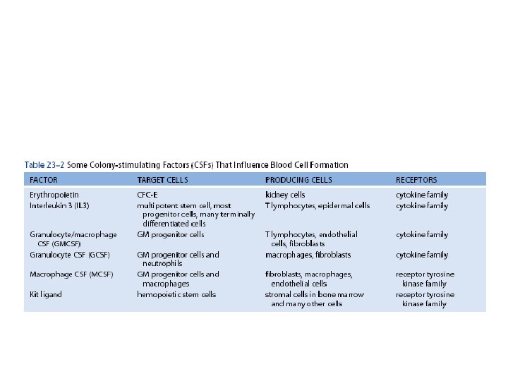
Hematopoietic hierarchy Downloaded from: Student. Consult (on 4 March 2010 09: 40 AM) © 2005 Elsevier
 -99% of the cells -oxygen")
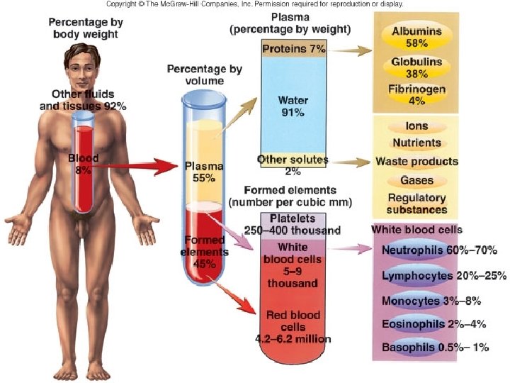
Composition of blood CELLULAR COMPONENTS Red blood cells (erythrocytes) -99% of the cells -oxygen and carbon dioxide transport White blood cells (leukocytes) -defence of the organism Platelets (thrombocytes) - blood clotting PLASMA -55 % of the blood -water -electrolytes -proteins albumin fibrinogen globulins -transported molecules (mostly transported bound to proteins) nutrients, vitamins, trace elements metabolic products hormones fatty substances

Difference between plasma and serum Figure 6 -1 Blood: Plasma, serum, and cells © 2005 Elsevier

Formed elements

Blood smear and May-Grünwald Giemsa staining to study the morphology of blood cells one drop of blood

Cells in the blood smear erythrocytes eosinophil neutrophil basophil granulocytes monocyte small medium lymphocyte neutrophil granulocyte
")
Red blood cells (erythrocytes)
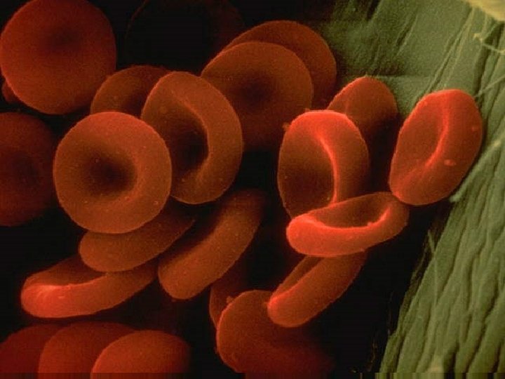
 -no cell nucleus -disk-shaped, concave on both sides, (special cytoskeleton")
Red blood cells (erythrocytes) -no cell nucleus -disk-shaped, concave on both sides, (special cytoskeleton with spektrin molecules) Function: O 2 and CO 2 transport lifespan: 120 days, mostly broken down in the spleen and in the liver (iron: stored, reused,
 and finally most")
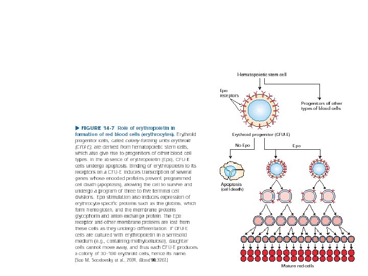
Erythrocytes develops from erythroblasts and lost the nucleus during differentiation (reticulocytes) and finally most of the cellular organels (erythrocytes) Specialisation = a cell filled with hemoglobin! Reticulocyte count in peripheral blood is increased after loss of blood.

Cell membrane of a red blood cell Downloaded from: Student. Consult (on 28 November 2010 05: 02 PM) © 2005 Elsevier
")
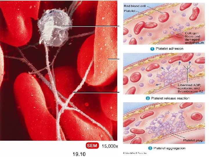
Platelets (thrombocytes)
 -cytoplasm fragments which derives from megakaryocytes (giant cells in the bone marrow)")
Platelets (thrombocytes) -cytoplasm fragments which derives from megakaryocytes (giant cells in the bone marrow) -lifespan: 5 -10 days 250 -300 000/mm 3 2 -3 mm Function: blood clotting

Megakaryocyte in the bone marrow

Cell biology of platelets Figure 6 -11 Platelets Downloaded from: Student. Consult (on 28 November 2010 05: 02 PM) © 2005 Elsevier

Blood clotting or hemostasis Downloaded from: Student. Consult (on 28 November 2010 05: 02 PM) © 2005 Elsevier
 relatively colorless cells Granulocytes Agranulocytes (polymorphonuclear leukocytes) neutrophils (neutrophil granulocytes)")
White blood cells (leukocytes) relatively colorless cells Granulocytes Agranulocytes (polymorphonuclear leukocytes) neutrophils (neutrophil granulocytes) lymphocytes eosinophils (eosinophil granulocytes) monocytes basophils (basophil granulocytes) Their lifespan varies, can be from a few hours to years. Together with the lymphoid organs the white cells form the immune system.
 -small granules in")
Neutrophil granulocyte -size: 10 -15 mm -segmented nucleus (3 -5 lobes) -small granules in the citoplasm 60 -70% of white blood cells also called phagocytes because they phagocytose foreign material (first cells to reach the site of an inflammation) part of the nonspecific immune system Neutrophil Chase https: //www. youtube. com/watch? v=i. F Ous 8 ehx. Uc

Primary granules contain elastase and myeloperoxidase, secondary granules contain lysozyme and proteases in neutrophils Figure 6 -4 Neutrophil Downloaded from: Student. Consult (on 28 November 2010 05: 02 PM) © 2005 Elsevier

Neutrophils migrate across the vessel wall

Molecular mechanisms accompanying neutrofil migration Figure 6 -9 Homing and inflammation Downloaded from: Student. Consult (on 28 November 2010 05: 02 PM) © 2005 Elsevier

Eosinophil granulocyte -size: 10 -15 mm; -two-lobed nucleus -eozinophil granules containing histamin, crystals can be detected by EM -1 -6% of white blood cells -are capable of phagocytosis (Ag-Ab complexes), in allergic reactions they bound to and inactivate excess histamine (from mast cells or basophils)
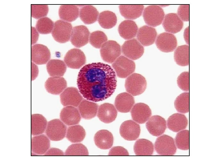

Figure 6 -10 Mast cell-eosinophil interaction in asthma Downloaded from: Student. Consult (on 28 November 2010 05: 02 PM) © 2005 Elsevier

Basophil granulocyte - size: 10 -15 mm -segmented nucleus (not seen because of the granules) -many, large, basophil cytoplasmic granules (heparin, histamine). 0 -1% of white blood cells effector cells in allergy, immediate hypersensitivity

Heparin and histamin containing granules of basophils Figure 6 -6 Basophil Downloaded from: Student. Consult (on 28 November 2010 05: 02 PM) © 2005 Elsevier

Comparison of granulocytes

Mononuclear leukocytes
 -ovale or kidney-shaped nucleus, numerous lysosomes in")
Monocytes -size: 15 -20 mm (largest WBC!) -ovale or kidney-shaped nucleus, numerous lysosomes in the cytoplasm -precursors of tissue macrophages 2 -6 % of white blood cells Function: coordination of cellular and humoral immune response

Figure 6 -8 Monocyte Downloaded from: Student. Consult (on 28 November 2010 05: 02 PM) © 2005 Elsevier

Lymphocytes -size: 8 -10 mm -round-shaped nucleus, organellum rich citoplasm -20 -40 % of whit blood cells Functions: cells of the specific (humoral and cellular) immunity Subtypes: B- and T-lymphocytes NK cells (against tumor cells) activated lymphocytes increase their cytoplasm and r. ER

Figure 6 -7 Lymphocyte Downloaded from: Student. Consult (on 28 November 2010 05: 02 PM) © 2005 Elsevier

Morphological differences between resting and activated lymphocytes

Comparison of white blood cells by electron microscopy

Summary of main functions of blood cells

Thank you for attention!

References: The human body, Thieme Kierszenbaum: Histology and Cell Biology, Elsevier Alberts et al. : Molecular biology of the cell Lodish et al. : Molecular cell biology Co-author of the lecture: Dr. Nándor Nagy, Dr. Tibor Wenger
- Slides: 47