Blood Blood Vessels Circulation 21 1 Physical Characteristics

Blood, Blood Vessels & Circulation 21 -1
 than water and flows more slowly")
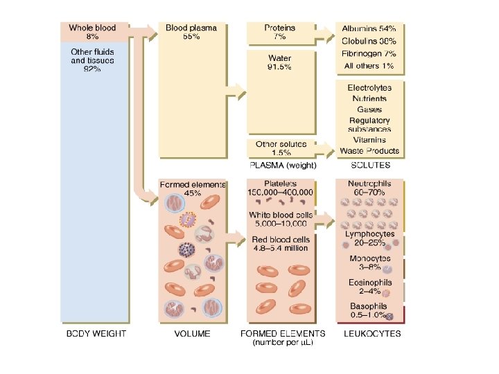
Physical Characteristics of Blood • Thicker (more viscous) than water and flows more slowly than water • Temperature of 100. 4 degrees F • p. H 7. 4 (7. 35 -7. 45) • 8 % of total body weight • Blood volume – 5 to 6 liters in average male – 4 to 5 liters in average female – hormonal negative feedback systems maintain constant blood volume and osmotic pressure
 – about 2% are dissolved")
Blood components • 55% = plasma: mainly water (~91%) – about 2% are dissolved substances = sugars, amino acids, lipids & vitamins, ions, dissolved gases, hormones & nutrient proteins – 7 to 8% are plasma proteins: provide a role in balancing osmotic pressure and water flow between the blood and extracellular fluid/tissues – most common plasma proteins: albumins, globulins, clotting protein (fibrinogen) plasma proteins fluid moved from low protein concentration to high protein concentration – loss of plasma proteins from blood – increases osmotic pressure in the surrounding tissues and results in water flow out of blood into tissues swelling

Hematopoiesis HSC

Blood: Cellular elements • 45% of blood is the cellular elements or formed elements • 99% of blood cells are erythrocytes or RBCs – most numerous cell type in the body – 4 to 6 million per ul blood – flat, biconcave discs • provides a larger surface area for diffusion of oxygen across their membrane • thinness of the membrane allows rapid diffusion • very flexible membrane that allows their deformation for travel through thin capillaries
 in the")
Erythrocytes • RBCs: – formed by differentiation of hematopoietic stem cells (HSCs) in the red bone marrow into myeloid stem cells erythrocytes – red bone marrow makes about 2 million RBCs per second! – first formed as an immature RBC = reticulocyte • still possess a nucleus and organelles • maturation of the reticulocyte causes loss of nucleus and organelles and the filling of the RBC with close to 250 million Hb molecules – RBCs have no nuclei or organelles but contain crucial enzymes to keep them alive for ~ 120 days – 1. glycolytic enzymes –for energy production (i. e. ATP) - RBCs rely on glycolysis and the enzymes of this pathway exclusively – 2. carbonic anhydrase – enzyme on RBC surface » plays role in dissolving of CO 2 in blood plasma

Erythrocytes: Hemoglobin • pigment – naturally colored that is red due to its iron content – composed of a: – 1. globin portion • four, highly folded protein chains • 2 alpha chains & 2 beta chains • change their shapes as oxygen binds Hb

Erythrocytes: Hemoglobin – 2. heme component • one molecule of iron-based heme bound to each globin protein chain = 4 total • each heme can bind one oxygen – total binding capacity of 4 oxygen molecules per Hb • binding of the 1 st O 2 increases the binding efficiency of the other three O 2 molecules • at sea level - Hb is 98% saturated with oxygen • saturation level can be affected by temperature (increase temp, decrease saturation) • saturation level can be affected by atmospheric pressure (decrease pressure, decrease saturation)

Cells of Immunity: WBCs • cells of the lymphoid lineage – T and B lymphocytes (lymphocytes) • cells of the myeloid lineage – neutrophils, basophils, mast cells, eosinophils and monocytes, macrophages • BUT WBCs can also be classified based on appearance of the cytoplasm after staining – A. Granulocytes: neutrophils, macrophages, other phagocytes, eosinophils, basophils – B. Agranular: T and B cells WBCs can enter tissues where they function

Immunity • Immunity: ability of the body to defend itself from infectious agents, foreign cells, cancer cells • immune system has two functional divisions • innate immune system: non-specific immunity – – – divided into: cell-mediated and humoral (secreted) mediated includes chemical and physical barriers defensive chemicals: complement and inflammation no memory all forms of life immune system recognizes some king of general molecular property marking the invader as foreign • usually a foreign carbohydrate or lipid in the cell wall – includes the response known as inflammation

Immunity • adaptive immune system: pathogen and antigen specific response – – also divided into cell-mediated and humoral mediated chemical defenses and lymphocytes memory results found only in jawed vertebrates

https: //www. youtube. com/watch? v=k. TGz. VUj. FLNE 21 -13

Anatomy of Blood Vessels • Closed system of tubes that carries blood • Arteries carry blood away from heart to tissues – elastic arteries – muscular arteries – arterioles • Capillaries are thin enough to allow exchange • Venules merge to form veins that bring blood back to the heart 21 -14
 – innermost layer - simple squamous epithelium known as")
• Tunica interna (intima) – innermost layer - simple squamous epithelium known as endothelium Arteries • Tunica media – major component - circular smooth muscle fibers – smooth muscle is innervated by sympathetic nervous system – relaxation of this muscle causes vasodilation • increases diameter of vessel contraction of this muscle causes or vasoconstriction • decreases diameter of vessel • Tunica externa – elastic & collagen fibers 21 -15

Veins • Proportionally thinner walls than same diameter artery – tunica media less muscle – lack external & internal elastic lamina • Still adaptable to variations in volume & pressure • Valves are thin folds of tunica interna designed to prevent backflow • Venous sinus has no muscle at all – coronary sinus that drains the heart muscle or dural venous sinuses that drain the brain 21 -16

Capillaries • Microscopic vessels that connect arterioles to venules • Found near every cell in the body but more extensive in highly active tissue (muscles, liver, kidneys & brain) • Function is exchange of nutrients, gases & wastes between blood and tissue fluid • Structure is single layer of simple squamous epithelium (endothelium) and its basement membrane • different types of capillaries – depending on how permeable they are 21 -17

The Lymphatic System • Lymphocytes travel throughout the body in spaces between the cells and are carried in the blood and lymphatic system. • the lymphatic system = system of lymphatic vessels + lymph nodes + lymphatic tissues (spleen, thymus, tonsils) that filter lymph and circulate WBCs • lymph = yellow-colored fluid that is produced from your blood plasma at capillary beds – produced when plasma filters out of your blood and into your tissues – some of that filtrate drains from tissues and becomes lymph

The Lymphatic System • blood enters into a capillary • blood pressure forces some of the blood plasma into the tissues – known as ultrafiltration • blood osmotic pressure causes the reabsorption of some of this fluid back into the capillary to become plasma again • the fluid that remains in the tissues and is not reabsorbed = interstitial fluid • excess interstitial fluid drains away as lymph excess IF is Lymph

Lymph Nodes • lymph must by returned to your blood stream – via the subclavian veins • along the way it flows through lymph nodes which house T and B lymphocytes and macrophages • these immune cells clean the lymph of bacteria • so what gets returned to your blood is cleaned • lymph is the way we “launder” our blood

The Lymphatic System • the lymphatic system is made of numerous lymphatic organs designed to either clear the lymph of foreign invaders or your blood http: //www. niaid. nih. gov/topics/immun e. System/Pages/structure. Images. aspx
 Lymph nodes: various locations in the body 2) Tonsils: lymphatic tissue")
Lymphoid Organs 1) Lymph nodes: various locations in the body 2) Tonsils: lymphatic tissue located in the pharynx (adenoids) or oral cavity (palatine tonsils) -defense against pathogens brought in through food and drink 3) Spleen: upper left region of the abdomen -cleanses the blood plasma directly -made of white and red pulp -white pulp contains lymphocytes -red pulp contains red blood cells & macrophages
 Bone marrow (red): adult - within the spongy bone of the")
Lymphoid Organs 4) Bone marrow (red): adult - within the spongy bone of the epiphyses, pelvis, skull, clavicle, sternum -site of blood plasma cleansing -also site of origin for all blood cells (RBCs, WBCs) 5) Thymus gland: located below the trachea, on top of the heart -production of T lymphocytes

Velocity of Blood Flow • Speed of blood flow in cm/sec is inversely related to cross-sectional area – blood flow is slows in the arterioles • slowest rate in capillaries allows for exchange • Blood flow becomes faster when vessels merge to form veins • blood returning to the heart = Venous Return • Venous return depends on: – 1. skeletal contraction – 2. valves preventing backflow 21 -24

Blood Pressure • Pressure exerted by blood on walls of a vessel – caused by contraction of the ventricles – highest in aorta • 120 mm Hg during systole & 80 during diastole • If heart rate increases, BP rises • Pressure falls steadily in systemic circulation with distance from left ventricle • Regulation of Blood Pressure: – 1. role of cardiovascular center in the medulla – 2. sensory receptors – proprioceptors (physical activity), baroreceptors, chemoreceptors (oxygen, carbon dioxide) – 3. hormones – epinephrine, anti-diuretic hormone – 4. local factors – local physical and chemical factors – 5. higher centers of the brain, limbic system, hypothalamus 21 -25

Vascular Pathways 1. Systemic 21 -26

Vascular Pathways 2. Pulmonary 21 -27

Arterial Branches of Systemic Circulation • All are branches from aorta supplying arms, head, lower limbs and all viscera with O 2 from the lungs • Aorta arises from left ventricle (thickest chamber) – 4 major divisions of aorta • • ascending aorta arch of aorta thoracic aorta abdominal aorta 21 -28

Aorta and Its Superior Branches • Aorta is largest artery of the body – ascending aorta • 2 coronary arteries supply myocardium – arch of aorta -- branches to the arms & head • brachiocephalic trunk branches into right common carotid and right subclavian • left subclavian & left carotid arise independently – descending aorta – divides into thoracic and abdominal 21 -29

Left Common Carotid Brachiocephalic Trunk Left Subclavian Superior Vena Cava Aortic Arch Ascending Aorta Pericardium Diaphragm 21 -30

Major Systemic Veins • All veins empty into the right atrium of the heart – superior vena cava drains the head and upper extremities – inferior vena cava drains the abdomen, pelvis & lower limbs – coronary sinus is large vein draining the heart muscle back into the heart 21 -31

Common Carotid Branches Circle of Willis • • External carotid arteries – supplies structures external to skull as branches of maxillary and superficial temporal branches – “Seven little fairies ascended over Polly’s super mums” Internal carotid arteries (contribute to Circle of Willis) – supply eyeballs and parts of brain 21 -32

posterior auricular superficial temporal maxillary occipital internal carotid external carotid sinus facial lingual superior thyroid 21 -33

Veins of the Head and Neck • External and Internal jugular veins drain the head and neck into the superior vena cava • Blood from the brain drains into several large dural sinuses - empty into internal jugular vein – Transverse sinus – Sigmoid sinus – Sagittal sinus 21 -34

Subclavian Branches • Subclavian arteries pass superior to the 1 st rib – gives rise to vertebral a. that supplies part of the blood to the Circle of Willis on the base of the brain • Become the axillary artery in the armpit • Become the brachial artery in the arm – deep brachial artery – radial and ulnar collaterals • Divide into radial and ulnar branches in the forearm 21 -35

vertebral Ulnar collateral thyrocervical Radial collateral brachial suprascapular Common interosseous thoracoacromial axillary Common Carotid subscapular circumflex humeral ulnar deep brachal radial brachial interosseous radial collateral ulnar collateral Deep palmar arch Superficial palmar arch brachial Digital arteries ulnar radial

Arteries of the Lower Extremity • External iliac artery become femoral artery when it passes under the inguinal ligament & into the thigh – – femoral artery gives off deep femoral branch femoral artery becomes popliteal artery behind the knee gives off anterior tibial and fibular/peroneal branches continues on as posterior tibial artery (runs alongside the tibial nerve) 21 -37

Common iliac External iliac Internal iliac Deep femoral Ascending branch of Lateral femoral circumflex Obturator Deep femoral continues Descending branch of Lateral circumflex Femoral -lateral femoral circumflex wraps around head of femur – gives off ascending and descending branches – descending branch runs down lateral side of thigh -descending genicular runs down medial side of thigh

Common iliac External iliac Internal iliac Deep femoral Ascending branch of Lateral femoral circumflex Obturator Lateral femoral circumflex Femoral Descending Genicular Deep femoral continues Deep Femoral Descending br Of Lateral femoral circumflex Genicular Arteries of the Knee Anterior Tibial

Femoral Deep Femoral Descending Genicular Descending br Of Lateral femoral circumflex Genicular Arteries of the Knee Anterior Tibial 21 -40

Veins of the Systemic Circulation • Drain blood from entire body & return it to right side of heart • Deep veins parallel the arteries in the region • Superficial veins are found just beneath the skin • All venous blood drains to either superior or inferior vena cava or coronary sinus 21 -41

Abdominal Aorta and Its Branches • Supplies abdominal & pelvic viscera & lower extremities – – celiac trunk supplies liver, stomach, spleen & pancreas superior & inferior mesenteric arteries supply intestines renal arteries supply kidneys gonadal arteries supply ovaries and testes • Splits into common iliac arteries at 4 th lumbar vertebrae – external iliacs supply lower extremity – internal iliacs supply pelvic viscera 21 -42

Inferior Vena Cava Celiac Superior Mesenteric Renal Gonadal Inferior mesenteric Common Iliac 21 -43

Visceral Branches off Abdominal Aorta • Celiac trunk is first branch inferior to diaphragm – left gastric artery, splenic artery, common hepatic artery (gives rise to right gastric) • Superior mesenteric artery lies in mesentery of intestines – supplies upper intestines (small and large) – jejunal, ileals, ileocolic, right & middle colic arteries • Inferior mesenteric artery – supplies descending colon, sigmoid colon & rectum – left colic, sigmoid and rectal branches 21 -44

Left Gastric Hepatic Proper Common Hepatic Splenic Vein Celiac trunk Left Colic Artery Inferior Mesenteric Sigmoid Superior Rectal
- Slides: 45