Blood Blood n The only fluid tissue in

Blood

Blood n The only fluid tissue in the human body n Classified as a connective tissue n Components of blood – Living cells – Non-living matrix § Plasma
 sink to the")
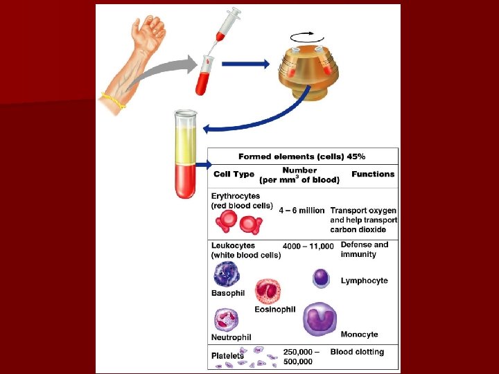
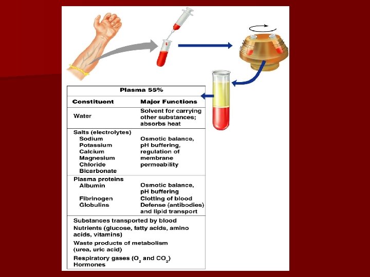
Blood n If blood is centrifuged – Erythrocytes (Red Blood Cells) sink to the bottom (45% of blood, a percentage known as the hematocrit) – Buffy coat contains leukocytes and platelets (less than 1% of blood) § Buffy coat is a thin, whitish layer between the erythrocytes and plasma – Plasma rises to the top (55% of blood)

Physical Characteristics of Blood n Color range – Oxygen-rich blood is scarlet red – Oxygen-poor blood is dull red n p. H must remain between 7. 35– 7. 45 n Blood temperature is slightly higher than body temperature at 100. 4°F n In a healthy man, blood volume is about 5 – 6 liters or about 6 quarts n Blood makes up 8% of body weight

Blood Plasma n Composed of approximately 90% water n Includes many dissolved substances – Nutrients – Salts (electrolytes) – Respiratory gases – Hormones – Plasma proteins (most abundant) – Waste products ******When the clotting factors are taken out of the plasma it is called SERUM.

Blood Plasma n Plasma proteins – Most abundant solutes in plasma – Most plasma proteins are made by liver – Various plasma proteins include § Albumin—regulates osmotic pressure § Clotting proteins—help to stem blood loss when a blood vessel is injured n Antibodies—help pathogens protect the body from

Blood Plasma n Acidosis – Blood becomes too acidic n Alkalosis – Blood becomes too basic n In each scenario, the respiratory system and kidneys help restore blood p. H to normal
 n Leukocytes – White blood cells (WBCs)")
n Erythrocytes – Red blood cells (RBCs) n Leukocytes – White blood cells (WBCs) n Platelets

Photomicrograph of a Blood Smear Figure 10. 2
 – Main function is to carry oxygen")
n Erythrocytes (red blood cells or RBCs) – Main function is to carry oxygen – Anatomy of circulating erythrocytes § Biconcave disks § Essentially bags of hemoglobin § Anucleate (no nucleus) § Contain very few organelles n Wear out in 100 to 120 days – 5 million RBCs per cubic millimeter of blood

n Hemoglobin – Iron-containing protein – Binds strongly, but reversibly, to oxygen – Each hemoglobin molecule has four oxygen binding sites – Each erythrocyte has 250 million hemoglobin molecules – Normal blood contains 12– 18 g of hemoglobin per 100 m. L blood

n Homeostatic imbalance of RBCs – Anemia is a decrease in the oxygen-carrying ability of the blood – Sickle cell anemia (SCA) results from abnormally shaped hemoglobin – Polycythemia is an excessive or abnormal increase in the number of erythrocytes

Table 10. 1

Figure 10. 3
 – Crucial in the body’s defense against")
n Leukocytes (white blood cells or WBCs) – Crucial in the body’s defense against disease – These are complete cells, with a nucleus and organelles – Able to move into and out of blood vessels (diapedesis) – Can move by ameboid motion – Can respond to chemicals released by damaged tissues – 4, 000 to 11, 000 WBC per cubic millimeter of blood

n Abnormal numbers of leukocytes – Leukocytosis § WBC count above 11, 000 leukocytes/mm 3 § Generally indicates an infection – Leukopenia § Abnormally low leukocyte level § Commonly caused by certain drugs such as corticosteroids and anticancer agents – Leukemia § Bone marrow becomes cancerous, turns out excess WBC

Types of leukocytes – Granulocytes § Granules in their cytoplasm can be stained § Possess lobed nuclei § Include neutrophils, eosinophils, and basophils – Agranulocytes § Lack visible cytoplasmic granules § Nuclei are spherical, oval, or kidney-shaped § Include lymphocytes and monocytes

n List of the WBCs from most to least abundant – – – Neutrophils Lymphocytes Monocytes Eosinophils Basophils n Easy way to remember this list – – – Never Let Monkeys Eat Bananas
")
Table 10. 2 (2 of 2)
")
Table 10. 2 (1 of 2)
 Neutrophils -Multilobed nucleus with fine granules - Act as phagocytes")
Types of granulocytes 1) Neutrophils -Multilobed nucleus with fine granules - Act as phagocytes at active sites of infection 2) Eosinophils -Large brick- red cytoplasmic granules -Found in response to allergies and parasitic worms
 3) Basophils § Have histamine-containing granules § Initiate inflammation")
n Types of granulocytes (continued) 3) Basophils § Have histamine-containing granules § Initiate inflammation

Types of agranulocytes n Lymphocytes -Nucleus fills most of the cell - Play an important role in the immune response Monocytes -Largest of the white blood cells -Function as macrophages -Important in fighting chronic infection n
 – Needed for the clotting")
n Platelets – Derived from ruptured multinucleate cells (MEGAKARYOCYTES) – Needed for the clotting process – Normal platelet count = 300, 000/mm 3 Vitamin K plays an important role in blood clotting

Hematopoiesis n Blood cell formation n Occurs in red bone marrow epiphyseal plates, epiphyses of femur and humerus, flat bones of skull & pelvis. n All blood cells are derived from a common stem cell (hemocytoblast) n Hemocytoblast differentiation – Lymphoid stem cell produces lymphocytes – Myeloid stem cell produces all other formed elements

Hematopoiesis Hemocytoblast stem cells Lymphoid stem cells Myeloid stem cells Secondary stem cells Basophils Erythrocytes Platelets Lymphocytes Monocytes Eosinophils Neutrophils Figure 10. 4

Formation of Erythrocytes n Unable to divide, grow, or synthesize proteins n Wear out in 100 to 120 days n When worn out, RBCs are eliminated by phagocytes in the spleen or liver n Lost cells are replaced by division of hemocytoblasts in the red bone marrow
 n Kidneys")
Control of Erythrocyte Production n Rate is controlled by a hormone (erythropoietin) n Kidneys produce most erythropoietin as a response to reduced oxygen levels in the blood n Homeostasis is maintained by negative feedback from blood oxygen levels

Control of Erythrocyte Production Imb ala nce Normal blood oxygen levels Imb ala Increased O 2 - carrying ability of blood nce Stimulus: Decreased RBC count, decreased availability of O 2 to blood, or increased tissue demands for O 2 Reduced O 2 levels in blood More RBCs Kidney releases erythropoietin Enhanced erythropoiesis Erythropoietin stimulates Red bone marrow Figure 10. 5

Formation of White Blood Cells and Platelets n Controlled by hormones – Colony stimulating factors (CSFs) and interleukins prompt bone marrow to generate leukocytes – Thrombopoietin stimulates production of platelets

Hemostasis n Stoppage of bleeding resulting from a break in a blood vessel n Hemostasis involves three phases – Vascular spasms – Platelet plug formation – Coagulation (blood clotting)

Hemostasis Figure 10. 6

Hemostasis n Vascular spasms – Vasoconstriction causes blood vessel to spasm – Spasms narrow the blood vessel, decreasing blood loss

Hemostasis n Platelet plug formation – Collagen fibers are exposed by a break in a blood vessel – Platelets become “sticky” and cling to fibers – Anchored platelets release chemicals to attract more platelets – Platelets pile up to form a platelet plug

Hemostasis Step 1: Vascular Spasms Step 2: Platelet Plug Formation Injury to lining of vessel exposes collagen fibers; platelets adhere Collagen fibers Platelet plug forms Platelets Figure 10. 6, step 3
 – PF 3 (a")
Hemostasis n Coagulation – Injured tissues release tissue factor (TF) – PF 3 (a phospholipid) interacts with TF, blood protein clotting factors, and calcium ions to trigger a clotting cascade – Prothrombin activator converts prothrombin to thrombin (an enzyme)
 – Thrombin joins fibrinogen proteins into hairlike molecules of insoluble")
Hemostasis n Coagulation (continued) – Thrombin joins fibrinogen proteins into hairlike molecules of insoluble fibrin – Fibrin forms a meshwork (the basis for a clot)

Hemostasis Figure 10. 7

Hemostasis n Blood usually clots within 3 to 6 minutes n The clot remains as endothelium regenerates n The clot is broken down after tissue repair

Undesirable Clotting n Thrombus – A clot in an unbroken blood vessel – Can be deadly in areas like the heart n Embolus – A thrombus that breaks away and floats freely in the bloodstream – Can later clog vessels in critical areas such as the brain

Bleeding Disorders n Thrombocytopenia – Platelet deficiency – Even normal movements can cause bleeding from small blood vessels that require platelets for clotting n Hemophilia – Hereditary bleeding disorder – Normal clotting factors are missing

Blood Groups and Transfusions n Large losses of blood have serious consequences – Loss of 15– 30% causes weakness – Loss of over 30% causes shock, which can be fatal n Transfusions are the only way to replace blood quickly n Transfused blood must be of the same blood group

Human Blood Groups n Blood contains genetically determined proteins n Antigens (a substance the body recognizes as foreign) may be attacked by the immune system n Antibodies are the “recognizers” n Blood is “typed” by using antibodies that will cause blood with certain proteins to clump (agglutination)

ABO Blood Groups n Based on the presence or absence of two antigens – Type A – Type B n The lack of these antigens is called type O

ABO Blood Groups n The presence of both antigens A and B is called type AB n The presence of antigen A is called type A n The presence of antigen B is called type B n The lack of both antigens A and B is called type O

ABO Blood Groups n Blood blood type AB can receive A, B, AB, and O – Universal recipient n Blood type B can receive B and O blood n Blood type A can receive A and O blood n Blood type O can receive O blood – Universal donor

ABO Blood Groups Table 10. 3

Rh Blood Groups n Named because of the presence or absence of one of eight Rh antigens (agglutinogen D) that was originally defined in Rhesus monkeys n Most Americans are Rh+ (Rh positive) n Problems can occur in mixing Rh+ blood into a body with Rh– (Rh negative) blood

Rh Dangers During Pregnancy n Danger occurs only when the mother is Rh – and the father is Rh+, and the child inherits the Rh+ factor n Rho. GAM shot can prevent buildup of anti-Rh+ antibodies in mother’s blood

Rh Dangers During Pregnancy n The mismatch of an Rh– mother carrying an Rh+ baby can cause problems for the unborn child – The first pregnancy usually proceeds without problems – The immune system is sensitized after the first pregnancy – In a second pregnancy, the mother’s immune system produces antibodies to attack the Rh+ blood (hemolytic disease of the newborn)

In the old days the child would be born dead- stillborn Disorder- erythroblastosis fetalis

Blood Typing n Blood samples are mixed with anti-A and anti-B serum n Coagulation or no coagulation leads to determining blood type n Typing for ABO and Rh factors is done in the same manner n Cross matching—testing for agglutination of donor RBCs by the recipient’s serum, and vice versa

Blood Typing Figure 10. 8

Developmental Aspects of Blood n Sites of blood cell formation – The fetal liver and spleen are early sites of blood cell formation – Bone marrow takes over hematopoiesis by the seventh month n Fetal hemoglobin differs from hemoglobin produced after birth n Physiologic jaundice results in infants in which the liver cannot rid the body of hemoglobin breakdown products fast enough
 is transmitted through saliva, so you")
Mononucleosis n Infectious n The virus (Epstein Barr) is transmitted through saliva, so you can get it through kissing, but you can also be exposed through a cough or sneeze, or by sharing a glass or food utensils with someone who has mono n Usually symptomatic in adolescents & young adults, but young children have a few symptoms and often goes unrecognized
 n Sore throat, perhaps")
Mono symptoms n Fatigue n General feeling of unwellness (malaise) n Sore throat, perhaps a strep throat that doesn't get better with antibiotic use n Fever n Swollen tonsils as well as lymph nodes in your neck and armpits n Headache n Skin rash n Soft, swollen spleen

n Incubation period of approximately 4 to 6 weeks, although in young children this period may be shorter. n Symptoms usually lessen within a couple of weeks, although fatigue, enlarged lymph nodes and a swollen spleen may last for a few weeks longer

Treatment n no specific therapy available to treat infectious mononucleosis. Antibiotics don't work against viral infections such as mono. Treatment mainly involves bed rest and drinking plenty of fluids
- Slides: 60