Blood Anna L Kiss Department of Anatomy Histology

Blood Anna L. Kiss Department of Anatomy, Histology and Embryology Semmelweis University Budapest 2017

Blood § Cells + „exracellular matrix” : plasma § Cells: red blood cells white blood cells granulocytes • Mesoderm agranulocytes
")
Erythrocytes d: 7µm biconcave, no nucleus, limited life time (about 120 days)

Haemoglobin: haem+globin protein • transport of molecular oxygen • reverseble binding • hem: iron 2+ containing molecule

Erythrocytes sickle-cell disease: 1 amino acid is changed in the globin molecule: genetic mutation

Leukocytes Granulocytes: segmented nucleus granules inside of the cytoplasm neutrophyl basophyl Agranulocytes: monocytes lymphocytes eosinophyl

Granulocytes: neutrophyls segmented nucleus, granules in the cytoplasm they are ~75% of leukocytes

Extravasatio: white blood cells migrate out from the blood vessels

Granulocytes: basophils Bilobed nucleus Granulse: stained with basic dye

Granulocytes: eosinophils allergy, parasitism nucleus: bilobed granules: stained by basic dye

Agranulocytes: monocytes professional phagocytes; monocytes/macrophages: monocyte/derived cells in the connective tissue and in different organs

Agranulocytes: lymphocytes small cells (d: 7μm, with large nucleus T cells: (thymus derived: killer, helper, supressor etc. ) B cells: • plasma cells: antibody production; • memory cells

Haemopoesis
 cell debris: containing secretory granules")
Platelets (trombocytes) cell debris: containing secretory granules

Platelet

Blood clothing: chain reaction Factors: many factors are involved, most of them come from platelets • protrombin (produced in the liver • fibrinogen (globular, water soluble protein, produced in the liver • fibrin: fibrillar proteins, water insoluble Injury of the blood vessels: chain reaction starts, Ca ion prersent

Blood clothing protrombin Ca 2+ fibrinogen fibrin
 • T lymphocytes: killer, helper, supressor •")
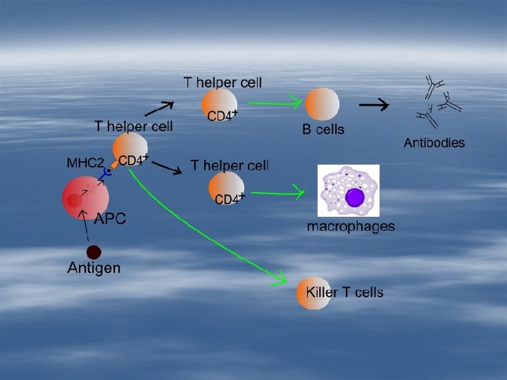
Immunresponse Cells: • lymphocytes (T and B) • T lymphocytes: killer, helper, supressor • B lymphocytes: plasma cells (antibody production) memory cells • antigen presenting cells: dendritic cells, macrophages

Antigen presenting cells

Dendritic cells

• T lymphocytes: • killer cells express antibody on their plasma membrane cellular immunresponse humoral immunresponse: helper cells (T lymphocytes) plasma cells (B lymphocytes) • helper cells: secret interleukins

Cellular imune response: Natural killer cells

Humoral immune response

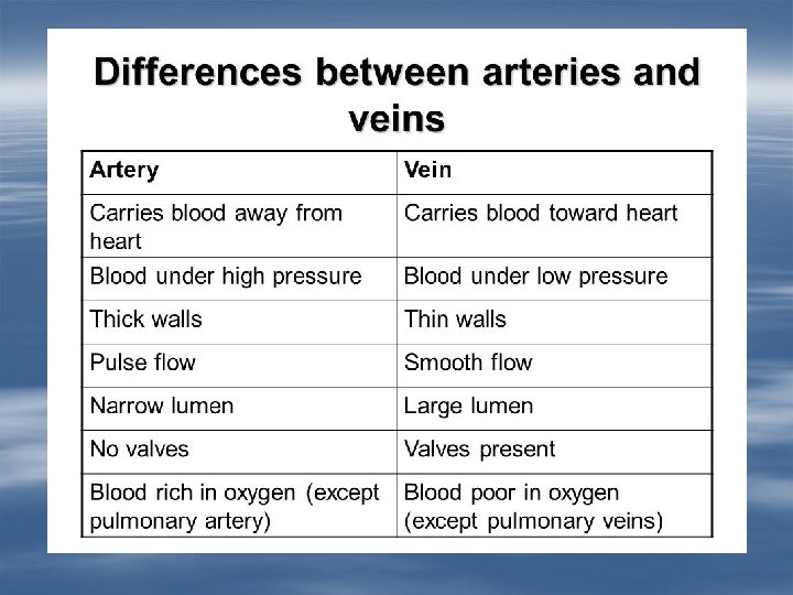
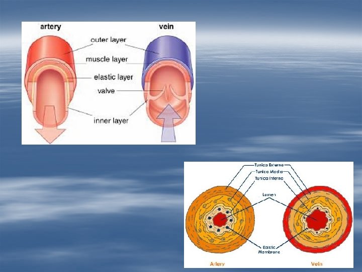
Blood vessels § 3 layers: – t. intima: endothel+subendothelial conn, tissue internal elastic membrane – t. media: smooth muscle +elastic fibers external elastic membrane – t. adventitia: connective tissue

Blood vessels

Aorta elastic fibers are the dominant in the t. media

Vein with valve Valves. t. intima

Artery and vein more muscle cells in the t. media

Small artery and vein

Arterioles, venules. capillaries Smooth muscle cells V A

Capillaries and venules

Röhlich Pál: Szövettan. Budapest, 1999 A szövettani képek a Humánmorfológiai és Fejlődésbiológiai Intézet gyűjteményéből származnak. Carola R, Harley JP, Noback CR: Human Anatomy & Physiology, Mc. Graw-Hill Inc. , USA, 1990 Wikipedia
- Slides: 35