Blistering Diseases Abdulmajeed Alajlan Associate Professor Hair Transplant

Blistering Diseases Abdulmajeed Alajlan Associate Professor Hair Transplant & Laser Department of Dermatology- KSU


Blistering Diseases Objectives • To know the definition & classification of Blistering diseases • To recognize the primary presentation of different types of main blistering diseases • To understand the possible pathogenesis of the main types of blistering diseases • To have an overview about managements lines of these diseases


definition • Vesicles and bullae are raised lesions that contain fluid. • A vesicle is less than 0. 5 cm in diameter. • A bulla is larger than 0. 5 cm in diameter.

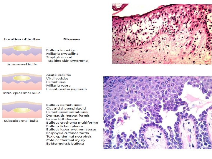
CLASSIFICATION OF VESICULOBULLOUS DISEASES INTRA EPIDERMAL BLISTERS The lesion is formed within the epidermis SUB EPIDERMAL BLISTERS : Lesions formed between the epidermis and the dermis

1. PEMPHIGUS VULGARIS 2. BULLOUS PEMPHIGOID 3. CHRONIC BULLOUS DISEASE OF CHILDHOOD 4. PARANEOPLASTIC PEMPHIGUS

Blistering Diseases Objectives • To know the definition & classification of Blistering diseases • To recognize the primary presentation of different types of main blistering diseases • To understand the possible pathogenesis of the main types of blistering diseases • To have an overview about managements lines of these diseases


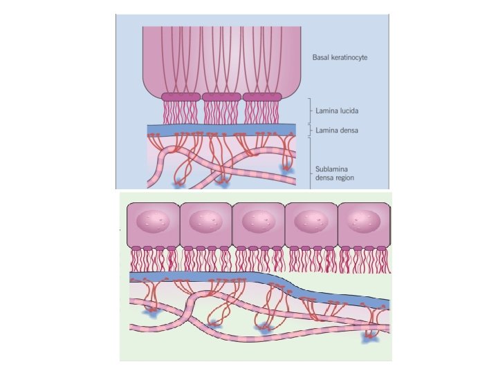
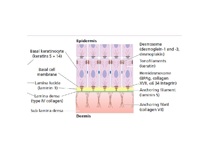
Dermo-edidermal junction

1. PEMPHIGUS VULGARIS 2. BULLOUS PEMPHIGOID 3. CHRONIC BULLOUS DISEASE OF CHILDHOOD 4. PARANEOPLASTIC PEMPHIGUS


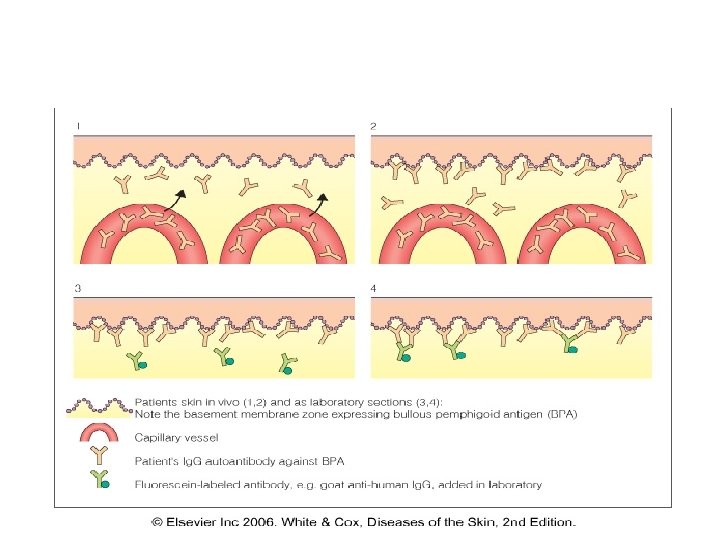
• Accurate pathological diagnosis requires 2 biopsies of a small newly formed lesion and perilesional skin for immunopathological studies.

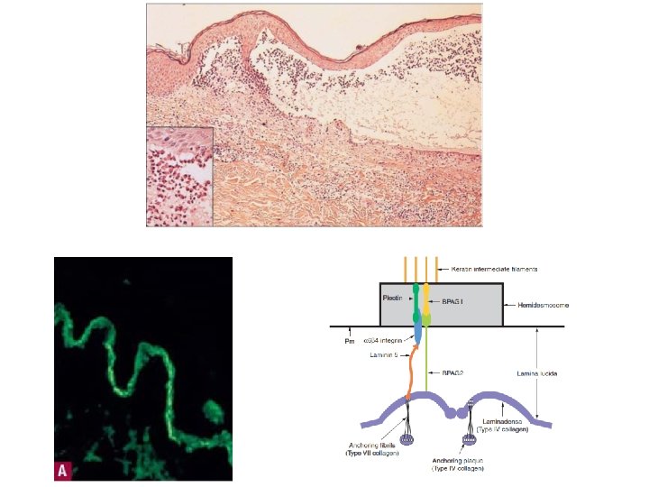
Diagnostic tests 1. Routine histology – Lesional sample –small bulla or edge of large one. 2. Direct immunofluorescence – Perilesional sample 3. Indirect immunofluorescence – Patient’s serum is added to specific substrates that express antigen of interest. 4. Electron microscopy.

1. Routine histology

immunofluorescence Ig. G C 3 Ig. A


1. PEMPHIGUS VULGARIS 2. BULLOUS PEMPHIGOID 3. CHRONIC BULLOUS DISEASE OF CHILDHOOD 4. PARANEOPLASTIC PEMPHIGUS

Blistering Diseases PEMPHIGUS VULGARIS

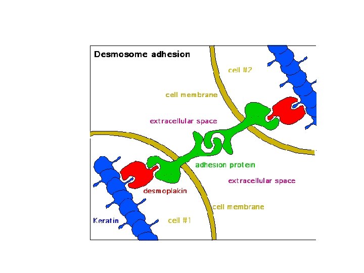
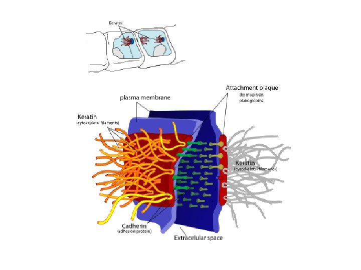
Pemphigus • Pemphigus is a group characterized by blistering of the skin and mucous membranes. • Auto-antibodies against DESMOSOMES in epidermis and mucosal surface.

Four sub-clinical varients : Pemphigus Vulgaris: is the most common Pemphigus variant, and the form usually responsible for oral lesions Folacious, vegetens, erythematosus
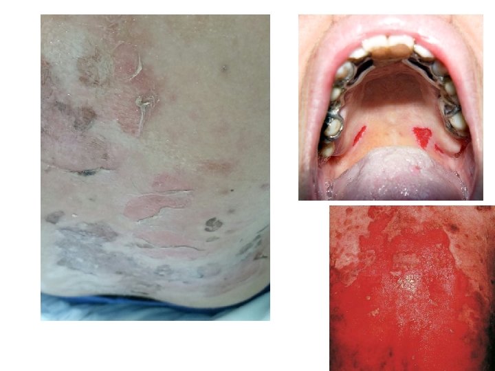

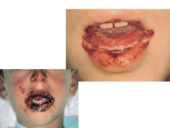
Pemphigus Vulgaris • Begins with erosions on mucous membrane then other skin areas. • Very painful. • +ve Nikolsky’s sign

+ve Nicholsky sign – Twisting pressure on normal skin shears skin.

Pemphigus Vulgaris • Begins with erosions on mucous membrane then other skin areas. • Very painful. • +ve Nikolsky’s sign. • Age: middle-age 40 -60 years. • Secondary infection and disturbance of fluid and electrolyte balance are common complications.

Pathology and immunopathology

Acantholysis Immunofluorescence Ig. G and C 3

Treatment • High dose systemic steroids 60 -100 mg of prednisolone. • Immunosuppressive agent such as azathioprine cyclophosphamide , Methotrexate or mycophenolate • Patient will probably have to remain on systemic steroids for long time. • Antibiotics; to treat superinfection

Biological Rx: 1 - Rituximab • IV 86% free of disease after 3 y 2 -IVIG (intravenous immunoglobulin)

Drug induced blistering diseases

Drug-induced PV Drugs can induce PV Drugs reported most significantly in association with PV are; Penicillamine Captopril Anti epiliptic phenytoin and carbamazepine

1. PEMPHIGUS VULGARIS 2. BULLOUS PEMPHIGOID 3. CHRONIC BULLOUS DISEASE OF CHILDHOOD 4. PARANEOPLASTIC PEMPHIGUS

Paraneoplastic pemphigus • The least common and most severe type of pemphigus is paraneoplastic pemphigus (PNP). This disorder is a complication of cancer, • usually lymphoma and Castleman's disease. It may precede the diagnosis of the tumor. Painful sores appear on the mouth, lips, and the esophagus. • Complete removal and/or cure of the tumor may improve the skin disease,


immunofluorescence Ig. G C 3 Ig. A

1. PEMPHIGUS VULGARIS 2. BULLOUS PEMPHIGOID 3. CHRONIC BULLOUS DISEASE OF CHILDHOOD 4. PARANEOPLASTIC PEMPHIGUS

Bullous pemphigoid • Characterized by large blisters on an erythematous base. • Mainly in older age group more than 60 y. • The prognosis is usually good.

Clinical features • Elderly patents. • Large tense blisters on upper arms and thighs. • Eczematous base. • Itch rather than pain. • Oral lesions are less frequent than pemphigus.


Pathology • Sub epidermal between epidermis and dermis the epidermis forms the roof of the blister. • Antigens identified are BP 1 and BP 2. • Immunoglobulin and complement are deposited in the lamina lucida of the basement membrane in a linear band.

Treatment • Mild may also respond very well to potent or moderately potent topical steroids alone. • Severe pemphigoid : Systemic steroids , but unlike pemphigus, it may be possible to discontinue. • The addition of either azathioprine enable the oral steroid dose to be reduced more rapidly.

1. PEMPHIGUS VULGARIS 2. BULLOUS PEMPHIGOID 3. CHRONIC BULLOUS DISEASE OF CHILDHOOD 4. PARANEOPLASTIC PEMPHIGUS
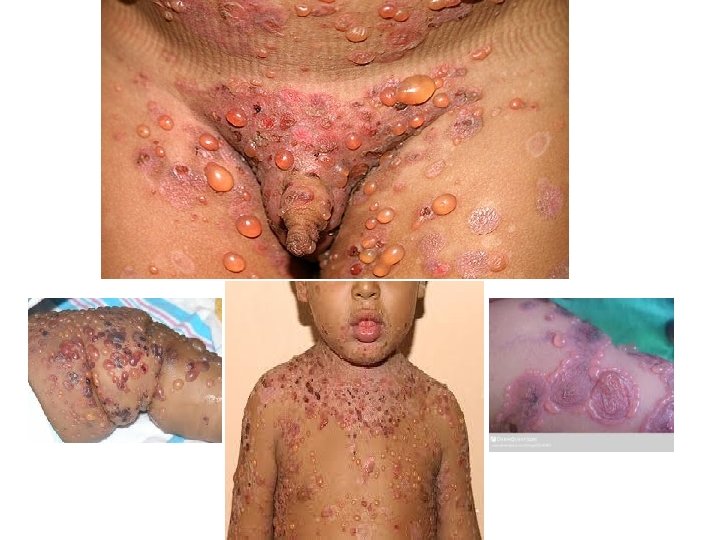

CLINICAL FEATURES • Circular clusters of large blisters like the type seen in pemphigoid (cluster of jewels) • It involves the perioral area, lower trunk, inner thighs and genitalia • Blistering may spread all over the body

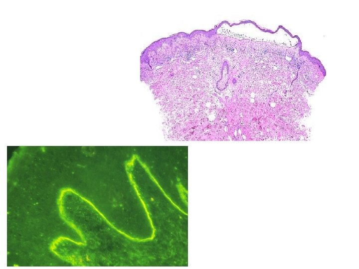
INVESTIGATION • Skin Biopsy will show subepidermal splits • Direct IF reveals Ig. A along the BM of the epidermis in a linear pattern


 Sulphonamides and immunosupressants Erythromycine")
TREATMENTS • • Oral dapsone 50 -200 mg daily (Ad/E) Sulphonamides and immunosupressants Erythromycine Flucloxacillin : 7 cases reported from KKUH

1. PEMPHIGUS VULGARIS 2. BULLOUS PEMPHIGOID 3. CHRONIC BULLOUS DISEASE OF CHILDHOOD 4. PARANEOPLASTIC PEMPHIGUS

Blistering Diseases Objectives • To know the definition & classification of Blistering diseases • To recognize the primary presentation of different types of main blistering diseases • To understand the possible pathogenesis of the main types of blistering diseases • To have an overview about managements lines of these diseases

Thank you
- Slides: 62