BLEEDING TENDENCY Hemostatic mechanism The normal haemostatic response

BLEEDING TENDENCY

Hemostatic mechanism The normal haemostatic response to vascular damage depends on closely linked interaction between the blood vessel wall, circulating platelets and blood coagulation factors. An efficient and rapid mechanism for stopping bleeding from sites of blood vessel injury is clearly essential for survival. Nevertheless, such a response needs to be tightly controlled to prevent extensive clots developing and to break down such clots once damage is repaired. The haemostatic system thus represents a delicate balance between procoagulant and anticoagulant mechanisms allied to a process for fibrinolysis. So, hemorrhage may result from deficiency or dysfunction of any of these elements. (Vasoconstriction, Platelet plug formation, and Fibrin thrombus formation)

The clinical and laboratory evaluation of a patient with bleeding tendency:
History: -site or sites of bleeding -severity and duration of bleeding -age of onset")
A)History: -site or sites of bleeding -severity and duration of bleeding -age of onset -was bleeding spontaneous or after trauma? -family history of similar problem -surgical history -drug history e. g aspirin, NSAID
Physical Examination: -site of bleeding: platelet and blood vessel usually have mucous memberane bleeding")
B)Physical Examination: -site of bleeding: platelet and blood vessel usually have mucous memberane bleeding e. g epistaxis, hematuria, GIT bleeding, and menorrhagia, while clotting factor deficiency(factor VIII, IX) cause muscle and joint bleeding with much more extensive ecchymoses and hematoma formation.

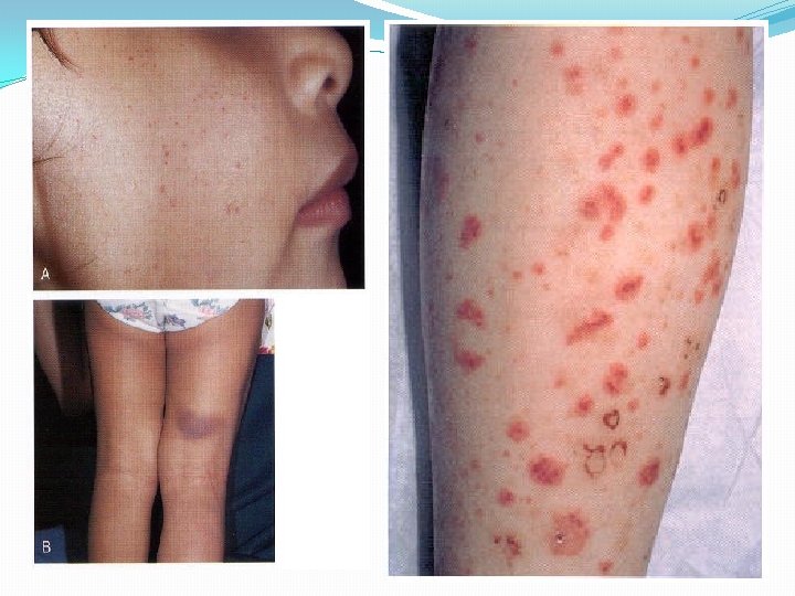
*Haemorrhages into the skin do not blanche with pressure, which helps distingushe these from telangiectasia and erythema, which are due to dilated skin blood vessels. • Petechiae : <1 mm in diameter • Purpuric spots: 1 -10 mm in diameter - palpable: vasculitis(e. g. Henoch-Schonlein purpura - non palpable: thrombocytopenia • Ecchymoses(larger bruises) • Haematoma(tender elevation)
Laboratory tests: 1)Bleeding time: assesse the function of platelets and their interaction with the")
C)Laboratory tests: 1)Bleeding time: assesse the function of platelets and their interaction with the vascular wall(bleeding stops within 4 -8 min) 2)Platelet count: >50000/mm 3 rarely have significant clinical bleeding. 3)Activated partial thromboplastin time(a. PTT): normal level 30 -40 sec, it measures the intrinsic clotting system. 4)Prothrombin time(PT): Normal range 10 -13 sec, it measures the extrinsic clotting system. 5)Clotting factor assays: normal range 50 -150 U/dl(50 -150%) Mild deficiency >5 U/dl (but below the normal range)after significant trauma Moderate deficiency 1 -5 U/dl(mild trauma induce bleeding) Severe deficiency <1 U/dl (spontaneous bleeding)
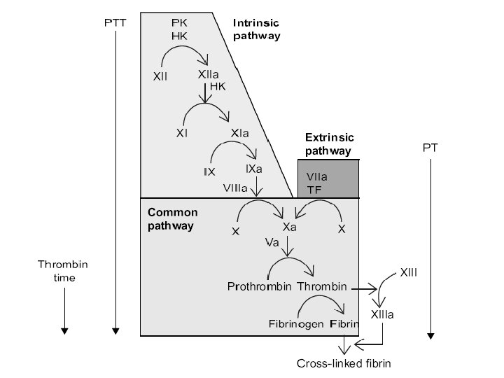

*Laboratory Findings in Coagulation Disorders: Disorder a. PTT PT Bleeding time Platelet count Factor VIII, IX prolonged normal deficiency Von Willebrand prolonged normal Thrombocytopenia normal prolonged low Platelet function defect normal prolonged normal Vitamin K deficiency prolonged normal DIC prolonged low
")
(Clotting Factors Disorders)
 and Factor IX(Hemophilia B) deficiency")
Congenital clotting factor disorders Factor VIII(Hemophilia A) and Factor IX(Hemophilia B) deficiency

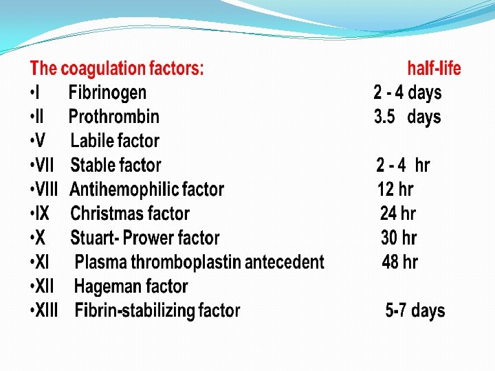
Genetics and classification: Is x-linked recessive, with 85% having factor VIII deficiency and 10 -15% having factor IX deficiency. Some female carries of hemophilia A or B will have sufficient reduction of their factor VIII or IX through lyonization of the X chromosome to produce mild bleeding.

Clinical manifestations: Both factors not crosses the placenta, thus bleeding symptoms may be present from birth or occur in the fetus. Only about 30% of patients bleed with circumcision. When a child begins to crawl and walk causes bruising, intramuscular hematoma and hemarthrosis. The hallmark of hemophilia is the hemarthrosis (ankle, knee and the elbow joints) Iliopsoas muscle bleeding leading to hypovolemic shock and inability to extend the hip. Life- threating bleeding in the hemophilic patient is CNS and upper air way bleeding.
prolonged a. PTT (usually 2 -3 times the upper limits of")
Laboratory findings : 1)prolonged a. PTT (usually 2 -3 times the upper limits of normal) 2)platelet count, bleeding time, PT, TT, are normal. 3)The specific assay for factor VIII, IX will confirm the diagnosis: Normal range 50 -150 U/dl(50 -150%) Mild deficiency >5 U/dl (but below the normal range)after significant trauma Moderate deficiency 1 -5 U/dl(mild trauma induce bleeding) Severe deficiency <1 U/dl (spontaneous bleeding)
prevention of trauma. Although it is easy to advise parents")
Treatment: A. General measures 1)prevention of trauma. Although it is easy to advise parents that their child should avoid trauma, this advice is practically useless. Toddlers are active, and injure themselves easily. Effective measures include anticipatory guidance, including the use of car seats, seatbelts, and bike helmets, and the importance of avoiding high-risk behaviors. 2)avoid aspirin and other NSAID. 3)immunization against hepatitis B virus.

B. Replacement therapy *In Mild factor VIII deficiency, the patient’s endogenously produced factor VIII can be released by the administration of desmopressin acetate(by intranasal inhaler). *Moderate and Severe factor VIII deficiency recombinant factor VIII and recombinant factor IX. Factor replacement therapy is the mainstay of hemophilia treatment. The degree of factor correction required to achieve hemostasis is largely determined by the site and nature of the particular bleeding episode. C. PROPHYLAXIS Is usually provided every 2– 3 days to maintain a measurable plasma level of clotting factor (1– 2%) when assayed just before the next infusion.
 • FFP effective in treating")
• Cryoprecipitate contains: (1, 8, 13, and VWF) • FFP effective in treating factors 2, 5, 10, 11 deficiency not effectine in treating factors 7, 8, 9 deficiency
chronic joint destruction. 2)risk of transfusion-transmitted infections (HIV, Hepatitis B or C).")
Chronic complications: 1)chronic joint destruction. 2)risk of transfusion-transmitted infections (HIV, Hepatitis B or C). 3)development of an inhibitors to either factor VIII or IX.
 Is autosomal disease with mild to moderate bleeding, more in")
Factor XI deficiency(Hemophilia C) Is autosomal disease with mild to moderate bleeding, more in Ashkenazi Jews. -PTT is longer than it is in hemophilia A or B. -treated with FFP.

Von Willebrand disease Is the most common hereditary bleeding disorder(1 -2% of the general population). Is inherited autosomally, but is more in women (menorrhagia is a major symptom). Pathophysiology: VWF is a large multimeric glycoprotein that is synthesized in megakaryocytes and endothelial cells. It responsible for the normal interaction with the subendothelial matrix and platelets, and serves as the carrier protein for plasma factor VIII.

Classification: Type 1 VWD: the protein is reduced but not absent. Typw 2 VWD: qualitatively abnormal. Type 3 VWD: the protein is absent. Clinical manifestations: Mucocutaneous bleeding(bruising, epistaxis, menorrhagia • and postoperative hemorrhage particularly after mucosal surgery such as tonsillectomy.
prolong bleeding time. 2)prolong PTT. - Both may be normal in Type")
Laboratory findings: 1)prolong bleeding time. 2)prolong PTT. - Both may be normal in Type 1 VWD. 3)quantitative assay for VWF antigen, VWF activity. 4)plasma factor VIII activity. 5)determination of VWF structure. 6)platelet count.
 induces the release of VWF from the endothelial cells.")
Treatment: Type 1 VWD: desmopressin(DDAVP) induces the release of VWF from the endothelial cells. In some patients with type 2 VWD, desmopressin may be similarly effective, but in other circumstances the released VWF is dysfunctional. Replacement therapy: plasma derived VWF containing concentrates that also contain factor VIII.

Acquired clotting factor disorders

Vitamin K deficiency: Causes: 1 -lack of oral intake of vit. K. 2 -alteration in the gut flora due to long term use of broad-spectrum antibiotics. 3 -malabsorption of vitamin K(cystic fibrosis or biliary atresia). 4 -hemorrhagic disease of the newborn.

Hemorrhagic Disease of the Newborn classic form A moderate decrease in factors II, VII, IX, and X normally occurs in all newborn infants by 48 -72 hr after birth, rare in term infants and more frequent in premature infants, accentuation and prolongation of this deficiency between the 2 nd and 7 th days of life result in spontaneous and prolonged bleeding with a gradual return to birth levels by 7 -10 days of age. This transient deficiency of vitamin K–dependent factors is probably due to lack of free vitamin K from the mother and absence of the bacterial intestinal flora normally responsible for the synthesis of vitamin K and is responsive to and prevented by vitamin K therapy. Breast milk is a poor source of vitamin K, so hemorrhagic complications are more frequent in breast-fed than in formulafed infants.
 life-threatening vitamin K deficiency–induced bleeding also occurs")
Early-onset (onset from birth to 24 hr) life-threatening vitamin K deficiency–induced bleeding also occurs if the mother has been treated with drugs (phenobarbital, phenytoin) that interfere with vitamin K function. Late onset (>2 wk) is often associated with vitamin K malabsorption, as noted in neonatal hepatitis or biliary atresia.

Liver disease: Since all the clotting factors are produced exclusively in the liver(except factor VIII), coagulation abnormalities are very common in patients with severe liver disease. Treatment: FFP or cryoprecipitate+Vitamin K.
 Are a heterogeneous group of conditions including DIC that")
Disseminated Intravascular Coagulation (consumptive coagulopathy) Are a heterogeneous group of conditions including DIC that result in consumption of clotting factors, platelets, and anticoagulant proteins. Consequences of this process include widespread intravascular deposition of fibrin leading to tissue ischemia, necrosis, generalized hemorrhagic state and hemolytic anemia.
infection: e. g meningococcemia. 2)Tissue injury: like massive burn. 3)Malignancy: acute promyelocytic leukaemia.")
Causes: 1)infection: e. g meningococcemia. 2)Tissue injury: like massive burn. 3)Malignancy: acute promyelocytic leukaemia. 4)Venom or toxin: snake bites. 5)GIT disorders: fulminant hepatitis.
prolongation of PT, PTT, and TT. 2)decrease factor II, V, VIII, fibrinogen")
Laboratory findings: 1)prolongation of PT, PTT, and TT. 2)decrease factor II, V, VIII, fibrinogen and platelet. 3)Blood smear: fragmented , burr and helmetshaped RBC (schistocytes). 4)FDP(fibrin degredation product), D-dimers are appear in the blood.
general measures: -treat the trigger that caused the DIC -restore normal hemostasis by")
Treatment: A)general measures: -treat the trigger that caused the DIC -restore normal hemostasis by correcting the shock, acidosis and hypoxia. B)replacement therapy: platelet infusions, cryoprecipitate, and /or FFP.
")
(Platelet Disorders)

*Platelets circulate with a life-span 10 -14 days. *Normal platelet count is(150 -450 X 109/L) Causes of thrombocytopenia(<150 X 109/L) 1)decreased production (congenital or acquired). 2)increased destruction (immune or nonimmune).
 Is the most common cause of acute onset of thrombocytopenia.")
IMMUNE THROMBOCYTOPENIC PURPURA (ITP) Is the most common cause of acute onset of thrombocytopenia.
,")
Etiology: 1 -4 weeks after exposure to a common viral infection(e. g. Epstein-Barr virus), a small number of children develop an autoantibody directed against the platelet surface.

Clinical manifestations: *there is a history of a preceding viral infection. *a healthy (1 -4 yr)old child who has a sudden onset of generalized petechiae, purpura, and mucous membrane bleeding. Prognosis: *70 -80% of children with acute ITP have a spontaneous resolution within 6 months. *10 -20% develop chronic ITP. * less than 1% develop intracranial hemorrhage.
severe thrombocytopenia(<20 X 109/L). 2)Hb, WBC, are normal. (Hb may be decreased")
Laboratory findings: 1)severe thrombocytopenia(<20 X 109/L). 2)Hb, WBC, are normal. (Hb may be decreased due to profuse bleeding). 3)B. M: increase number of megakaryocytes.
intravenous")
Treatment: *Platelet transfusion in ITP is usually contraindicated unless life-threatening bleeding is present. 1)intravenous immunoglobulin(IVIG): dose 0. 8 -1 g/kg/day(1 -2 days). 2)prednisone: 1 -4 mg/kg/day(for 2 -3 weeks). 3)i. v anti-D therapy: dose 50 Mg/kg. 4)splenectomy: indicated in a- older child >4 year with severe ITP last >1 year. b- life threatening hemorrhage(intracranial hemorrhage).
 1)Henoch-Schonlein purpura : an Ig. A-mediated vasculitis, presents with palpable")
(Disorders of Blood Vessels) 1)Henoch-Schonlein purpura : an Ig. A-mediated vasculitis, presents with palpable purpura on the lower extremities and buttocks, renal insufficiency, arthritis, and abdominal pain. platelet count is normal. 2)Scurvy(vit. C deficiency): causes impaired collagen synthesis that results in weakened blood vessels. 3)Inherited disorders of collagen synthesis(e. g. Ehlers -Danlos syndrome). 4)Malnutrition and corticosteroids may weaken the collagen supporting vessels.
- Slides: 42