
BLEEDING DISORDERS By 6 th year M S
BLEEDING DISORDERS By 6 th year M. S Ghaida’a AL-Araj
Definition: defects in hemostasis that lead to an increased susceptibility of bleeding (also known as hemorrhagic diathesis). Caused by: 1 - Platelet disorders (primary hemostasis defect), ITP, TTP , HUS , Bernard-Soulier Glanzmann’s Thrombasthenia 2 - Coagulation disorders (secondary hemostasis defect), further divided into either intrinsic or extrinsic defects Hemophilia, Vitamin K deficiency 3 - A combination of both Von Willebrand Disease, DIC may be inherited or acquired
Types of bleeding 1 - Mucocutaneous bleeding (e. g. , epistaxis, petechiae, gastrointestinal bleeding) • Platelet disoeders • 2 - Bleeding into potential spaces (e. g. , hemarthrosis, muscular bleeding) • In coagulation disorder •
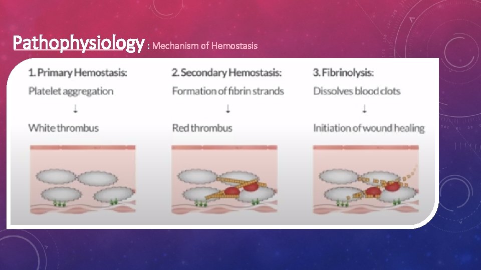
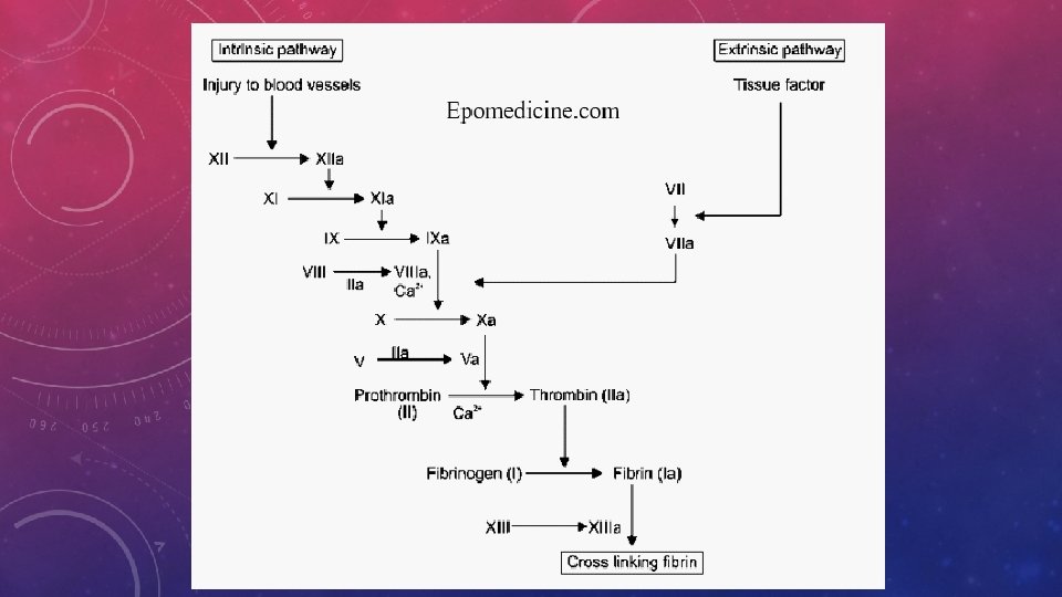
Pathophysiology : Mechanism of Hemostasis

Primary Hemostasis

Secondary Hemostasis A B
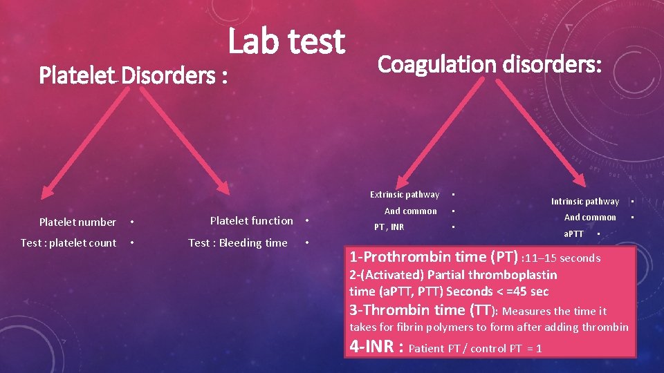
Lab test Platelet Disorders : Platelet number • Test : platelet count • Platelet function • Test : Bleeding time • Coagulation disorders: Extrinsic pathway • And common • PT , INR Intrinsic pathway • And common • • a. PTT • 1 -Prothrombin time (PT) : 11– 15 seconds 2 -(Activated) Partial thromboplastin time (a. PTT, PTT) Seconds < =45 sec 3 -Thrombin time (TT): Measures the time it takes for fibrin polymers to form after adding thrombin 4 -INR : Patient PT / control PT = 1
Platelet Disorders
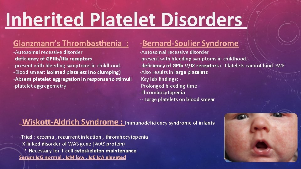
Inherited Platelet Disorders Glanzmann’s Thrombasthenia : -Autosomal recessive disorder -deficiency of GPIIb/IIIa receptors -present with bleeding symptoms in childhood. -Blood smear: Isolated platelets (no clumping) -Absent platelet aggregation in response to stimuli -platelet aggregometry - Wiskott-Aldrich -Bernard-Soulier Syndrome -Autosomal recessive disorder -present with bleeding symptoms in childhood. -deficiency of GPIb V/IX receptors : - Platelets cannot bind v. WF -Also results in large platelets Key lab findings: Prolonged bleeding time -Thrombocytopenia -- Large platelets on blood smear Syndrome : Immunodeficiency syndrome of infants -Triad : eczema , recurrent infection , thrombocytopenia - X linked disorder of WAS gene (WAS protein) * Necessary for T-cell cytoskeleton maintenance Serum Ig. G normal. Ig. M low , Ig. E Ig. A elevated
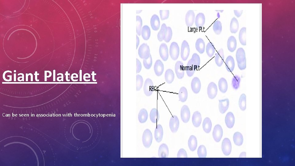
Giant Platelet Can be seen in association with thrombocytopenia
Acquired platelet disorder 1 - ITP (Immune thrombocytopenic purpura ) immune-mediated destruction of platelets and possibly inhibition of platelet release from the megakaryocyte caused by anti-GPIIB/IIIA antibodies : Leads to Consumption in splenic macrophages Splenomegaly Thrombocytopenia Types : acute (less than 6 months) or chronic (more than 6 months ) In children, it is usually an acute disease, most commonly following an infection , and with a self-limited course. In adults, it is a more chronic disease, although in some adults, spontaneous remission occurs,
Diagnosis of exclusion • Rule out other causes of bone marrow suppression Clinical presentation : mucocutaneous bleeding Test : often very low, platelet count, with an otherwise normal peripheral blood cells and smear Large plts on perepheral smear - Laboratory testing for antibodies (serologic testing) is usually not helpful due to the low sensitivity and specificity of the current tests • Treatment: According to platelet count and degree of bleeding Some of them require careful follow up (check platelet count ) with no specific therapy mostly in children the standard of practice among many physicians is to initiate treatment in adult patients with ITP when the platelet count is less than 30, 000, . - Steroids - IVIG (blocks Fc receptors in macrophages) - Splenectomy
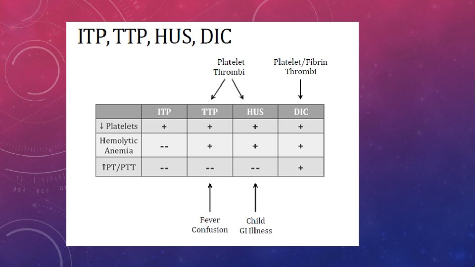
2 - TTP Thrombotic thrombocytopenic purpura is a thrombotic microangiopathy, a condition in which microthrombi that form in small vessles and sometimes cause occlusion of the microvasculature (i. e. , the arterioles and capillaries) Etiology : 95% acquired / 5% congenital due to acquired autoantibodies against a proteolytic enzyme congenital there is mutation of this enzyme. ADAMTS 13 acti vityless <10% → ↓ Breakdown of v. WF multimers → v. WF multimers accumulate on endothelial cell surfaces → microthrombus formation → blockage of small vessels → RBC fragmentation (hemolysis) and end-organ damage Risk factors Drugs (chemotherapeutic agent , cyclosporine ) Pregnancy Systemic disease: cancer, HIV, SLE
Clinical symptoms : Diagnosis : 1 -Hematology the pentad of clinical findings consists of: ↓ Platelets ↓ Hemoglobin ↑ Reticulocytes↓ Haptoglobin 1 -Fever Normal or mildly elevated (PT) and (a. PTT) D-dimer levels Negative Coombs test 2 -Neurological signs and symptoms 2 -Peripheral blood smear Altered mental status, delirium Large number of schistocytes (up to 10% of RBCs) Seizure, focal defects, stroke Low number of platelets 3 -Serum chemistry Headache, dizziness ↑ LDH, ↑ indirect bilirubin (hemolytic anemia) 3 -Low platelet count (i. e. thrombocytopenia) ↑ BUN and ↑ creatinine (impaired renal function) Petechiae, purpura 4 -Urinalysis Hematuria, proteinuria Mucosal bleeding 5 -ADAMTS 13 activity Prolonged bleeding after minor cuts ↓ ADAMTS 13 activity 4 -Microangiopathic hemolytic anemia Not a routinely available test and should be used for confirmation only Fatigue, dyspnea, and pallor 6 -Identification of secondary causes) Jaundice 5 -Impaired renal function Treatment : acquired or congenital Hematuria, • -Plasma exchange: removes antibodies with immunosuppressant Oliguria, anuria • -congenital FFP Platelet counts monitored to determine efficacy and prevent relapses
3 -HUS Hemolytic Uremic Syndrome thrombotic microangiopathy -Similar to TTP The two conditions have same pathophysiology and clinical findings in common, but different etiologies It is caused by bacterial toxins, most commonly the Shigalike toxin in E. coli or Shegilla 2 -Presentation : A diarrheal illness (usually bloody) for the past 5– 10 days precedes the onset of HUS symptoms in many children. The triad of clinical findings occurring in HUS consists of: : Low platelets (i. e. , thrombocytopenia) Treatment : Petechiae, purpura 1 -Primarily supportive. Mucosal bleeding Prolonged bleeding after minor cuts 2 -dialysis as indicated for AKI: up to 50% of HUS patients require dialysis. Microangiopathic hemolytic anemia 3 -only in refractory cases and a. HUS: plasma exchange therapy Fatigue, dyspnea, and pallor -typical HUS plasma infusion or plasma exchange has not been shown to alter the Jaundice overall course. ADAMTS 13 levels are generally reported to be normal in hus Impaired renal function Hematuria, Oliguria, anuria 3 -Eculizumab monoclonal antibody to complement factor c 5; blocks complement activation FDA a. HUS
DIC Disseminated Intravascular Coagulation • is a disorder characterized by systemic activation of the clotting cascade with microthrombi formation, platelet consumption, and subsequent exhaustion of all clotting factors. MANIFESTATIONS OF BLEEDING Petechiae, purpura, ecchymosis Hematuria Hematemesis, hematochezia Collection of blood in body cavities: hemoperitoneum, hemothorax MANIFESTATIONS OF THROMBOSIS Acute renal failure: oliguria Hepatic dysfunction: jaundice ARDS: dyspnea, Pulmonary thromboembolism dyspnea, chest pain, hemoptysis Deep vein thrombosis lower limb edema Neurological dysfunction: altered mental status, stroke • The diagnosis of DIC 1 -Elevated PT/PTT - Consumption of factors 2 - Low platelets - Consumption of platelets 3 - Low fibrinogen (consumption) 4 -elevated D-dimer FDP 5 - Microangiopathic hemolytic anemia • Low RBC (anemia) • Schistocytes on blood smear
Causes of DIC The Mnemonic “STOP Making Trouble!”- helps recall the etiology of DIC. M - malignancy, T transfusion, S - sepsis/snakebites, Ttrauma (acute traumatic coagulopathy), O- obstetric complications, P - pancreatitis 1 - Obstetrical emergencies : ( Amniotic fluid contains tissue factor) : abruptio placenta , major haemorrhagic bleeding , can develop DIC. 2 -Sepsis : 1 - Endotoxin –> activates coagulation cascade , 2 - Cytokines 3 - Cancer: (well-described hypercoagulable state) Excess coagulation: DIC 4 - snake bites : Thrombin-like glycoproteins within venom
Treatment 1 - Treatment of the underlying disorder 2 - Fresh frozen plasma: replace clotting factors if PT or a. PTT > 1. 5 times the • normal value 3 - RBCs Hb < 7 g/d. L • 4 -platelets Platelet count < 10, 000 in asymptomatic individuals • Platelet count < 50, 000 in patients with active bleeding or if a surgical • procedure is planned 5 -Cryoprecipitate (for low fibrinogen) fibrinogen < 150 mg/d. L 6 -Organ failure type: natural protease inhibitor (e. g. , antithrombin) • 7 -Non-symptomatic type: heparin • •
Von Willebrand Disease
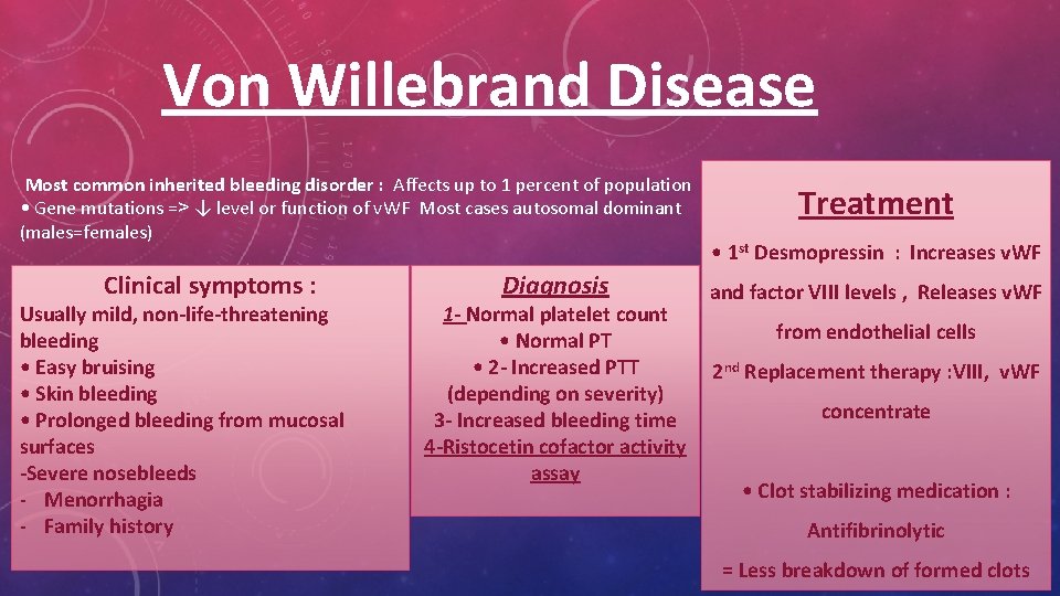
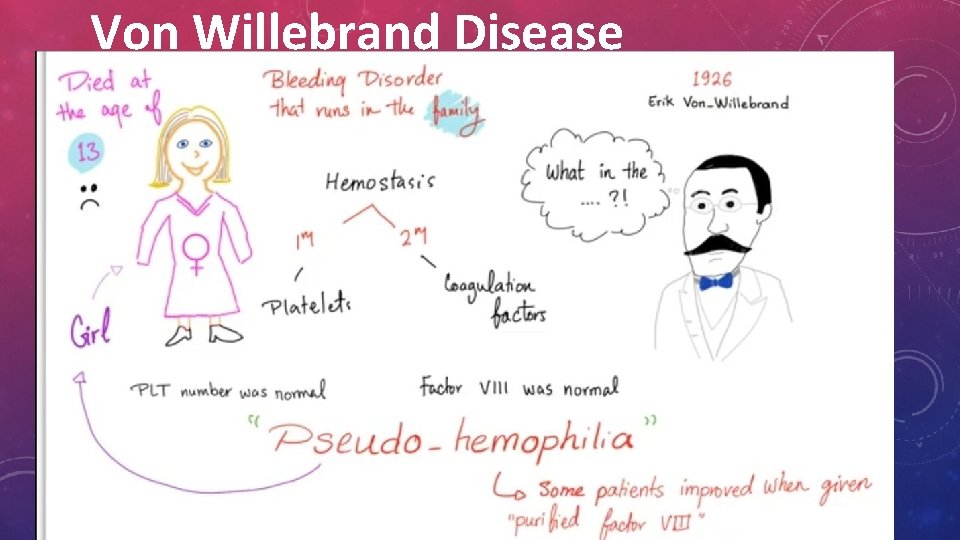
Von Willebrand Disease Most common inherited bleeding disorder : Affects up to 1 percent of population • Gene mutations => ↓ level or function of v. WF Most cases autosomal dominant (males=females) Clinical symptoms : Usually mild, non-life-threatening bleeding • Easy bruising • Skin bleeding • Prolonged bleeding from mucosal surfaces -Severe nosebleeds - Menorrhagia - Family history Diagnosis 1 - Normal platelet count • Normal PT • 2 - Increased PTT (depending on severity) 3 - Increased bleeding time 4 -Ristocetin cofactor activity assay Treatment • 1 st Desmopressin : Increases v. WF and factor VIII levels , Releases v. WF from endothelial cells 2 nd Replacement therapy : VIII, v. WF concentrate • Clot stabilizing medication : Antifibrinolytic = Less breakdown of formed clots
Coagulation disorders:
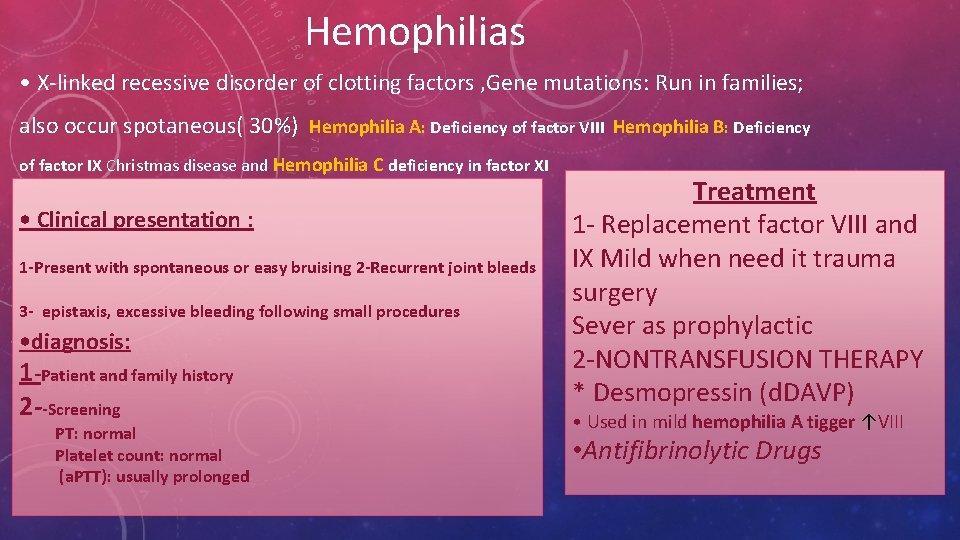
Hemophilias • X-linked recessive disorder of clotting factors , Gene mutations: Run in families; also occur spotaneous( 30%) Hemophilia A: Deficiency of factor VIII Hemophilia B: Deficiency of factor IX Christmas disease and Hemophilia C deficiency in factor XI • Clinical presentation : 1 -Present with spontaneous or easy bruising 2 -Recurrent joint bleeds 3 - epistaxis, excessive bleeding following small procedures • diagnosis: 1 -Patient and family history 2 --Screening PT: normal Platelet count: normal (a. PTT): usually prolonged Treatment 1 - Replacement factor VIII and IX Mild when need it trauma surgery Sever as prophylactic 2 -NONTRANSFUSION THERAPY * Desmopressin (d. DAVP) • Used in mild hemophilia A tigger ↑VIII • Antifibrinolytic Drugs
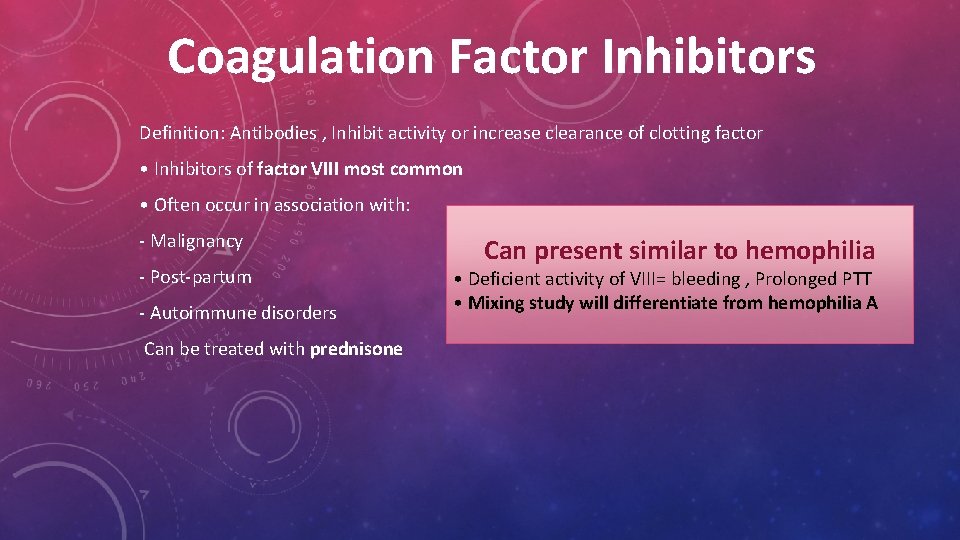
Coagulation Factor Inhibitors Definition: Antibodies , Inhibit activity or increase clearance of clotting factor • Inhibitors of factor VIII most common • Often occur in association with: - Malignancy - Post-partum - Autoimmune disorders Can be treated with prednisone Can present similar to hemophilia • Deficient activity of VIII= bleeding , Prolonged PTT • Mixing study will differentiate from hemophilia A
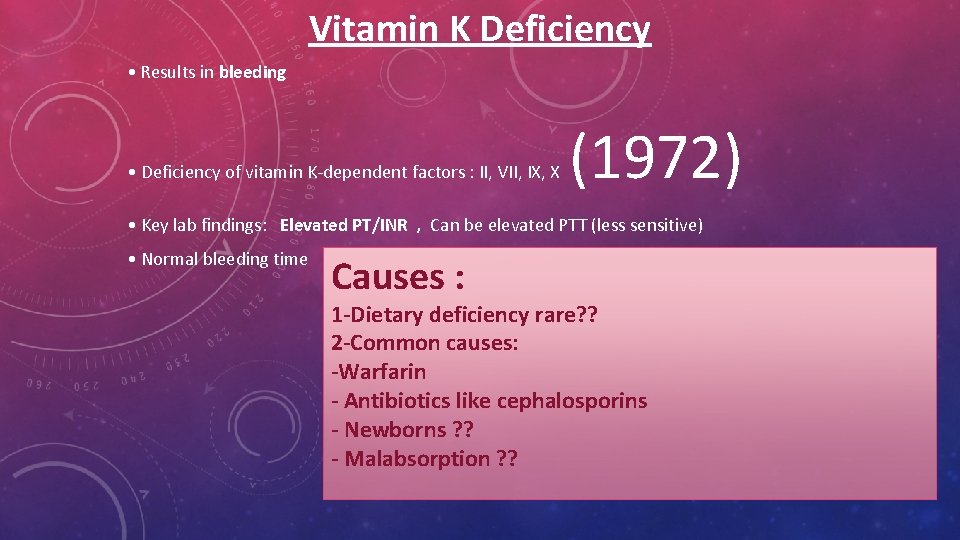
Vitamin K Deficiency • Results in bleeding • Deficiency of vitamin K-dependent factors : II, VII, IX, X (1972) • Key lab findings: Elevated PT/INR , Can be elevated PTT (less sensitive) • Normal bleeding time Causes : 1 -Dietary deficiency rare? ? 2 -Common causes: -Warfarin - Antibiotics like cephalosporins - Newborns ? ? - Malabsorption ? ?

- Slides: 27