Bladder physiology and incontinence Bladder physiology Lower urinary

Bladder physiology and incontinence

Bladder physiology Lower urinary tract is composed of bladder and urethra and prostate in male The two function of LUT are urinary storage and urinary emptying These function depend on integrated function of CNS, PNS, urinary bladder and urethra Urethral tone depends on urethral striated and smooth muscle and urethral lamina propria The vascular filling of lamina propria is important for urinary continence, estrogen is known to increase urethral blood flow and so distension of lamina propria

- filling phase requires increasing urine volume at low detrusor pressure, closed sphincters, absence of involuntary bladder contraction - In voiding phase: -coordinated contraction of bladder muscle with concomitant decrease of resistance at level of smooth and striated sphincters ( synergy)

• Afferent signal during bladder filling will relies to CNS which in turn inhibit spontaneous smooth muscle contraction • Pelvic nerves afferent consist of ag fibers or c unmylinated fibers during inflammation or neuropathic condition there is recruitement of c fibers • Linking the bladder filling with episodic bursts of sensation

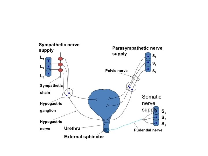
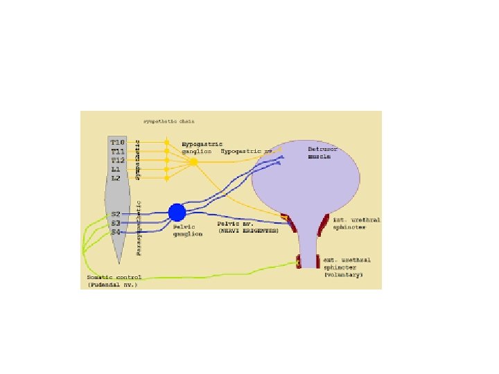
• Voiding when there is synergic contraction of bladder detrusor muscle and relaxation of the sphincter • The smooth muscle of the bladder (detrusor muscle) is innervated by parasympathetic • Afferent innervation which sense bladder filling ascend with parasympathetic to the cord and then to pontine micturition center (barringtons nucleus) • the pons center receive input from other brain area , if the time is appropriate for voiding it will stimulate voiding

• two Sphincters: 1 - internal sphincter ( smooth muscle of bladder neck and proximal urethra) , physiologic sphincter and not under voluntary control, it is supplied by sympathetic
: the external sphincter composed of two parts the striated muscle")
2 -external sphincter (rhabdosphincter): the external sphincter composed of two parts the striated muscle of pelvic floor and striated muscle of distal urethra (intrinsic rhabdosphincter. - it is supplied by pudendal nerve , and is under voluntary control
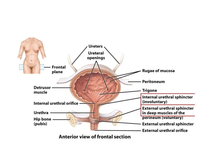

• External urethral sphincter is composed of two parts: periurethral striated muscle of pelvic floor (fast twitch and slow twitch fibers), the striated muscle of distal urethral sphincter (slow twitch fibers) • The normal bladder may be spontaneous active because of population of cells within the detrusal muscle (interstitial cell of myofibroblast) bladder pacemakers

Incontinence - definition: - involuntary leakage of urine, results from failure of urine storage during filling phase - It is more common in female and increase with age - Incontinence can be transient or chronic. Transient incontinence may occur after vaginal childbirth or during an acute lower urinary tract infection and usually resolves spontaneously. - Chronic incontinence can result from a multitude of causes and is often persistent and progressive

• 1 - urge incontinence: - involuntary leak of urine accompanied or immediately preceded by urgency it is due to bladder overactivity which • - Can be due to detrusor over-activity or low compliance or both

• Detrusal overactivity may be neurogenic (due to neurological cause • Or non neurogenic like BOO, inflammatory condition , stress urinary incontinence , aging, polyuria , constipation

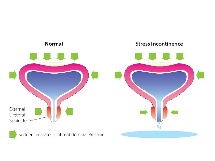
2 - stress incontinence: - involuntary leak of urine on effort or exertion or coughing or sneezing In men, stress incontinence is mostly due to surgery (eg, after radical prostatectomy) or trauma to the bladder neck or urethral sphincter. The causes of stress incontinence in women are more complicated , The vast majority of stress incontinence occurs in women after middle age (with history of vaginal deliveries and obstructed labor). It is usually a result of weakness or disruption of the pelvic floor muscle and ligaments leading to poor support of the vesicourethral sphincteric unit andso hypermobility of the bladder neck and urethra or weakness of the urethral sphincteric tissues

• During exertion, both passive pressure transmission from increased abdominal pressure and reflex-related contraction of the sphincteric muscles augment urethral resistance to prevent urine leakage. • urethral tone is maintained by smooth and striated muscle activity, and the cushioning effect of the soft, compressible submucosal vascular plexus. • During rises in intra-abdominal pressure, the urethra is compressed against the supporting structures, which act like a backboard


3 - overflow incontinence: -the involuntary loss of urine associated with bladder overdistension - Mainly in male with bladder outflow obstruction - Treatment is by relief of obstruction - Diabetic cystopathy can result in overflow incontinence, through decreasing detrusor contractility.

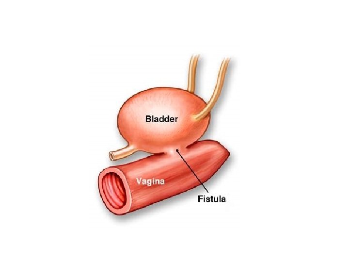
4 - continuous incontinence: - Leaking of urine continuously ( without aggravating factor and all the times) - Caused by fistula VVF ( extravesical incontinence) - Ectopic ureter in female.

5 - mixed incontinence: - More than one type in the same patient urge and stress incontinence.

assessment • History : the type of incontinence and if mixed if one component is more common or cause more bother • The leakage should be quantified( the nuber of pads, the frequency of leakage • The LUTS • Neurological symptom or bowl function • Female asked about prolapse symptom • Past medical and surgical history

• Complete physical examination is performed with emphasis on a neurologic assessment and the abdominal, pelvic and rectal examination • Examination of vaginal epithelium • Documentation of urethral leakage with cough • Presence of prolapse

• Voiding diaries: which is measurement of frequency and voided volume, episode of incontinence, and fluid intake in 24 hours • It should be completed for at least 3 days • Urine analysis: UTI may cause urinary incontinence or the existing UI may worsens with UTI • PVR volume : the volume of urine that remains in bladder after voiding, measured by catheter or U/S • PVR volume should be assessed in PTN with voiding symptom or before surgery for stress incontinence
 that")
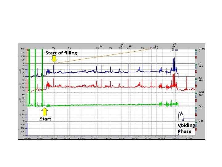
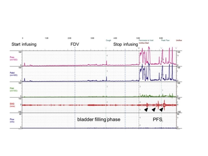
urodynamic • Urodynamic study: an interactive study of the lower urinary tract (LUT) that evaluates the transport, storage, and evacuation of urine. • obtain functional information about the LUT • The study can be divided up into two main phases: the storage (or filling) phase (cystometry) and the emptying (or voiding) phase (pressure flow study).

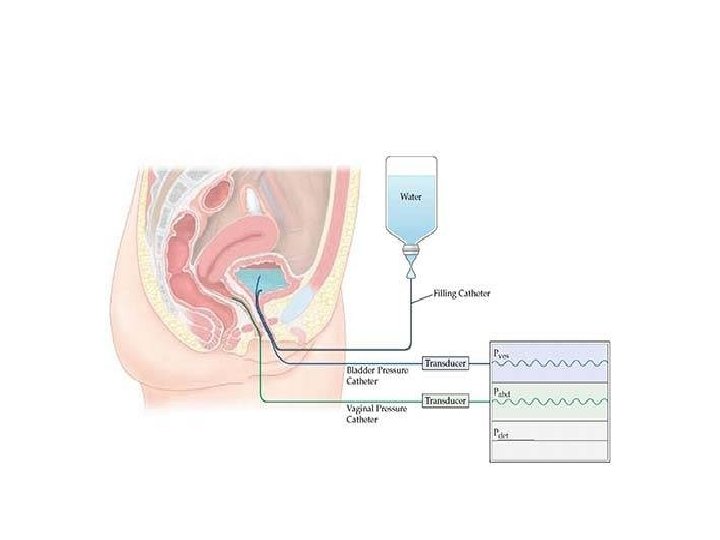
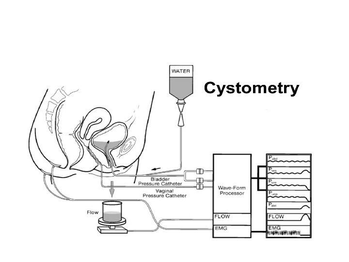
• Cystometry: is a method used to measure the pressure/volume relationship of the bladder during bladder filling. • Measurements routinely obtained during cystometry include bladder sensation, contractions, compliance, and capacity • A urethral catheter is required to perform cystometry and is typically 6– 8 Fr in size, this catheter has a pressure transducer on the end that measures bladder pressure (Pves).

• The urethral catheter often has a double lumen to allow for simultaneous pressure transduction and bladder filling with water, saline, or radiographic contrast • Some urethral catheters also have a second transducer that allows for the concurrent measurement of urethral pressures (Pura). • Since the bladder is considered an intra-abdominal organ and experiences pressure effects from the abdomen, a second catheter is often placed per rectum to measure abdominal pressure (Pabd).

• By urethral and rectal catheter it is possible to calculate the bladder or detrusor pressure (Pdet) by the following formula: Pdet = Pves – Pabd.

• Sensation is essential for normal bladder control and may be either reduced (ie, in certain neurologic conditions and diabetic cystopathy) or heightened (ie, in patients with bladder pain syndrome or cystitis • First sensation: The feeling that the patient becomes aware of bladder filling(40% of bladder capacity). • • First desire: The feeling to urinate at the next convenient time, but urination could be delayed(75% of bladder capacity • • Strong desire: The persistent desire to urinate without fear of leakage( maximum capacity).
, should be assessed during cystometry and")
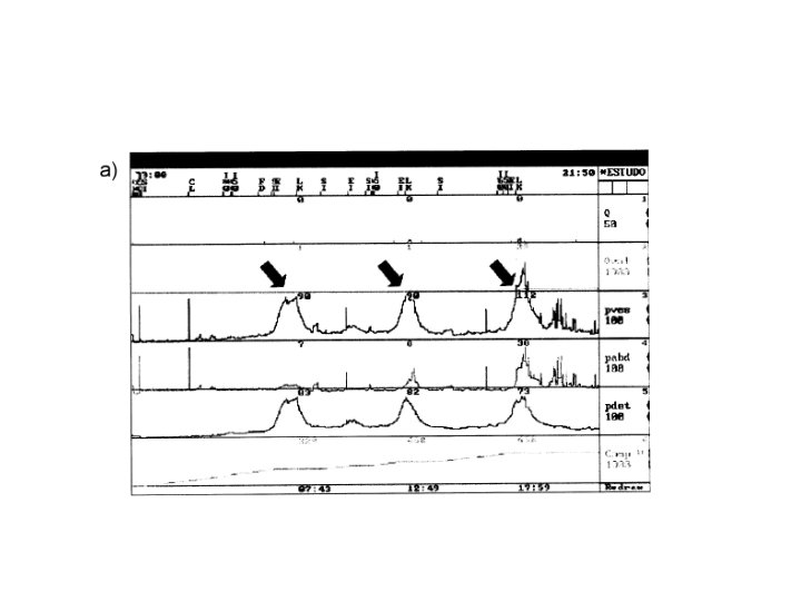
• Bladder contractions or detrusor overactivity (DO), should be assessed during cystometry and should not be present during normal bladder filling. • DO may be associated with a sense or urgency and/or urgency urinary incontinence and divided into neurogenic if associated with a neurologic condition (such as spinal cord injury or multiple sclerosis) or idiopathic when there is no clear cause

DOA

• During bladder filling the pressure remains low despite increase in volume because the bladder is highly compliant organ • Compliance is the change in volume/change in pressure • The bladder is compliant due to viscoelasticity of connective tissue and muscle bundles, where there will be rearrangement of these tissue to accommodate urine at low pressure

• Low compliance may be the result of : • Change of composition (more collagen and less elastin) like what happen in chronic inflammation, BOO, neurological decentralization • Hypertrophic muscle is less compliant.

in poorly compliant bladders, there is a rise in pressure as the bladder fills • there is no one cutoff value to describe “normal compliance. We usually use a value of 10 m. L/cm H 2 O to define abnormal compliance

Compiance - Change in vol / change in pressue

• Maximum cystometric capacity is the volume at which the individual feels that s/he can no longer delay micturition and is associated with a strong desire to void. • While there is no one “normal” bladder capacity, it is generally defined as 350– 500 m. L in adults. In children, the following formula can be used to calculate bladder capacity, Age plus two multiplied by thirty.

• VOIDING PRESSURE FLOW STUDIES: • Once the patient’s bladder is full and the patient is given permission to void, the emptying phase can begin. This is performed with both the urethral and rectal catheters in place • Pressure–flow studies can identify three fundamental voiding states: • 1. Low detrusor pressure and high flow rate (unobstructed) • 2. High detrusor pressure and low flow rate (obstructed) • 3. Low detrusor pressure with low flow rate (poor contractility

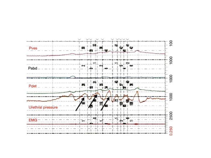
• Urethral Pressure Profilometry: • To maintain continence the pressure in the urethra should exceeds the pressure in the bladder. If not leakage of urine may result. • Urethral pressure profilometry (UPP) is the measurement of intraluminal pressure along the length of the urethra and provides a measure of the resistance exerted by the urethra

• The UPP is performed using a double-lumen urethral catheter that allows for the simultaneous recording of both bladder and urethral pressures • Urethral closure pressure profile: Measured by the subtraction of intravesical pressure from urethral pressure

Treatment of UUI • Lifestyle change: decrease fluid intake, caffien reduction. • Timed voiding: void every one to two hours during the day • Bladder training: e where the patient attempts to consciously delay voiding and to increase the interval between voids, last for minimum of 6 weeks

• Anticholinergic-antimuscarinics are the mainstay of medical therapy for urgency incontinence. example : solifenacin, tolterodine, , oxybutynin. • Side effect include urinary retention, dry mouth, constipation, blurred vision , confusion • Additional pharmaceutical options include beta 3 adrenergic receptor agonists (mirabegron)
 has also been")
• Injection of the detrusor muscle with botulinum toxin (Botox) has also been successfully utilized for idiopathic, medication refractory OAB • Neuromodulation therapies activate afferent inhibitory pathways • Augmentation cystoplasty

Treatment of SUI • Lifestyle modifications: Obesity and smoking has been shown to be associated with SUI. • Proper fluid management as well as restriction of alcohol and caffeine intake can reduce the leakage amount and episodes of SUI • Pelvic floor muscle training : at least 8 contractions , 3 times per day for 3 monthes • Alpha-agonists such as pseudoephedrine may be used for the treatment of stress incontinence
 is to fix the")
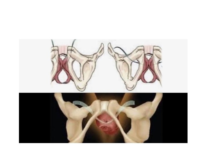
• Surgical therapy : which include retropubic suspension(Burch procedure) is to fix the anterior vaginal wall to Cooper’s ligament, • sling procedure: Tension-free vaginal tape (TVT)and many modifications (eg, TVT-O, placed through obturator foramen, or the minislings) are synthetic tapes consisting of propylene. The middle of the tape is placed under the midurethra, and the two arms are passed via retropubic space to lie above the rectus fascia •

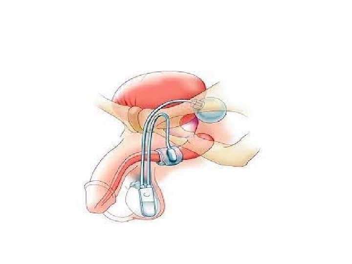
• artificial sphincter: This is a saline-filled implantable medical device that consists of three parts: a pressure-regulating balloon, a cuff that is placed around the urethra or bladder neck, and a pump placed in the labia in women and scrotum in men

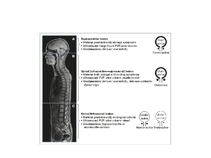
Voiding dysfunction according to spinal cord injury level 1 - suprapontine ( injury above brain stem) : - results in bladder overactivity with coordinated sphincter ( synergy) - Sensation usually preserved but it may be deficient - So patient usually presents with urinary incontinence

- Treatment by anticholinergic, botox injection or surgical.

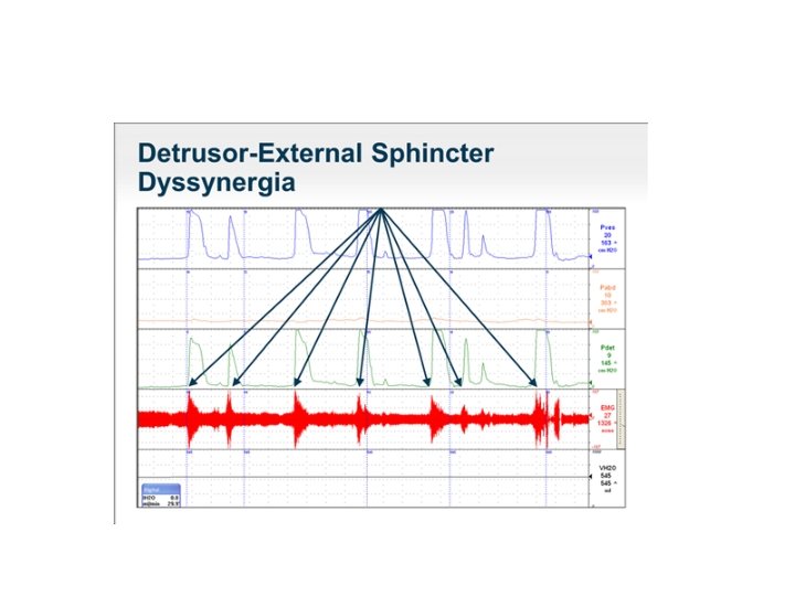
2 - spinal cord injury infrapontine but above spinal cord level T 6 : - Detrusal overactivity - Smooth and striated sphincter dyssynergy - Autonomic hyperreflexia (dysreflexia) - Impaired sensation - So patient presents with incontinence or retention

-Treatment: - to decrease outlet resistance by using baclofen and botox injection - Or use ISC to drain bladder regularly

Autonomic Dusreflexia - Usually in spinal cord injury between brainstem and spinal cord level T 6, so imbalanced reflex sympathetic discharge - Triggered by: - pelvic autonomic afferent activity ( as bladder or bowel distension , erection and ejaculation)

-Patient will develop hypertension, bradycardia , headache , sweating and flushing above spinal cord injury -treatment: -It is considered a medical emergency, If left untreated can cause seizures, retinal hemorrhage, myocardial infarction, cerebral hemorrhage, and, ultimately, death
 are mainstays in")
-Proper bladder and bowel care (ie, preventing fecal impaction, bladder distention) are mainstays in preventing episodes of autonomic dysreflexia.

-if patient becomes hypertensive , should place the patient in an upright position immediately, this takes advantage of an orthostatic response and helps with the pooling of blood in the lower extremities. -most commonly used agents for hypertension are nifedipine and nitrates

3 - spinal cord injury between spinal cord level T 6 and S 2 : -DOA -smooth sphincter synergy but striated sphincter dyssynergy -impaired sensation -patient presents with incontinence or obstructive symptoms and rarely retention

4 - injury below spinal cord level S 2: - detrusor areflexia with open smooth sphincter and striated sphincter retains a residual resting sphincter tone and is not under voluntary control. - Patient presents with retention

• Treatment • The primary aims and their prioritisation when treating neuro-urological disorders are: • 1. protection of the upper urinary tract; • 2. improvement of urinary continence; • 3. restoration of (parts of) the LUT function; • 4. improvement of the patient’s Qo. L. •
- Slides: 67