BIRADS classification in mammography Dr Egle Jonaitiene Dr


BI-RADS classification in mammography Dr. Egle Jonaitiene, Dr. Laima Grinyte 2016 09 19 -23 Dr. Ruta Briediene 2016 11 23

Radiologist report: • …a mass 9 mm size in the right breast , a lot of calcifications in the left breast… • Conclusion: what kind of lesion? Malignant? Bening? What next steps to do?

BI-RADS Breast imaging reporting and data system • Product of ACR • Tool for quality control • To standartise report • To make less interpretation possibility • Monitoring MG -1993 US - 2003

Report • Indication • Breast density • Findings • Dynamics, compare with previous • Assessment • Malignancy suspition • Recommendations
 1 – normal")
Assessment • • 0 – additional information needed (not full exam) 1 – normal (negative) 2 – benign changes 3 – possibly benign 4 – suspicious 5 – very suspicious 6 – proved malignancy

BI-RADS classification BI-RADS Findings Further management 0 Incomplete assessment Need of additional imaging or prior examinations 1 Negative Routine screening 2 Benign Routine screening 3 Probably benign - risk of malignancy is lower than 2%, Ultrasound imaging is necessary or a control mammography imaging and examination within 6 months 4 Suspicious - risk of malignancy is 2 -94%, Further cytology of pathohistology investigation is necessary 5 Highly suspicious - risk of malignancy is higher than 94% Referral to a surgeon is necessary

Density

PHT 2000

2002

PHT 2004

Density

Density 3 -4 Dense breast – higher breast cancer risk! • Inform women about low sensitivity of MG in dense breasts • Use digital MG • Ultrasound in screening ? • Ultrasound as further recommendation

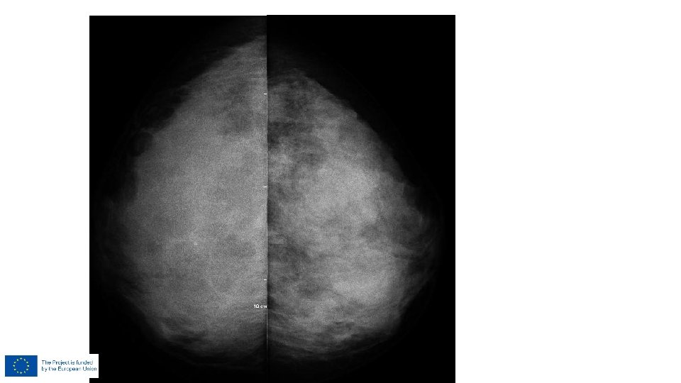
BI-RADS 1 – negative finding Symetrical fibroglandular and/or fatty tissue and no masses, architectural distortion or suspicious calcifications are present. If there is palpable abnormality?

Findings • Mass • Size • Form, margins, density • Localisation • Calcifications • Type, form • Distribution • Localisation • Architectural distortion • Other

A mass Size of a mass Form, margins, density, calcs

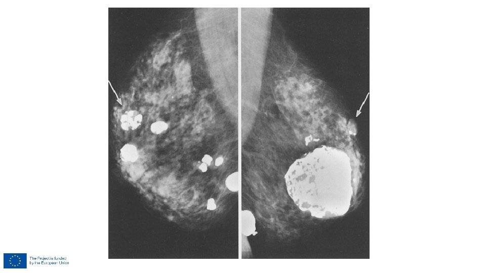
BI-RADS 2 – benign finding • Completely or partialy calcified fibroadenomas

BI-RADS 2 – benign finding • Fat necrosis calcifications

BI-RADS 2 – benign finding • Intramammary lymph nodes

BI-RADS 2 – benign finding • Fat containing formations – lipomas and fibroadenolipomas, oil cysts and galactoceles

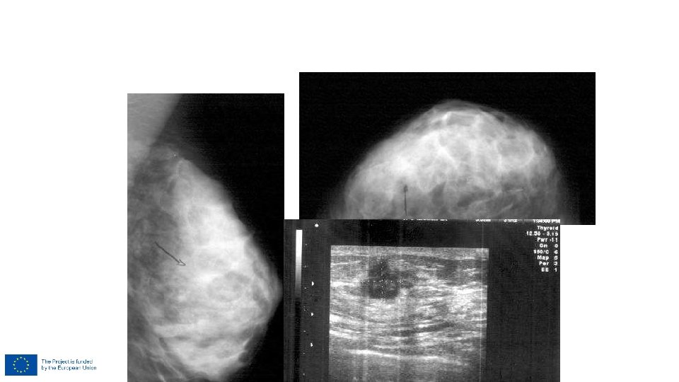
BI-RADS 3 – probably benign finding • Circumscribed, noncalcified masses

BI-RADS 3 – probably benign finding After FU – final assessment could be changed to BI-RADS 2

BI-RADS 3 US and biopsy: TN cancer US and biopsy: FA

Why BIRADS 3? • I don’t know • It is strange • I had similar cancer case • I am not sure about it. . • I want to follow-up • Well….

Oval, round shape circumscribed margins – BIRADS 2

Irregular form spiculated margins – BIRADS 5

Shape – oval margins – obscured BIRADS 3? BIRADS 4?
 • Less than")
BIRADS 3: Probably benign finding ( initial short-interval follow-up suggested ) • Less than 2% risk of malignancy • Non-palpable ( < 2 cm ) • Stable • Only after complete imaging evaluation 3 type of findings: • Cluster of round (punctate) calcifications • Noncalcified circumscribed solid mass • Focal asymmetric density

Classification • “The main potential pitfall is incorrect classification, ” Dr. Luis J. Pina, University Clinic of Navarra, Spain • "Specifically, the BI-RADS 3 category can become a 'holding tank' for problematic lesions which are so categorized without further diagnostic procedures. • This typically occurs when inexperienced radiologists feel uncertain about diagnosis. By Frances Rylands-Monk, Aunt. Minnie. Europe. com staff writer March 4, 2011

• BIRADS 3 is a temporary statement until the lesion is definitely classified as category 2 or 4

• Some malignant tumors can show a benign appearance • Some benign lesions have unusual signs • BIRADS 3 – in between benign and unclear lesions • Not in between benign and malignant lesions!

Probably Benign Finding BIRADS 3 Initial Short-Interval Follow-Up Suggested: • A finding placed in this category should have less than a 2% risk of malignancy. • It is not expected to change over the follow-up interval, but the radiologist would prefer to establish its stability.
 or")
• Don't use if unsure whether to render a benign (Category 2) or suspicious (Category 4) assessment. Then use Category 4. • Don't use in a screening examination • Don't use in a diagnostic examination if additional imaging is required to make a final assessment • Don't use if a lesion, previously assessed as Category 3 has increased in size or extent, like a mass on US with an increase of 20% or more of longest dimension. Then use category 4. • Don't recommend MRI to further evaluate a probably benign finding
 • Non-palpable lesions with microcalcifications categorized as")
Biopsy of BI-RADS 3 lesions (BI-RADS 4) • Non-palpable lesions with microcalcifications categorized as BI-RADS 3 (probably benign) should undergo a biopsy procedure until a more reliable system for description and classification of microcalcifications is available. R M PIJNAPPEL , The British Journal of Radiology, 77 (2004)
 • Planned pregnancy •")
Biopsy in specific situations • Palpable/symptomatic • Planned surgery (augmentation/reduction) • Planned pregnancy • High risk patients • Synchronous cancer • Size of the lesion • Patient´s decision

How can we decrease the BI-RADS 3 lesions? Reclassifying them as: • BIRADS 2 • BIRADS 4

How to reclassify BI-RADS 3 lesions? • Additiona. I imaging techniques • Compare with previous exams • Radiologist´s experience • Biopsy in specific situations

Additional imaging techniques In a clinical setting we can use all the imaging and biopsy techniques to re-classify a BI-RADS 3 lesion • • • Complemmentary mammographic views Tomosynthesis US Elastography MRI

TN invasive ca 2007 2008

Invasive ductal ca 2011 03 2011 12

2011 03 2011 12
 • Corellate")
Recommendations • Use all imaging modalities for lesion characterisation (not in screening!) • Corellate findings • Use different features for lesion characterisation in one imaging modality • Follow-up if no risk for cancer • Discuss with collegues

• In an ideal world there would be no BI-RADS 3 category, according to Dr. Luis J. Pina, University Clinic of Navarra, Spain

BI-RADS 4 – suspicious abnormality • Partially circumscribed mass, nondescript solid mass with indistinct margins, new indistinct, irregular solitary mass

BI-RADS 4

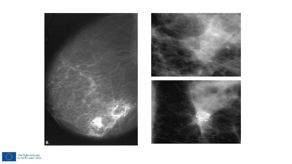
Growing mass/density 2008 2010 2011
")
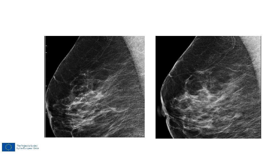
• Continuity in screening rounds • Prior MG (digital)

BI-RADS 5 – highly suspicious of malignancy • Spiculated, irregular highdensity mass, irregular spiculated mass with associated pleomorphic calcifications

Calcifications • Type, form • Distribution • Localisation Distribution • Diffuse • Regional • Linear • Segmental • Groups Obenauer et al. Eur Radiol 2005 Lazarus et al. Radiology 2006

BI-RADS 2 – benign finding • Large, rod like intraductal and periductal calcifications

BI-RADS 2 – benign finding • Simple cyst with mineralized wall

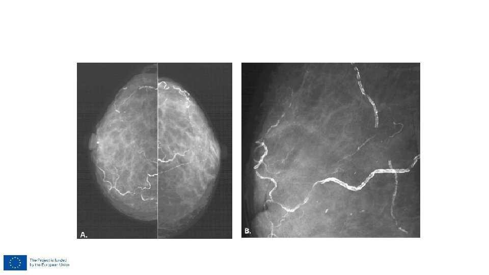
BI-RADS 2 – benign finding • Vascular calcifications

")
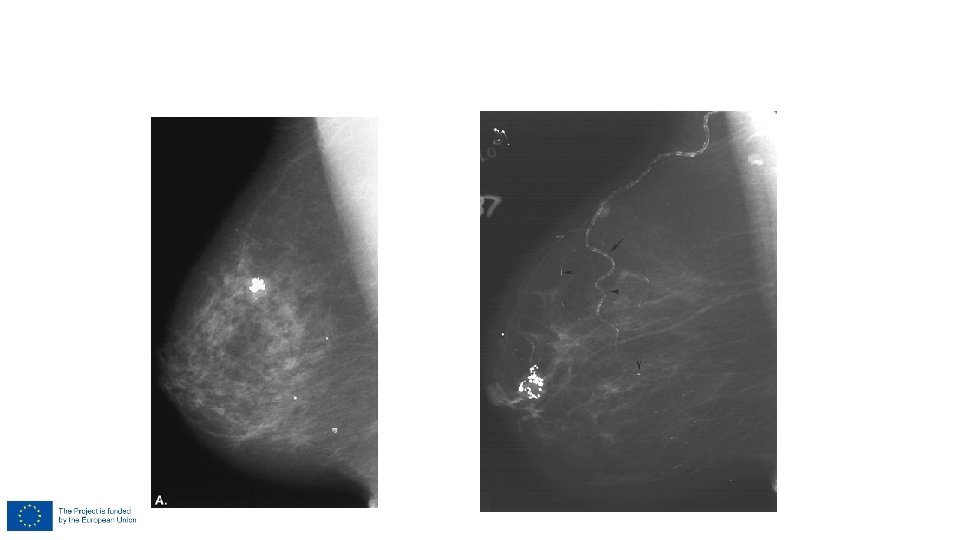
BI-RADS 3 – probably benign finding • Punctate microcalcifications (scattered or clustered)

BI-RADS 3 – probably benign finding If a BI-RADS 3 lesion shows any change during FU, it will change into a BI-RADS 4 or 5 and biopsy should be performed.

BI-RADS 4 – suspicious abnormality • Group amorphous or fine pleomorphic calcifications The pathologist could report sclerosing adenosis or ductal carcinoma in situ. Both diagnoses are concordant with the mammographic findings.

BI-RADS 4 – suspicious abnormality • Punctate microcalcifications with background density

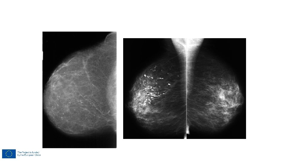
BI-RADS 5 – highly suspicious of malignancy • Segmental or linear arrangement of fine linear, branching calcifications

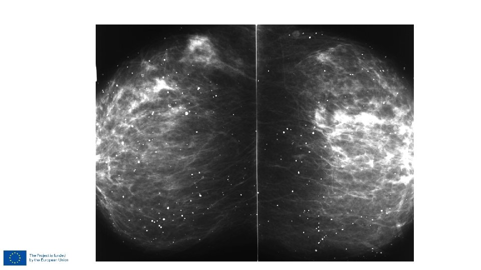
Pleomorfous calcs BI-RADS 5

BI-RADS 5 – ductal

Architectural distortion assimetry density

BI-RADS 2 – benign finding • Architectural distortion clearly related to prior surgery

BI-RADS 3 – probably benign finding • Focal asymmetry which becomes less dense on spot compression view

BI-RADS 4 – suspicious abnormality • Radial architectural distortion without central density

BI-RADS 4 – suspicious abnormality • Asymetrical density with architectural distortion

BI-RADS 4 Assimetry/distortion

BI-RADS 5 – highly suspicious of malignancy • Radial architectural distortions with central density

BI-RADS 0 – incomplete assessment • Additional mammographic imaging is needed: additional mammographic views, spot compression • Additional US or (complete) mammography is needed ONLY if equipment or personnel is not available or patient is unable to wait • Prior mammography or US are required to make a final assessment and issue an addendum including a revised assessment

BI-RADS and screening • Diagnostic MG • Complaints, clinical evaluation • BI-RADS • Diagnostic work-up • Follow-up • Screening MG • Yes/no (no BI-RADS) • Suspicious for cancer/ no suspicion • No BI-RADS 3! • BIRADS 1, 2 – routine screening • BIRADS 4, 5, 0 - recall

Radiologist report • To the woman? • To the GP? • To the specialist? Letter for Woman • Cancer was not found • Next screening date Info for GP

Using BI-RADS in screening Report of benign findings More info for woman/GP Less interval cancers? Economy/finances Must be prepared to examine all women, who got questions about her report • Proper training of radiologists • Training of GP • Info for woman • Proper system of breast services in the country • Centralised data and women recall system
- Slides: 80