
Biosynthesis of steroid hormones Synthesis and hydroxylation of

Biosynthesis of steroid hormones Synthesis and hydroxylation of calciols Biochemistry II Lecture 2 2009 (J. S. )
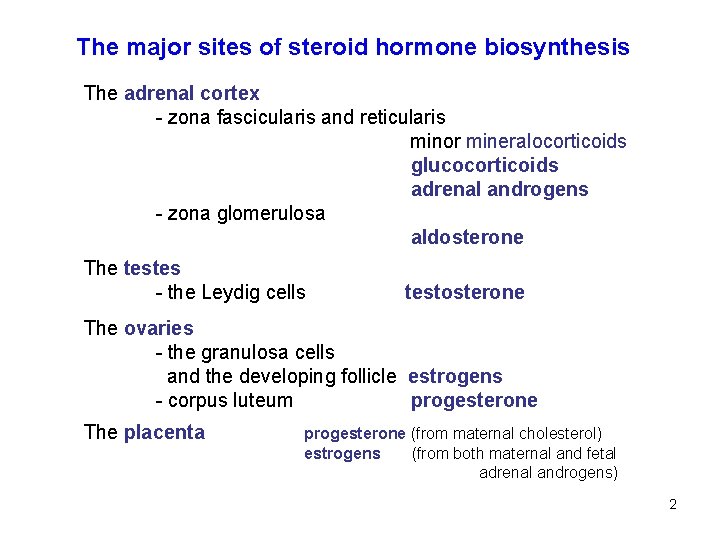
The major sites of steroid hormone biosynthesis The adrenal cortex - zona fascicularis and reticularis minor mineralocorticoids glucocorticoids adrenal androgens - zona glomerulosa aldosterone The testes - the Leydig cells testosterone The ovaries - the granulosa cells and the developing follicle estrogens - corpus luteum progesterone The placenta progesterone (from maternal cholesterol) estrogens (from both maternal and fetal adrenal androgens) 2
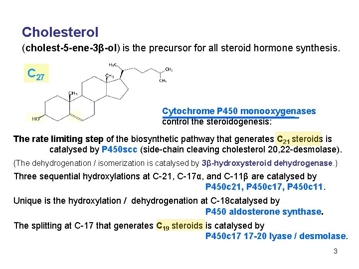
Cholesterol (cholest-5 -ene-3β-ol) is the precursor for all steroid hormone synthesis. C 27 Cytochrome P 450 monooxygenases control the steroidogenesis: The rate limiting step of the biosynthetic pathway that generates C 21 steroids is catalysed by P 450 scc (side-chain cleaving cholesterol 20, 22 -desmolase). (The dehydrogenation / isomerization is catalysed by 3β-hydroxysteroid dehydrogenase. ) Three sequential hydroxylations at C-21, C-17α, and C-11β are catalysed by P 450 c 21, P 450 c 17, P 450 c 11. Unique is the hydroxylation / dehydrogenation at C-18 catalysed by P 450 aldosterone synthase. The splitting at C-17 that generates C 19 steroids is catalysed by P 450 c 17 17 -20 lyase / desmolase. 3
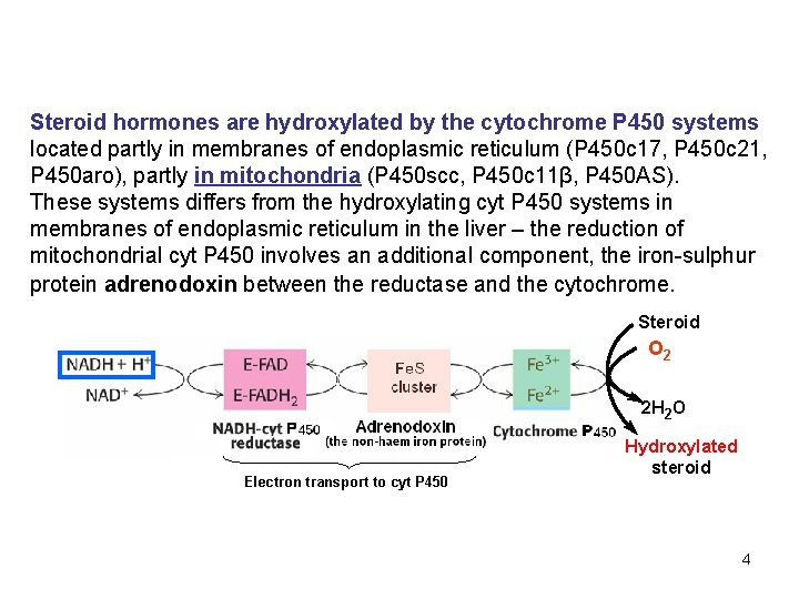
Steroid hormones are hydroxylated by the cytochrome P 450 systems located partly in membranes of endoplasmic reticulum (P 450 c 17, P 450 c 21, P 450 aro), partly in mitochondria (P 450 scc, P 450 c 11β, P 450 AS). These systems differs from the hydroxylating cyt P 450 systems in membranes of endoplasmic reticulum in the liver – the reduction of mitochondrial cyt P 450 involves an additional component, the iron-sulphur protein adrenodoxin between the reductase and the cytochrome. Steroid O 2 2 H 2 O Electron transport to cyt P 450 Hydroxylated steroid 4
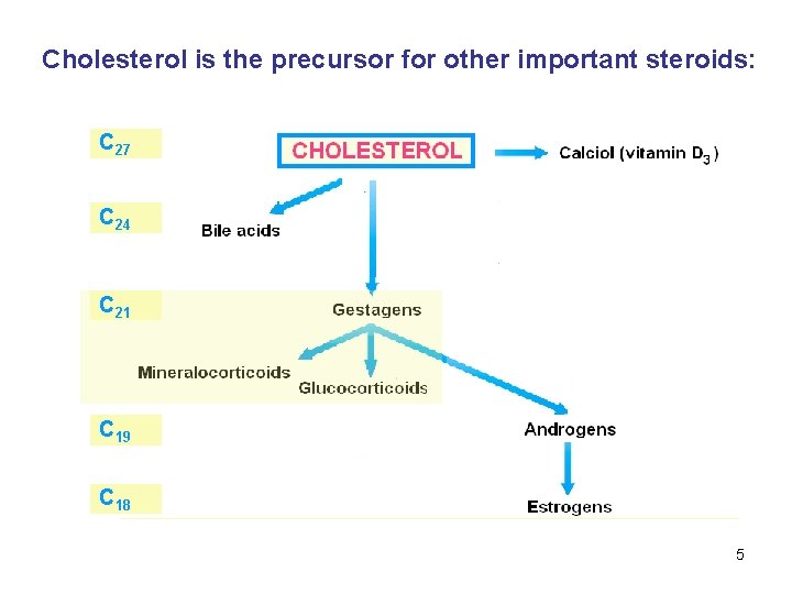
Cholesterol is the precursor for other important steroids: C 27 C 24 C 21 C 19 C 18 5
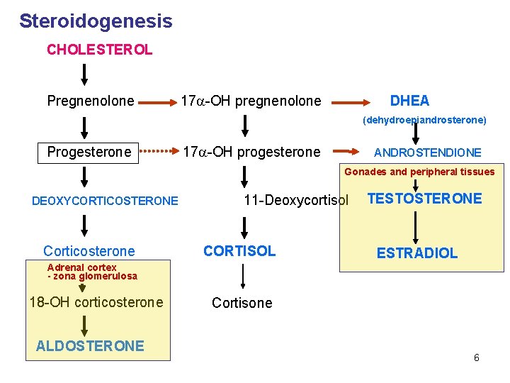
Steroidogenesis CHOLESTEROL Pregnenolone 17 -OH pregnenolone DHEA (dehydroepiandrosterone) Progesterone 17 -OH progesterone ANDROSTENDIONE Gonades and peripheral tissues DEOXYCORTICOSTERONE Corticosterone 11 -Deoxycortisol CORTISOL TESTOSTERONE ESTRADIOL Adrenal cortex - zona glomerulosa 18 -OH corticosterone ALDOSTERONE Cortisone 6
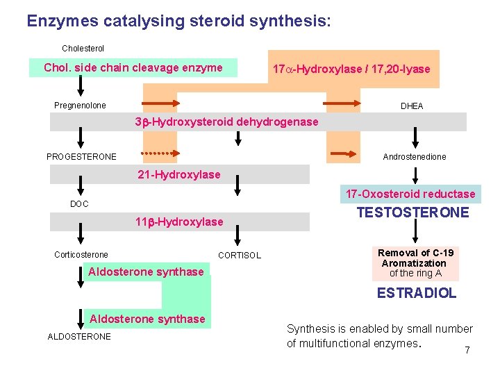
Enzymes catalysing steroid synthesis: Cholesterol Chol. side chain cleavage enzyme 17 -Hydroxylase / 17, 20 -lyase Pregnenolone DHEA 3 -Hydroxysteroid dehydrogenase PROGESTERONE Androstenedione 21 -Hydroxylase 17 -Oxosteroid reductase DOC 11 -Hydroxylase Corticosterone Aldosterone synthase CORTISOL TESTOSTERONE Removal of C-19 Aromatization of the ring A ESTRADIOL Aldosterone synthase ALDOSTERONE Synthesis is enabled by small number of multifunctional enzymes. 7
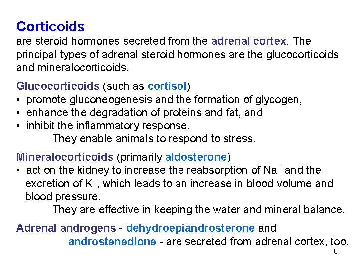
Corticoids are steroid hormones secreted from the adrenal cortex. The principal types of adrenal steroid hormones are the glucocorticoids and mineralocorticoids. Glucocorticoids (such as cortisol) • promote gluconeogenesis and the formation of glycogen, • enhance the degradation of proteins and fat, and • inhibit the inflammatory response. They enable animals to respond to stress. Mineralocorticoids (primarily aldosterone) • act on the kidney to increase the reabsorption of Na+ and the excretion of K+, which leads to an increase in blood volume and blood pressure. They are effective in keeping the water and mineral balance. Adrenal androgens - dehydroepiandrosterone androstenedione - are secreted from adrenal cortex, too. 8
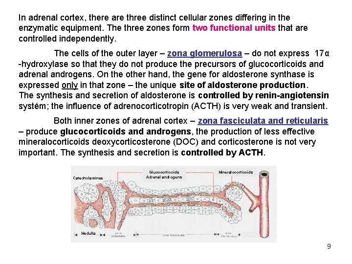
In adrenal cortex, there are three distinct cellular zones differing in the enzymatic equipment. The three zones form two functional units that are controlled independently. The cells of the outer layer – zona glomerulosa – do not express 17α -hydroxylase so that they do not produce the precursors of glucocorticoids and adrenal androgens. On the other hand, the gene for aldosterone synthase is expressed only in that zone – the unique site of aldosterone production. The synthesis and secretion of aldosterone is controlled by renin-angiotensin systém; the influence of adrenocorticotropin (ACTH) is very weak and transient. Both inner zones of adrenal cortex – zona fasciculata and reticularis – produce glucocorticoids androgens, the production of less effective mineralocorticoids deoxycorticosterone (DOC) and corticosterone is not very important. The synthesis and secretion is controlled by ACTH. Catecholamines Glucocorticoids Adrenal androgens Mineralocorticoids Medulla 9
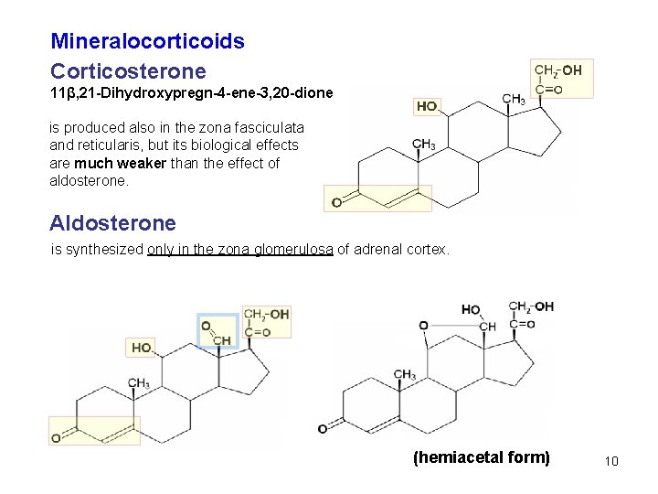
Mineralocorticoids Corticosterone 11β, 21 -Dihydroxypregn-4 -ene-3, 20 -dione is produced also in the zona fasciculata and reticularis, but its biological effects are much weaker than the effect of aldosterone. Aldosterone is synthesized only in the zona glomerulosa of adrenal cortex. (hemiacetal form) 10
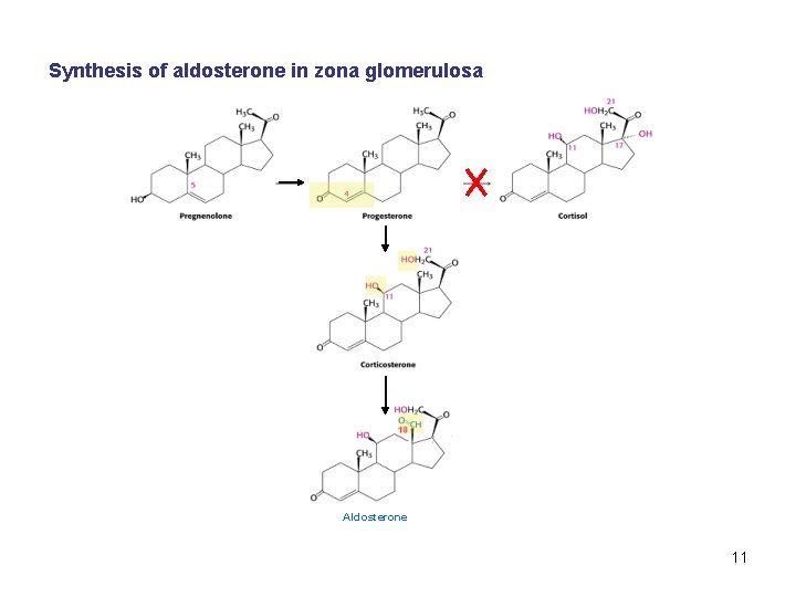
Synthesis of aldosterone in zona glomerulosa Aldosterone 11
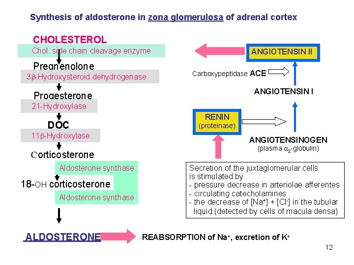
Synthesis of aldosterone in zona glomerulosa of adrenal cortex CHOLESTEROL Chol. side chain cleavage enzyme Pregnenolone 3 -Hydroxysteroid dehydrogenase ANGIOTENSIN II Carboxypeptidase ACE ANGIOTENSIN I Progesterone 21 -Hydroxylase DOC 11 -Hydroxylase Corticosterone Aldosterone synthase 18 -OH corticosterone Aldosterone synthase ALDOSTERONE RENIN (proteinase) ANGIOTENSINOGEN (plasma 2 -globulin) Secretion of the juxtaglomerular cells is stimulated by - pressure decrease in arteriolae afferentes - circulating catecholamines - the decrease of [Na+] + [Cl-] in the tubular liquid (detected by cells of macula densa) REABSORPTION of Na+, excretion of K+ 12
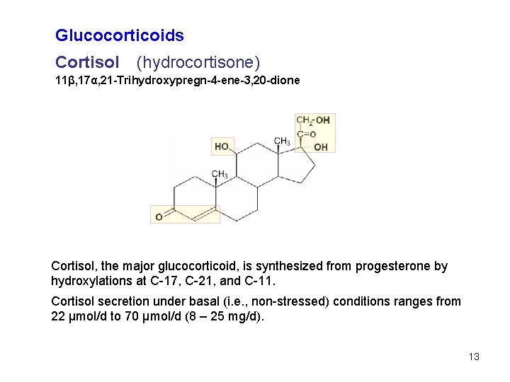
Glucocorticoids Cortisol (hydrocortisone) 11β, 17α, 21 -Trihydroxypregn-4 -ene-3, 20 -dione Cortisol, the major glucocorticoid, is synthesized from progesterone by hydroxylations at C-17, C-21, and C-11. Cortisol secretion under basal (i. e. , non-stressed) conditions ranges from 22 μmol/d to 70 μmol/d (8 – 25 mg/d). 13
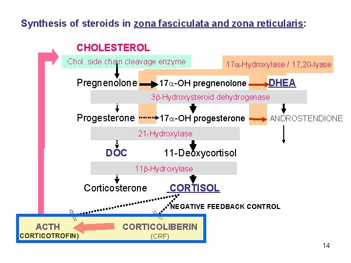
Synthesis of steroids in zona fasciculata and zona reticularis: CHOLESTEROL Chol. side chain cleavage enzyme Pregnenolone 17 -Hydroxylase / 17, 20 -lyase 17 -OH pregnenolone DHEA 3 -Hydroxysteroid dehydrogenase Progesterone 17 -OH progesterone ANDROSTENDIONE 21 -Hydroxylase DOC 11 -Deoxycortisol 11 -Hydroxylase Corticosterone CORTISOL NEGATIVE FEEDBACK CONTROL ACTH CORTICOLIBERIN (CORTICOTROFIN) (CRF) 14
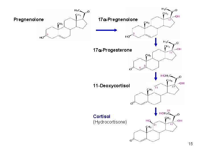
Pregnenolone 17 -Progesterone 11 -Deoxycortisol Cortisol (Hydrocortisone) 15
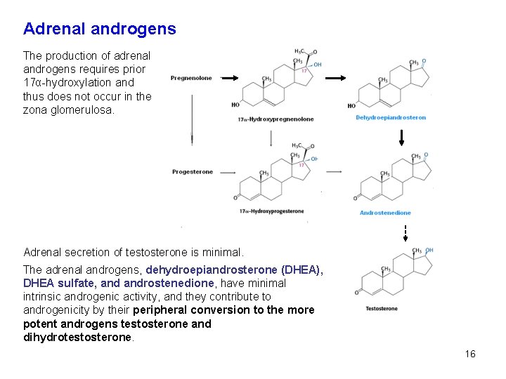
Adrenal androgens The production of adrenal androgens requires prior 17α-hydroxylation and thus does not occur in the zona glomerulosa. Adrenal secretion of testosterone is minimal. The adrenal androgens, dehydroepiandrosterone (DHEA), DHEA sulfate, androstenedione, have minimal intrinsic androgenic activity, and they contribute to androgenicity by their peripheral conversion to the more potent androgens testosterone and dihydrotestosterone. 16

Gonadal steroids In male reproductive function, the three steroids of primary importance are testosterone, dihydrotestosterone and estradiol. In the female reproductive system, the mature ovary synthesizes and secretes estrogens, progesterone, androgens, and their precursors. Testicular androgens C 19 are responsible for the development of male secondary sex characteristics. From a quantitative standpoint, the most important androgen is testosterone secreted by the testicular Leydig cells. Testosterone 17β-Hydroxyandrost-4 -ene-3 -one 17
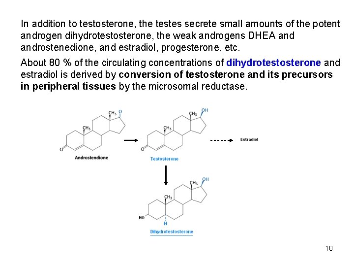
In addition to testosterone, the testes secrete small amounts of the potent androgen dihydrotestosterone, the weak androgens DHEA androstenedione, and estradiol, progesterone, etc. About 80 % of the circulating concentrations of dihydrotestosterone and estradiol is derived by conversion of testosterone and its precursors in peripheral tissues by the microsomal reductase. 18
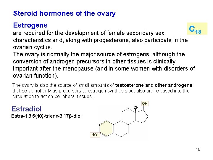
Steroid hormones of the ovary Estrogens C 18 are required for the development of female secondary sex characteristics and, along with progesterone, also participate in the ovarian cyclus. The ovary is normally the major source of estrogens, although the conversion of androgen precursors in other tissues is clinically important after the menopause (and in some women with disorders of ovarian function). The ovary is also the source of small amounts of testosterone and other androgens that serve not only as precursors to estrogen synthesis but also are released into the circulation to act on peripheral tissues. Estradiol Estra-1, 3, 5(10)-triene-3, 17β-diol 19
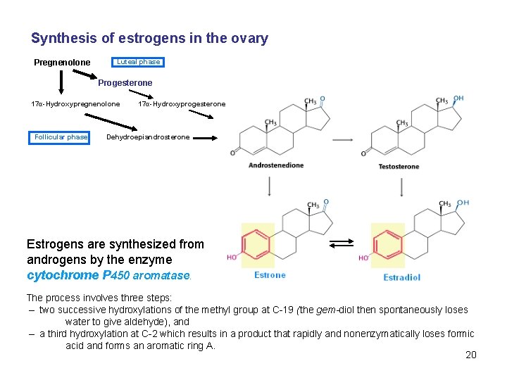
Synthesis of estrogens in the ovary Pregnenolone Luteal phase Progesterone 17α-Hydroxypregnenolone Follicular phase 17α-Hydroxyprogesterone Dehydroepiandrosterone Estrogens are synthesized from androgens by the enzyme cytochrome P 450 aromatase. The process involves three steps: – two successive hydroxylations of the methyl group at C-19 (the gem-diol then spontaneously loses water to give aldehyde), and – a third hydroxylation at C-2 which results in a product that rapidly and nonenzymatically loses formic acid and forms an aromatic ring A. 20
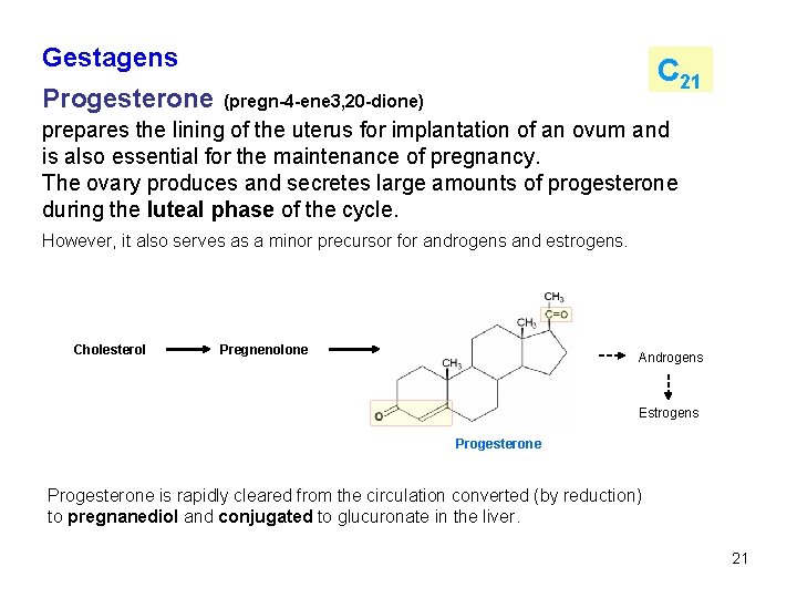
Gestagens Progesterone C 21 (pregn-4 -ene 3, 20 -dione) prepares the lining of the uterus for implantation of an ovum and is also essential for the maintenance of pregnancy. The ovary produces and secretes large amounts of progesterone during the luteal phase of the cycle. However, it also serves as a minor precursor for androgens and estrogens. Cholesterol Pregnenolone Androgens Estrogens Progesterone is rapidly cleared from the circulation converted (by reduction) to pregnanediol and conjugated to glucuronate in the liver. 21
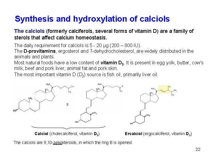
Synthesis and hydroxylation of calciols The calciols (formerly calciferols, several forms of vitamin D) are a family of sterols that affect calcium homeostasis. The daily requirement for calciols is 5 - 20 μg (200 – 800 IU). The D-provitamins, ergosterol and 7 -dehydrocholesterol, are widely distributed in the animals and plants. Most natural foods have a low content of vitamin D 3. It is present in egg yolk, butter, cow's milk, beef and pork liver, animal fat and pork skin. The most important vitamin D (D 2) source is fish oil, primarily liver oil. ≡ Calciol (cholecalciferol, vitamin D 3) Ercalciol (ergocalciferol, vitamin D 2) The calciols are 9, 10 -sekosteroids, in which the ring B is opened. 22
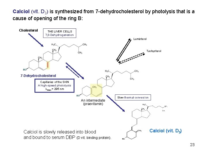
Calciol (vit. D 3) is synthesized from 7 -dehydrocholesterol by photolysis that is a cause of opening of the ring B: Cholesterol THE LIVER CELLS 7, 8 -Dehydrogenation Lumisterol Tachysterol 7 -Dehydrocholesterol Capillaries of the SKIN A high-speed photolysis λmax = 295 nm An intermediate (praevitamin) Calciol is slowly released into blood and bound to serum DBP (D vit. binding protein). Slow thermal conversion Calciol (vit. D 3) 23
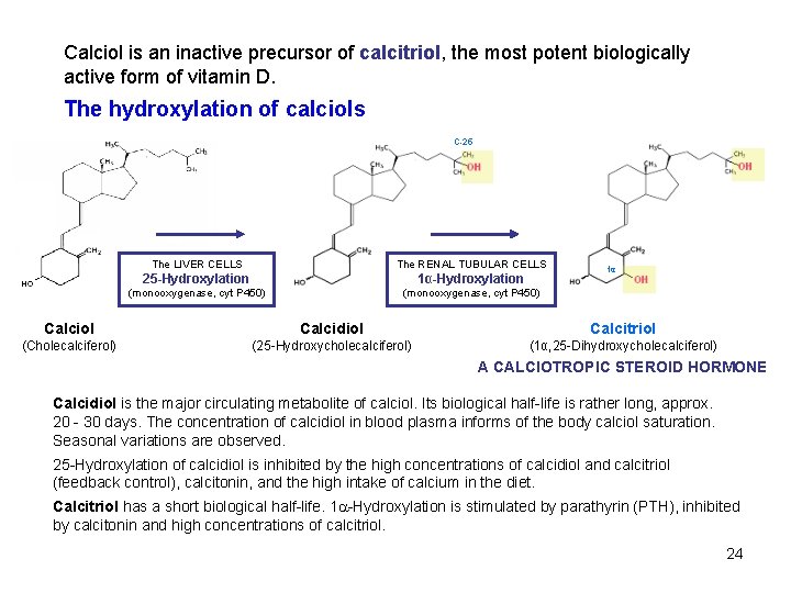
Calciol is an inactive precursor of calcitriol, the most potent biologically active form of vitamin D. The hydroxylation of calciols C-25 The LIVER CELLS The RENAL TUBULAR CELLS 25 -Hydroxylation 1α-Hydroxylation (monooxygenase, cyt P 450) 1α Calciol Calcidiol Calcitriol (Cholecalciferol) (25 -Hydroxycholecalciferol) (1α, 25 -Dihydroxycholecalciferol) A CALCIOTROPIC STEROID HORMONE Calcidiol is the major circulating metabolite of calciol. Its biological half-life is rather long, approx. 20 - 30 days. The concentration of calcidiol in blood plasma informs of the body calciol saturation. Seasonal variations are observed. 25 -Hydroxylation of calcidiol is inhibited by the high concentrations of calcidiol and calcitriol (feedback control), calcitonin, and the high intake of calcium in the diet. Calcitriol has a short biological half-life. 1 -Hydroxylation is stimulated by parathyrin (PTH), inhibited by calcitonin and high concentrations of calcitriol. 24
Biological action of calcitriol (1, 25 -(OH)2 D 3) In the intestine – increased absorption of Ca 2+ by enterocytes: - Induction of a change in conformation of the calmodulin in cytosol – transfer of Ca 2+ across the cytoplasmatic membrane is supported; - Induction of the calbindin synthesis in cytosol (Ca. BP, calcium-binding protein), Ca. BP enables the transport of Ca 2+ within the cell; - Induction of the Ca 2+-ATPase synthesis, that enables the export of Ca 2+ into the extracellular fluid. In the bone – it regulates both the resorption and regeneration of bone tissue - Induction of the osteocalcin synthesis (bone Gla protein, BGla. P), but the control mechanismus is not yet fully explained. 25
- Slides: 25