Bioseparation Lab 7 Ion Exchange Chromatography Dr Methak

Bioseparation Lab 7 Ion Exchange Chromatography Dr. Methak A ljboori
 is a technique that is commonly")
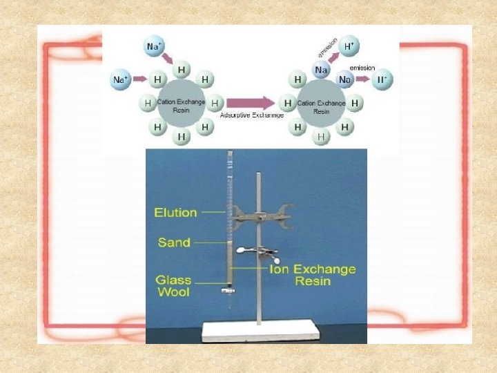
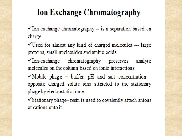
Ion Exchange Chromatography • Ion exchange chromatography (IEX) is a technique that is commonly used in biomolecule purification. It involves the separation of molecules on the basis of their charge. • This technique exploits the interaction between charged molecules in a sample and oppositely charged moieties in the stationery phase of the chromatography matrix.

Types of Ion Exchange Øanion exchangers Øcation exchangers

Cation exchange chromatography q. In cation exchange chromatography positively charged molecules are attracted to a negatively charged solid support. Commonly used cation exchange resins are sulfate derivatives(S-resin) and carboxylate derived ions (CM resins)

Anion exchange chromatography q. In anion exchange chromatography, negatively charged molecules are attracted to a positively charged solid support. Commonly used anion exchange resins are Quaternary amine (Q-resin); and Di. Ethyl Amino Ethane ( DEAE) resin.
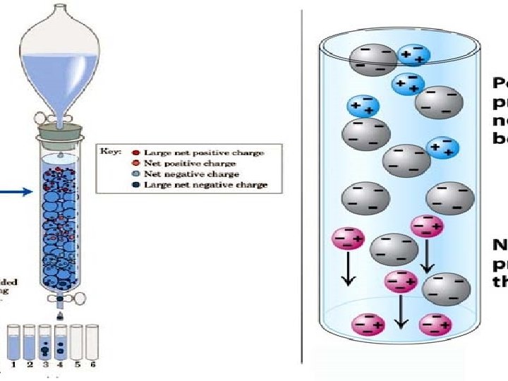

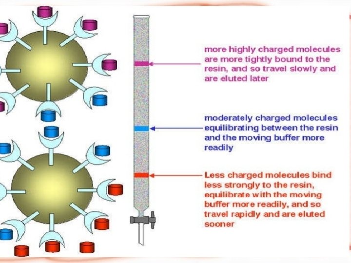
Principle of ion exchange chromatography ØIEX chromatography is used in the separation of charged biomolecules. ØThe crude sample containing charged molecules is used as the (mobile or liquid phase). ØWhen it passes through the chromatographic column, molecules bind to oppositely charged sites in the stationary phase.

ØThe molecules separated on the basis of their charge are eluted using a solution of varying ionic strength. ØBy passing such a solution through the column, highly selective separation of molecules according to their different charges takes place.

The reasons for the success of ion exchange are: 1 -Its widespread applicability 2 - High resolving power 3 - High capacity and the simplicity 4 -Controllability of the method.

Pros and Cons of Ion Exchange Chromatograph IEX Pros IEX Cons Permits high flow rate Sample must be loaded at low ionic strength Concentrates samples Clusters of positively charged residues can cause a netnegatively charged protein to bind a cation exchanger, and vice versa High yield Small changes in p. H can greatly alter binding profile of IEX resin Buffers are nondenaturing Particle size greatly influences resolution

p. H gradient ü A p. H gradient can also be applied to elute individual proteins on the basis of their isoelectric point (p. I) i. e. the point at which the amino acids in a protein carry neutral charge and hence do not migrate in an electric field. ü As amino acids are zwitter ionic compounds they contain groups having both positive and negative charges. ü Based on the p. H of the environment, proteins carry a positive, negative, or nil charge.

üAt their isoelectric point, they will not interact with the charged moieties in the column resin and hence are eluted. üA decreasing p. H gradient can be used to elute proteins using an anion exchange resin and an increasing p. H gradient can be used to elute proteins from cation exchange resins.

p. H gradient ü This is because increasing the buffer p. H of the mobile phase causes the protein to become less protonated (less positively charged) so it cannot form an ionic interaction with the negatively charged resin, allowing is elution. ü Conversely, lowering the p. H of the mobile phase will cause the molecule to become more protonated (less negatively charged), allowing its elution.

Resin Selection in Ion Exchange Chromatography ü Ion exchange resins have positively or negatively charged functional groups covalently linked to a solid matrix. Matrices are usually made of cellulose, polystyrene, agarose, and polyacrylamide. ü Some of the factors affecting resin choice are : 1 -Anion or cation exchanger 2 - Flow rate 3 - weak or strong ion exchanger 4 - particle size of the resin 5 - Binding capacity.

The Applications of Ion Exchange Chromatography 1 - Separation and Purification of blood components such as albumin, recombinant growth factors and enzymes. 2 - Biotechnology - Analytical applications such as quality control and process monitoring. 3 - Food and clinical research - to study wheat varieties and the correlation of proteinuria with different renal diseases. 4 - Fermentation - Cation exchange resins are used to monitor the fermentation process during ß-galactosidase production.

Advantages üIt is a non-denaturing technique. It can be used at all stages and scales of purification. üAn IEX separation can be controlled by changing p. H, salt concentration and/or the ion exchange media. üIt can serve as a concentrating step. A large volume of dilute sample can be applied to a media, and the adsorbed protein subsequently eluted in a smaller volume. üIt offers high selectivity; it can resolve molecules with small differences in charge.

Disadvantages ü Costly equipment and more expensive chemicals ü Turbidity 10 ppm. should be below

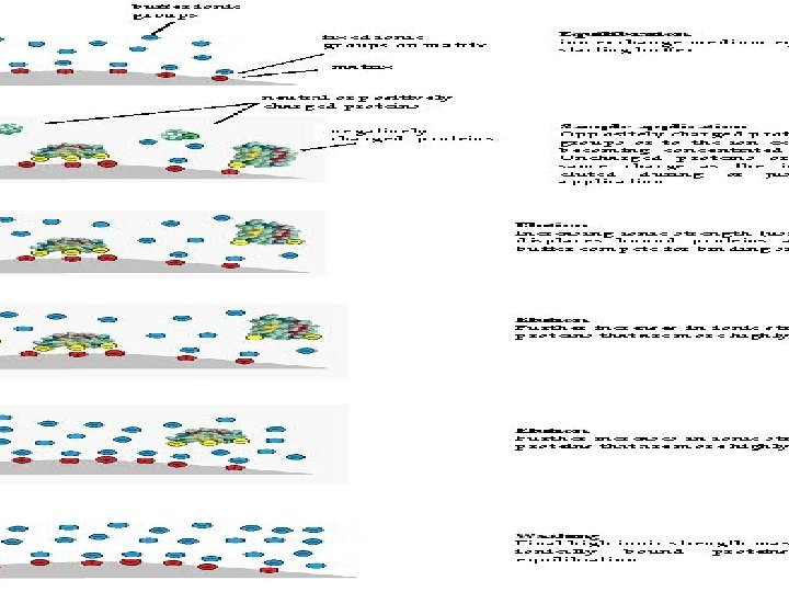
Stages in Ion Exchange Chromatography The Ion Exchange process can be separated into four basic stages: 1. Equilibration 2. Application of sample 3. Elution 4. Regeneration

Stages in Ion Exchange Chromatography

The Technique OF IEX: Key steps in the ion exchange chromatography procedure are: 1 -An crude protein sample is loaded into the ion exchange chromatography column at a particular p. H. 2 - Charged proteins will bind to the oppositely charged functional groups in the resin. 3 - A salt gradient is used to elute separated proteins. At low salt concentrations, proteins having few charged groups are eluted and at higher salt concentrations, proteins with several charged groups are eluted. 4 - Unwanted proteins and impurities are removed by washing the column.

Procedure Ion exchange chromatography column DEAE- Sephadex preparation DEAE- Sephadex gel have positive charged groups(Anion exchangers). 1 - Dissolve (10 g) of gel powder in 100 ml D. W 2 -Left the powder to settle down, remove the supernatant and repeat dissolving in water for several times until it was become completely clear. 3 - Degas the gel by using vacuum pump.

4 - Activate DEAE-Sephadex gel with 0. 25 M Na. Cl for 30 min and wash it with D. W. 5 - Pour the gel into column and left to package. 6 - Equilibrate the column with equilibration buffer.

Column preparation 1 - Apply the dialysed enzyme to DEAE –sephadex column then equilibrate the column and wash it with an equal volume of 0. 01 M phosphate buffer solution (p. H 7) to wash uncharged and positively charged proteins in the sample.
 by using gradient concentrations of sodium")
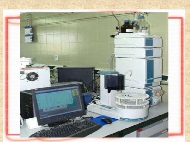
2 - Elution the bound proteins (negatively charged) by using gradient concentrations of sodium chloride ranged between 0. 1 and 0. 5 M. 3 - Detect all protein peaks by measuring the absorbance at 280 nm of each eluted fraction by using UV spectrophotometer.
: It was prepared by dissolving 1. 461 g of")
Na. Cl (0. 25 M): It was prepared by dissolving 1. 461 g of Na. Cl in 100 ml D. W. Ø Sodium chloride phosphate Solution It was prepared at different concentrations of Na. Cl ( 0. 1, 0. 2, 0. 3, 0. 4, and 0. 5 M) in 20 m. M Potassium phosphate buffer. 0. 1 M Na. Cl =0. 5844 gm in 100 ml DW 0. 2 M Na. Cl =1. 1688 gm in 100 ml DW 0. 3 M Na. Cl =1, 7532 gm in 100 ml DW 0. 4 M Na. Cl =2. 3376 gm in 100 ml DW 0. 5 M Na. Cl =2. 922 gm in 100 ml DW Ø
- Slides: 30