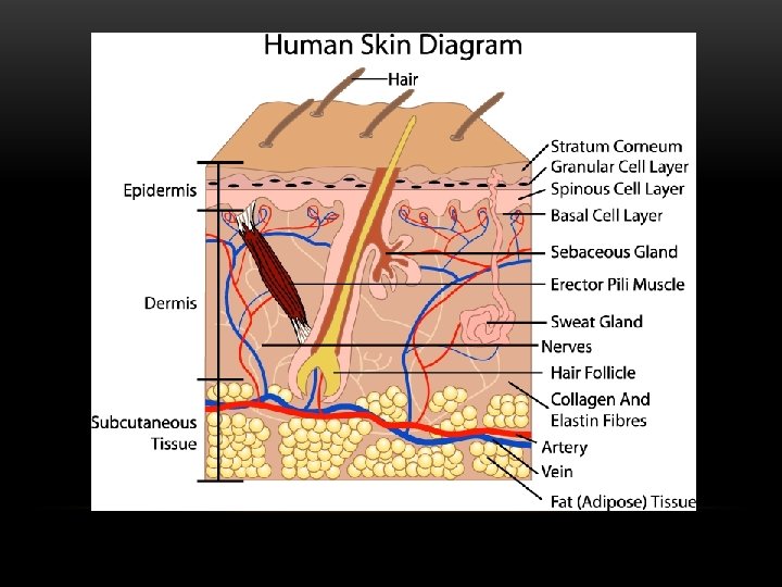
BIOMATERIAL DEVICES And the Epithelium Skin a A

BIOMATERIAL DEVICES And the Epithelium Skin

a. A deep wound ruptures blood vessels, and blood flows out and fills the wound. b. After a blood clot forms, a protective scab develops. Fibroblasts and white blood cells migrate to the wound site. c. New epidermis forms, and fibroblasts promote tissue regeneration. d. Freshly healed/remodeled skin. Quick Review of Wound Healing

First-degree burns affect only the outer layer of the skin. They cause pain, redness, and swelling. Second-degree (partial thickness) burns affect both the outer and underlying layer of skin. They cause pain, redness, swelling, and blistering. Third-degree (full thickness) burns extend into deeper tissues. They cause white or blackened, charred skin that may be numb.

The Gold Standard Partial thickness grafts Full thickness grafts
 Watson’s modification of")
FIGURE 6. The most popular tools for skin graft harvesting. (a) Watson’s modification of Humby knife. (b) Humeca Battery operated dermatome. (c) Padgett Dermatome. (d) Zimmer air dermatome. Tamer Seyhan (2011). Split-Thickness Skin Grafts, Skin Grafts - Indications, Applications and Current Research, Dr. Marcia Spear (Ed. ), ISBN: 978 -953 -307 -509 -9, In. Tech, DOI: 10. 5772/23658. Available from: http: //www. intechopen. com/books/skingrafts-indications-applications-and-current-research/split-thickness-skin-grafts
 is")
In a split thickness skin graft the surface layer of the skin (epidermis) is removed along with a portion of the deeper layer of the skin (dermis). Part of the dermis is left behind on the donor site and this enables the donor site to heal up in the same way as a graze Donor site pain and morbidity
 View of the traumatic clean soft tissue defect before split-thickness skin")
FIGURE 1. a) View of the traumatic clean soft tissue defect before split-thickness skin grafting. (b) Post-operative view of the patient after split-thickness skin grafting. Tamer Seyhan (2011). Split-Thickness Skin Grafts, Skin Grafts - Indications, Applications and Current Research, Dr. Marcia Spear (Ed. ), ISBN: 978 -953 -307 -509 -9, In. Tech, DOI: 10. 5772/23658. Available from: http: //www. intechopen. com/books/skingrafts-indications-applications-and-current-research/split-thickness-skin-grafts
 Hematoma, seroma Inadequate")
Causes of Split-Thickness Skin Graft Failure Inadequate recipient bed (poor vascularity) Hematoma, seroma Inadequate graft fixation and graft shearing Infection (in particular Streptococcus, which can “eat up” a graft within 24 hours) Technical errors (too thick or too thin graft, upsidedown graft. Systemic health problems and bad nutritional status Tamer Seyhan (2011). Split-Thickness Skin Grafts, Skin Grafts - Indications, Applications and Current Research, Dr. Marcia Spear (Ed. ), ISBN: 978 -953 -307 -509 -9, In. Tech, DOI: 10. 5772/23658. Available from: http: //www. intechopen. com/books/skingrafts-indications-applications-and-current-research/split-thickness-skin-grafts

In a full thickness skin graft the entire dermis and its overlying epidermis is removed. The donor site wound is closed with stitches.

https: //www. google. com/url? sa=i&rct=j&q=&esrc=s&source=images&cd=&cad=rja&do cid=DRUiwr 79 q. Ac 4 LM&tbnid=oi. Do. Uv. Ajk 6 Yo 9 M: &ved=0 CAQQj. B 0&url=http%3 A%2 F %2 Fbiotextiles 2012. wordpress. com%2 Fskingrafts%2 F&ei=1 j. Fa. Ua_h. IKm. M 0 QGDx 4 D 4 DA&bvm=bv. 44442042, d. dmg&psig=AFQj. C NGz. EEZ 4 n. Y 3 k. IR 4 g. O 7 GC 56 n. LSX 19 GQ&ust=1364951612794518

TYPES OF SKIN SUBSTITUTES • Acellular • Biobrane • Integra • Alloderm • Cellular • Transcyte • Dermagraft • Apligraft • Cultured Autografts • Cultured Skin Substitutes • Skin Gun

Acellular Substitutes

BIOBRANE https: //www. youtube. com/watch? v=KPFAPe. Akch. E Smith&nephew. com

INTEGRA DERMAL REGENERATION MATRIX The Dermal Replacement Layer is made of a 3 -dimensional porous matrix of cross-linked collagen and glycosaminoglycan (chondroiton-6 sulphate). The Temporary Epidermal Substitute Layer is made of silicone and functions to control fluid loss and serve as a bacterial barrier. Silicone layer is peeled away and an autograft is applied

ALLODERM • Acellular, non-living dermal replacement composed of human cadaveric skin in which the epidermis has been removed by salt processing. • Freeze-dried and has been used in both acute and chronic wounds. • Serves as a dermal scaffold and is accordingly intended for use in deeper wounds. • There is no epidermal component so clinicians commonly use the product with a split-thickness skin graft Dentistrytoday. com

Cellular Substitutes

APLIGRAF https: //www. youtube. com/watch? v=c 87 i. BUvxkb. M

DERMAGRAFT Autologous human fibroblasts, an extracellular matrix, and a bioabsorbable mesh scaffold Cryopreserved, 3 -dimensional, human dermal substitute Serial applications possible due to resorbable scaffold

CULTURED SKIN SUBSTITUTES AND AUTOLOGOUS SKIN GRAFTS http: //www. youtube. com/watch? v=Bz_FVDvgv. SE http: //www. sciencedirect. com/science/article/pii/S 1369703 X 03002535

THE SKIN GUN- RECELL https: //www. youtube. com/watch? v=em_I 33 KSEKQ
- Slides: 21