
Biology Of Cultured Cells Does Culturing Reflect Reality
Biology Of Cultured Cells
Does Culturing Reflect Reality • Culturing Deviates From In Vivo Environment – 3 -D matrix is disrupted (collagen, cell-cell contact) – Heterogeneity is changed – Local growth factors are removed • New Environment Promotes New Properties – Progenitors are encouraged to proliferate – Differentiated cells might not have the same function as starting differentiated cells
Adhesion • Majority Of Cells Adhere On Plastic (Treated) Provided They Are Not Transformed • It Was Observed That Cells Prefer –vely Charged Glass Surface • Plastic (polystyrene) Is Tissue Culture Treated – With High Energy Ionizing Radiation – Electric Ion Discharge • Adhesion Is Mediated By Surface Receptors And Matrix – Matrix Is Secreted By Cells, Adheres To Charged Plastic – Receptors Bind to Matrix
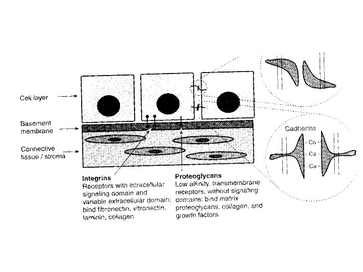
Cell Surface Adhesion Molecules • Three Major Classes – Cell-Cell Adhesion Molecules • • CAMs (Ca 2+ Independent) Cadherins (Ca 2+ Dependent) Primarily Between Homologous Cells Signaling occurs – Cell-Substrate Molecules • • Integrins Bind to fibronectin, entactin, laminin, collagen Bind the specific motif (RGD, arginine, glycine, aspratic) Comprised of and unit
Cell Surface Adhesion Molecules • 3 rd Class Is Proteoglycans – Also Binds Matrix or Other Proteoglycans – Not Via RGD Motif – Low affinity Growth Factor Receptors – May Aid Binding To Higher Affinity Receptors – No Signaling Capacity
Extracellular Matrix (ECM) • Spaces In Between Cells Filled With ECM – Common constituents: fibronectin, laminin, collagen, hyaluronan, proteoglycans, bound growth factors/cytokines • ECM Is Dependent On Cell Types – Fibrocytes secret collagen I and fibronectin – Epithelial cells secret laminin • In Most Cases Cell Lines Are Allowed To Make Their Own ECM • Sometimes We Provide ECM
Cell Proliferation
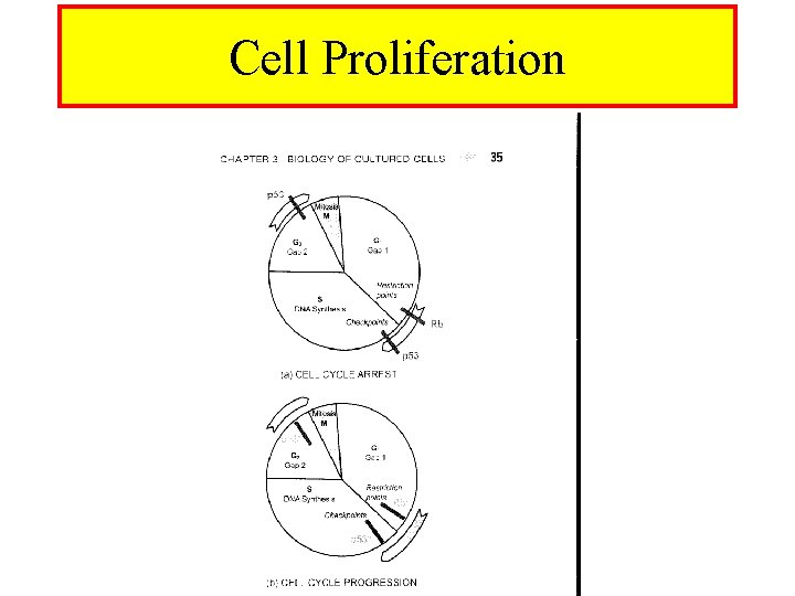
Cell Cycle • 4 Phases – M Phase, mitosis occurs • Chromatin condensation, sister chromatid separation • Daughter cells – G 1 Phase • Progression to DNA SYNTHESIS • Alternatively Go OR differentiation • Restriction Points – S Phase • DNA Synthesis • Progression to G 2 – G 2 Phase • Integrity of DNA Checkpoints • Apoptosis is an option – DNA fragmentation, cell shrinkage, formation of small vesicles
Control Of Cell Proliferation • Environment Regulates Entry Into Cell Cycle • External Growth Factors Promote Cell Proliferation – PDGF, EGF, FGF (+ve) – TGF- (-ve) – Interact with surface receptors • High Density Inhibits Proliferation (Contact Inhibition) • Inside The Cell Both Positive and Negative Factors – Positive, cyclins, Growth Factor Receptor Activation – Negative, p 53, Rb, Checkpoints
Proliferation vs Differentiation • Proliferation Does NOT Promote Differentiation • Differentiation Often Requires – High density – Cell-Cell Interaction – Cell-Matrix Interaction – Differentiation Factors • The Above Conditions Can Be Antagonistic To Proliferation
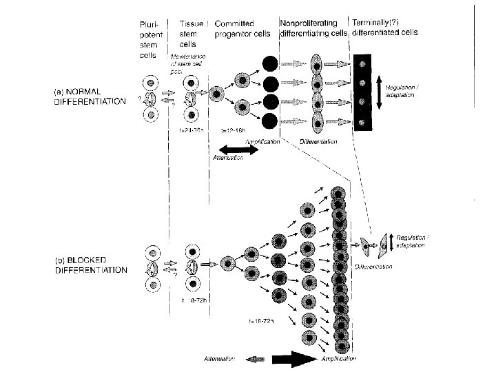
Tissue Retains Function Longer • 3 -D Tissue Retains Its Properties Longer But Can Not Be Propagated • To Overcome This Limitation – Cells Are Cultured On Matrices – Matrigel Is Commercially Available • Not Perfect But Promising – Heterotypic Cultures Are Promising – Pathological Behavior Can Be Studied
Dedifferentiation • Inability To Express In Vivo Phenotype Is Attributed To Dedifferentiation • Still Not Clear If Dedifferentiation Occurs – Wrong lineage expansion is a possibility – Undifferentiated cells dominate – Absence of appropriate inducers, hormones, matrix • Deadaptation vs Dedifferentiation – Deadaptation-enviroment suppresses phenotype, reversible – Dedifferentiation-conversion to primitive phenotype, irreversible
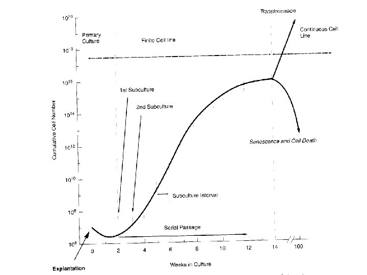
Evolution Of Cell Lines • After 1 st Passage Primary Culture Becomes Cell Line (note Between Finite and Continuous) • By 3 rd Passage Cell Line Stabilizes • Survival Of Stronger Might Not Necessarily Be The Objective • Mesenchymal Cells Usually Dominate – Ex. Fibroblasts • It Is Hard To Avoid Overgrowth Of Specialized Cells (Ex. Hepatic Parenchyma)
Evolution Of Cell Lines • Approximately 10 Passages • Senescence Follows – Thought To Be Due To Telomeres – Every Division Telomeres Shorten – Germ, Stem Cells Use Telomerase • Transformation Is Needed If Division Will Continue
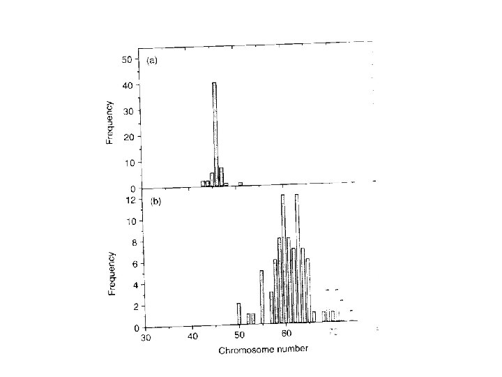
Continuous Cell Lines • • Finite Cell Lines Can Change To Continuous Often p 53 Mutation or Deletion Occurs Overexpression Of Telomerase Transformation vs Immortalization – Transformation-additional changes in growth characteristics – Immortalization-infinite lifespan • Aneuploidy Is A Characteristic Of Cont. Cell Lines – In between diploid and tetraploid – Heteroploidy is also observed • Most Cells Never Become Continuous Cell Lines
Cell Plasticity and Regenerative Medicine
Cell Types • Somatic cells share the same genome • Expression profile is what determines cell type. Examples of cell types: – Cardiac Muscle cells – Fibroblasts – Neurons – Stem Cells • Recent studies have shown that a small number of transcription factors can revert skin cells to Pluripotent Stem Cells – Oct 4, Sox 2, Nanog
Stem Cells Give Rise to Different Cell Types • Stem Cells Can Be Totipotent or Pluripotent • Totipotency is the ability to produce ALL the cell types in an organism (placenta, endoderm, mesoderm and ectoderm) – A zygote is a totipotent cell • Pluripotency is the ability to produce ALL types in an organism EXCEPT placenta (endoderm, mesoderm and ectoderm)
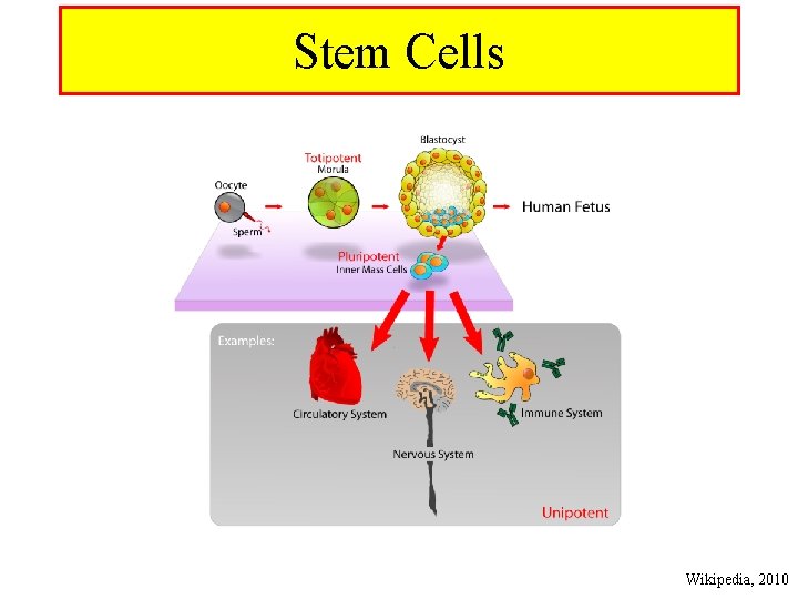
Stem Cells Wikipedia, 2010
Stem Cells and Regeneration • Stem Cells can regenerate damaged tissue • For example heart • Pluripotent Stem Cells (PSCs) hold great promise in regenerative medicine – A major obstacle is the danger of Stem Cells turning into tumors • Scientists are working on transient expression/suppression of key genes involved in inducible PSCs – A good approach is through use of si. RNA
Wound Healing and Regeneration • Humans have little regenerative capacity primarily due to tumor suppressing genes • Rb (retinoblastoma) is a key enzyme in tumor suppression. – If Rb gene is inactivated cells start acting as Stem Cells – This is a risky manipulation • Arf is another important tumor suppressing gene that if turned off regeneration is observed • Rb and Arf silencing was shown to result in muscle cell division and regeneration (Blau M, 2010) • The wound site is a unique site where cells start dividing to repair damage tissue – Adult cells are used in this process – They start acting as ‘younger’ cells for a relatively short period of time
Plasticity of Fibroblasts • Fibroblasts are a ubiquitous cell type • Recent study showed that heart fibroblasts can be turned into cardiomyocytes (Srivastava and colleagues, 2010) • Three transcription factors are needed for this transformation – Gata 4 – Mef 2 c – Tbx 5 • Expression of these transcription factors is most effectively achieved using genetically engineered retroviruses • Heart fibroblasts have the highest conversion efficiency into cardiomyocytes – Skin fibroblasts can also be converted with a lower efficiency
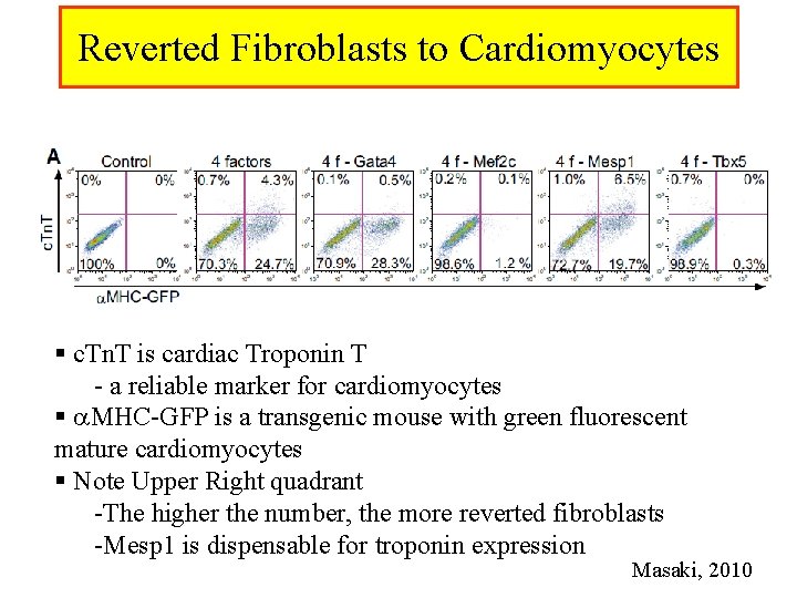
Reverted Fibroblasts to Cardiomyocytes § c. Tn. T is cardiac Troponin T - a reliable marker for cardiomyocytes § MHC-GFP is a transgenic mouse with green fluorescent mature cardiomyocytes § Note Upper Right quadrant -The higher the number, the more reverted fibroblasts -Mesp 1 is dispensable for troponin expression Masaki, 2010
- Slides: 27