
BIOLOGY Chapter 15 GENES AND PROTEINS Power Point

BIOLOGY Chapter 15 GENES AND PROTEINS Power. Point Image Slideshow
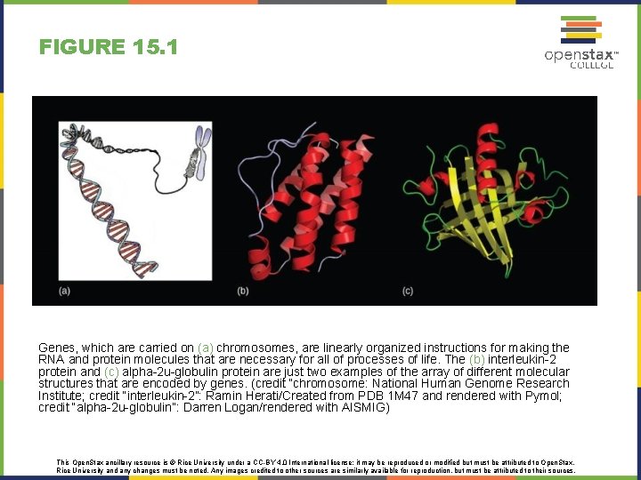
FIGURE 15. 1 Genes, which are carried on (a) chromosomes, are linearly organized instructions for making the RNA and protein molecules that are necessary for all of processes of life. The (b) interleukin-2 protein and (c) alpha-2 u-globulin protein are just two examples of the array of different molecular structures that are encoded by genes. (credit “chromosome: National Human Genome Research Institute; credit “interleukin-2”: Ramin Herati/Created from PDB 1 M 47 and rendered with Pymol; credit “alpha-2 u-globulin”: Darren Logan/rendered with AISMIG) This Open. Stax ancillary resource is © Rice University under a CC-BY 4. 0 International license; it may be reproduced or modified but must be attributed to Open. Stax, Rice University and any changes must be noted. Any images credited to other sources are similarly available for reproduction, but must be attributed to their sources.
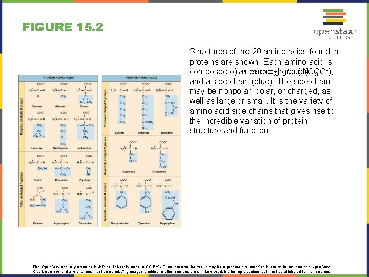
FIGURE 15. 2 Structures of the 20 amino acids found in proteins are shown. Each amino acid is + ( NH -), composed of), an a carboxyl amino group (COO 3 and a side chain (blue). The side chain may be nonpolar, or charged, as well as large or small. It is the variety of amino acid side chains that gives rise to the incredible variation of protein structure and function. This Open. Stax ancillary resource is © Rice University under a CC-BY 4. 0 International license; it may be reproduced or modified but must be attributed to Open. Stax, Rice University and any changes must be noted. Any images credited to other sources are similarly available for reproduction, but must be attributed to their sources.

FIGURE 15. 3 Instructions on DNA are transcribed onto messenger RNA. Ribosomes are able to read the genetic information inscribed on a strand of messenger RNA and use this information to string amino acids together into a protein. This Open. Stax ancillary resource is © Rice University under a CC-BY 4. 0 International license; it may be reproduced or modified but must be attributed to Open. Stax, Rice University and any changes must be noted. Any images credited to other sources are similarly available for reproduction, but must be attributed to their sources.
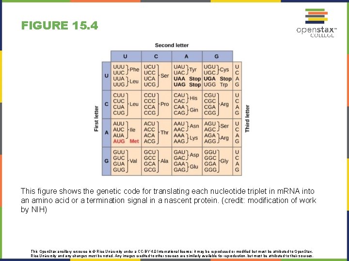
FIGURE 15. 4 This figure shows the genetic code for translating each nucleotide triplet in m. RNA into an amino acid or a termination signal in a nascent protein. (credit: modification of work by NIH) This Open. Stax ancillary resource is © Rice University under a CC-BY 4. 0 International license; it may be reproduced or modified but must be attributed to Open. Stax, Rice University and any changes must be noted. Any images credited to other sources are similarly available for reproduction, but must be attributed to their sources.
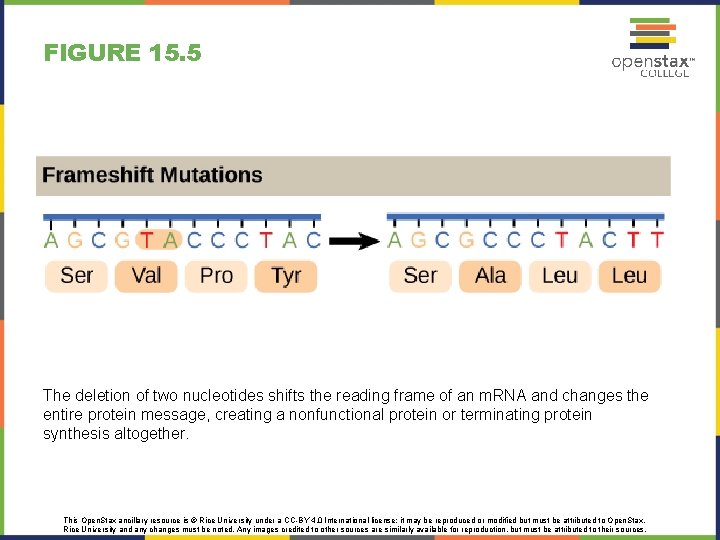
FIGURE 15. 5 The deletion of two nucleotides shifts the reading frame of an m. RNA and changes the entire protein message, creating a nonfunctional protein or terminating protein synthesis altogether. This Open. Stax ancillary resource is © Rice University under a CC-BY 4. 0 International license; it may be reproduced or modified but must be attributed to Open. Stax, Rice University and any changes must be noted. Any images credited to other sources are similarly available for reproduction, but must be attributed to their sources.
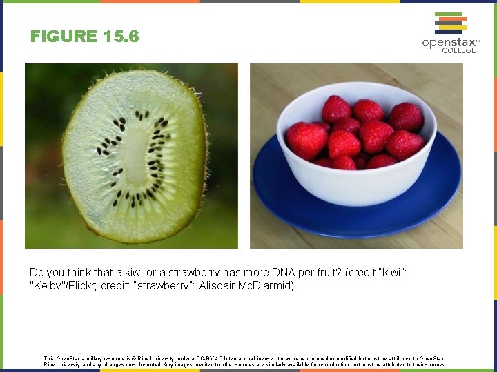
FIGURE 15. 6 Do you think that a kiwi or a strawberry has more DNA per fruit? (credit “kiwi”: "Kelbv"/Flickr; credit: “strawberry”: Alisdair Mc. Diarmid) This Open. Stax ancillary resource is © Rice University under a CC-BY 4. 0 International license; it may be reproduced or modified but must be attributed to Open. Stax, Rice University and any changes must be noted. Any images credited to other sources are similarly available for reproduction, but must be attributed to their sources.
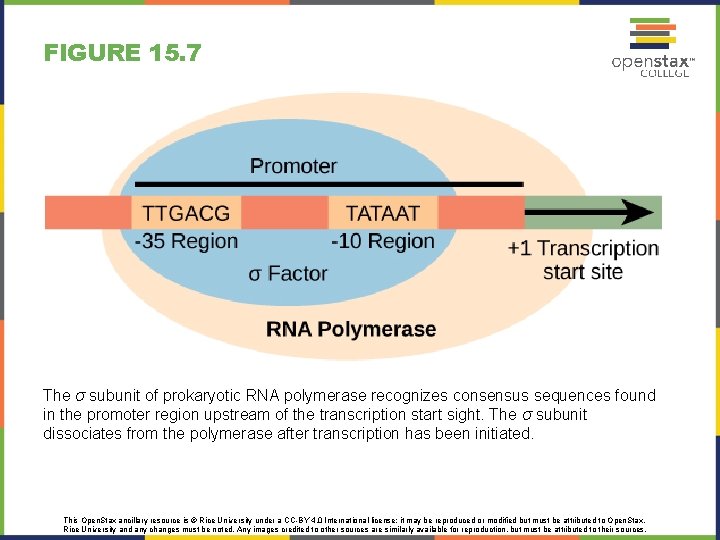
FIGURE 15. 7 The σ subunit of prokaryotic RNA polymerase recognizes consensus sequences found in the promoter region upstream of the transcription start sight. The σ subunit dissociates from the polymerase after transcription has been initiated. This Open. Stax ancillary resource is © Rice University under a CC-BY 4. 0 International license; it may be reproduced or modified but must be attributed to Open. Stax, Rice University and any changes must be noted. Any images credited to other sources are similarly available for reproduction, but must be attributed to their sources.
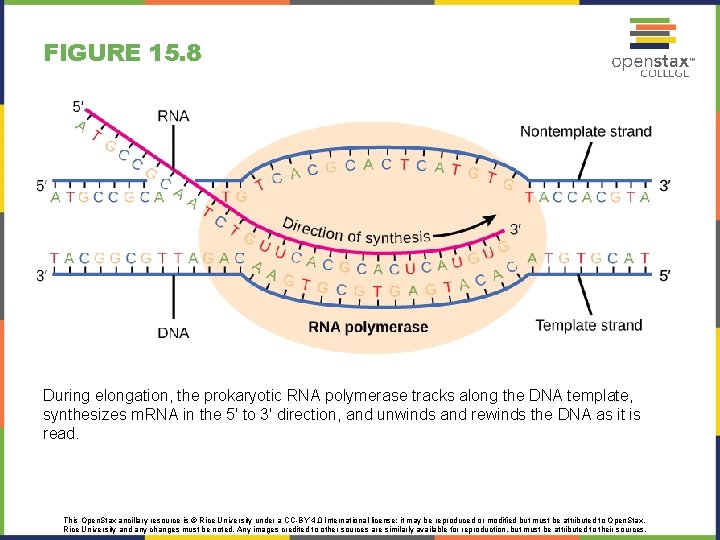
FIGURE 15. 8 During elongation, the prokaryotic RNA polymerase tracks along the DNA template, synthesizes m. RNA in the 5' to 3' direction, and unwinds and rewinds the DNA as it is read. This Open. Stax ancillary resource is © Rice University under a CC-BY 4. 0 International license; it may be reproduced or modified but must be attributed to Open. Stax, Rice University and any changes must be noted. Any images credited to other sources are similarly available for reproduction, but must be attributed to their sources.
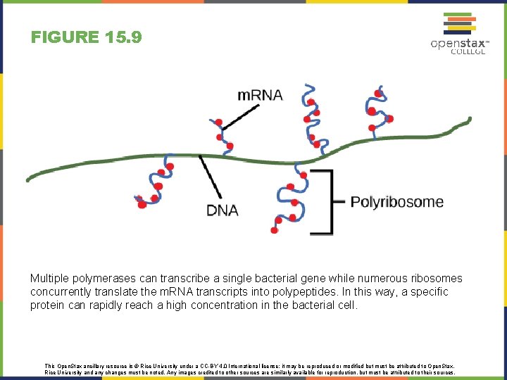
FIGURE 15. 9 Multiple polymerases can transcribe a single bacterial gene while numerous ribosomes concurrently translate the m. RNA transcripts into polypeptides. In this way, a specific protein can rapidly reach a high concentration in the bacterial cell. This Open. Stax ancillary resource is © Rice University under a CC-BY 4. 0 International license; it may be reproduced or modified but must be attributed to Open. Stax, Rice University and any changes must be noted. Any images credited to other sources are similarly available for reproduction, but must be attributed to their sources.
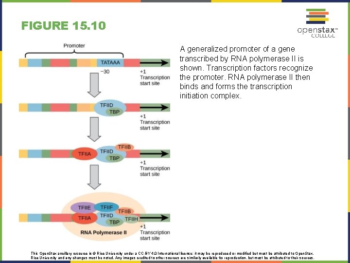
FIGURE 15. 10 A generalized promoter of a gene transcribed by RNA polymerase II is shown. Transcription factors recognize the promoter. RNA polymerase II then binds and forms the transcription initiation complex. This Open. Stax ancillary resource is © Rice University under a CC-BY 4. 0 International license; it may be reproduced or modified but must be attributed to Open. Stax, Rice University and any changes must be noted. Any images credited to other sources are similarly available for reproduction, but must be attributed to their sources.
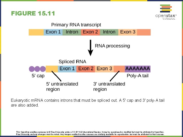
FIGURE 15. 11 Eukaryotic m. RNA contains introns that must be spliced out. A 5' cap and 3' poly-A tail are also added. This Open. Stax ancillary resource is © Rice University under a CC-BY 4. 0 International license; it may be reproduced or modified but must be attributed to Open. Stax, Rice University and any changes must be noted. Any images credited to other sources are similarly available for reproduction, but must be attributed to their sources.
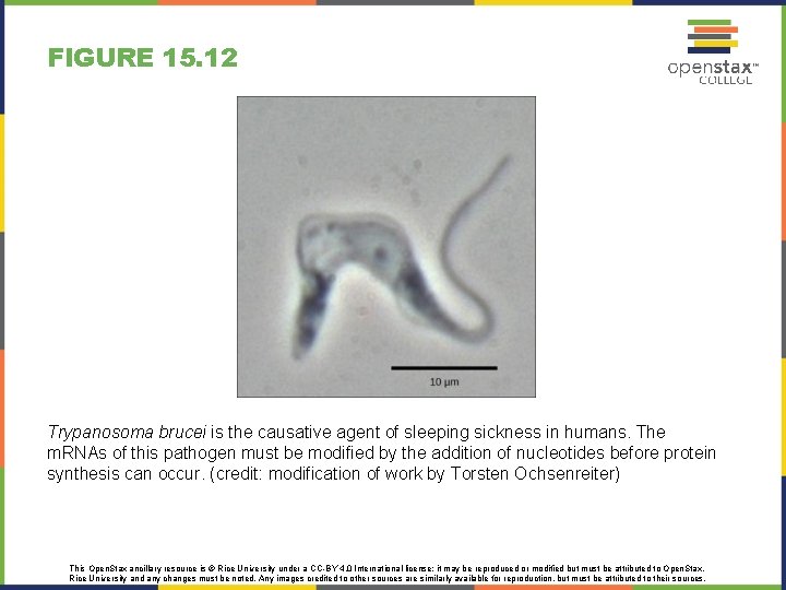
FIGURE 15. 12 Trypanosoma brucei is the causative agent of sleeping sickness in humans. The m. RNAs of this pathogen must be modified by the addition of nucleotides before protein synthesis can occur. (credit: modification of work by Torsten Ochsenreiter) This Open. Stax ancillary resource is © Rice University under a CC-BY 4. 0 International license; it may be reproduced or modified but must be attributed to Open. Stax, Rice University and any changes must be noted. Any images credited to other sources are similarly available for reproduction, but must be attributed to their sources.
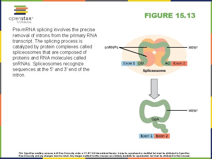
FIGURE 15. 13 Pre-m. RNA splicing involves the precise removal of introns from the primary RNA transcript. The splicing process is catalyzed by protein complexes called spliceosomes that are composed of proteins and RNA molecules called sn. RNAs. Spliceosomes recognize sequences at the 5' and 3' end of the intron. This Open. Stax ancillary resource is © Rice University under a CC-BY 4. 0 International license; it may be reproduced or modified but must be attributed to Open. Stax, Rice University and any changes must be noted. Any images credited to other sources are similarly available for reproduction, but must be attributed to their sources.
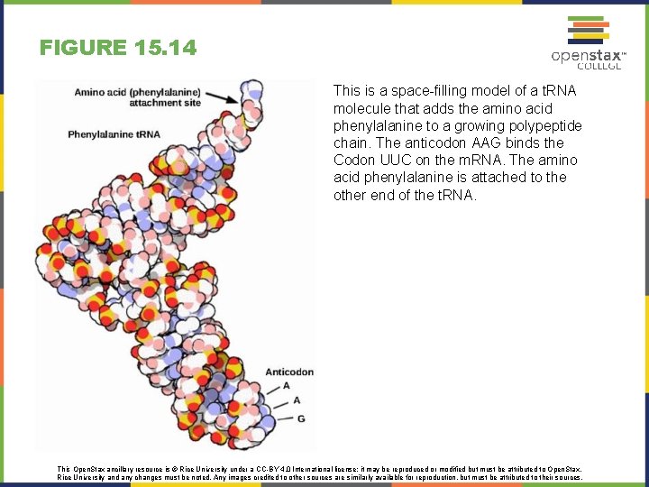
FIGURE 15. 14 This is a space-filling model of a t. RNA molecule that adds the amino acid phenylalanine to a growing polypeptide chain. The anticodon AAG binds the Codon UUC on the m. RNA. The amino acid phenylalanine is attached to the other end of the t. RNA. This Open. Stax ancillary resource is © Rice University under a CC-BY 4. 0 International license; it may be reproduced or modified but must be attributed to Open. Stax, Rice University and any changes must be noted. Any images credited to other sources are similarly available for reproduction, but must be attributed to their sources.
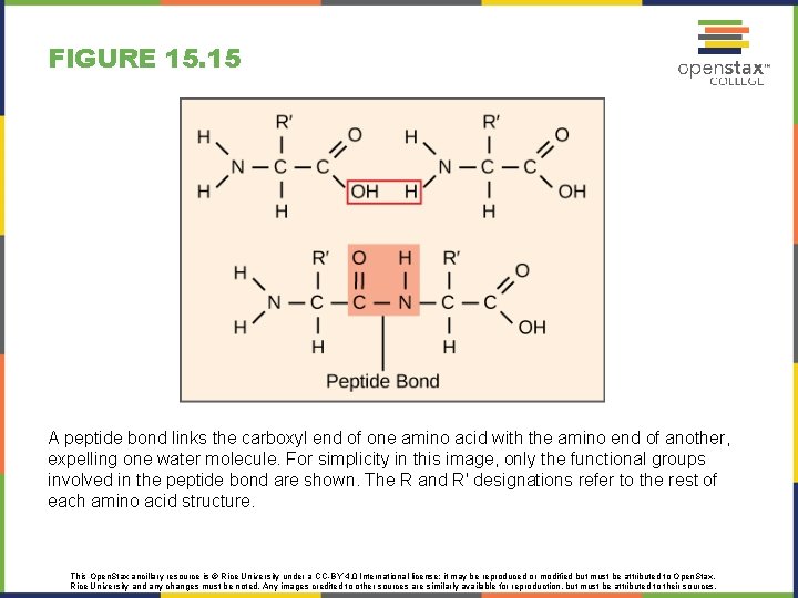
FIGURE 15. 15 A peptide bond links the carboxyl end of one amino acid with the amino end of another, expelling one water molecule. For simplicity in this image, only the functional groups involved in the peptide bond are shown. The R and R' designations refer to the rest of each amino acid structure. This Open. Stax ancillary resource is © Rice University under a CC-BY 4. 0 International license; it may be reproduced or modified but must be attributed to Open. Stax, Rice University and any changes must be noted. Any images credited to other sources are similarly available for reproduction, but must be attributed to their sources.
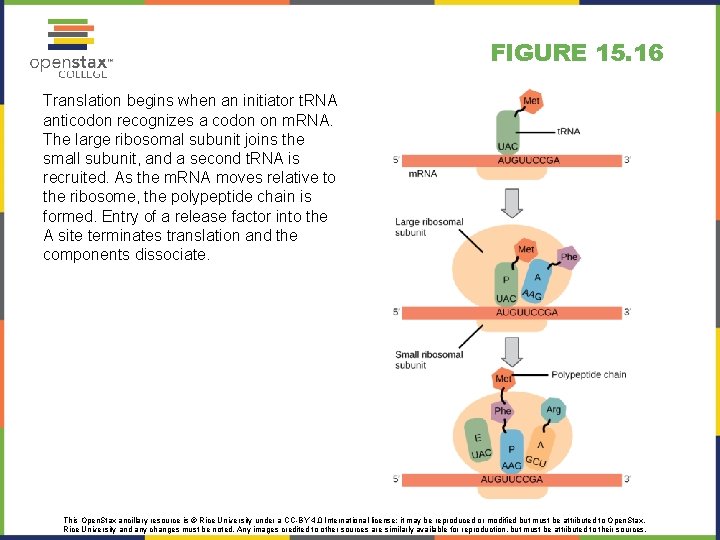
FIGURE 15. 16 Translation begins when an initiator t. RNA anticodon recognizes a codon on m. RNA. The large ribosomal subunit joins the small subunit, and a second t. RNA is recruited. As the m. RNA moves relative to the ribosome, the polypeptide chain is formed. Entry of a release factor into the A site terminates translation and the components dissociate. This Open. Stax ancillary resource is © Rice University under a CC-BY 4. 0 International license; it may be reproduced or modified but must be attributed to Open. Stax, Rice University and any changes must be noted. Any images credited to other sources are similarly available for reproduction, but must be attributed to their sources.
- Slides: 17