Biology and Behavior Neuron Individual nerve cell Soma

Biology and Behavior

Neuron: Individual nerve cell • Soma or cell body – Contains the nucleus and cell machinery • Most neurons have dendrites – Highly branched extensions that receive signals from other neurons • The axon is typically a much longer extension – Fibers that carries information away from the cell body – That may be covered with a myelin sheath Copyright © 2005 Pearson Education, Inc. publishing as Benjamin Cummings

Neuron Structure • Most of a neuron’s organelles – Are located in the cell body Dendrites Cell body Nucleus Synapse Signal Axon direction Axon hillock Presynaptic cell Postsynaptic cell Myelin sheath Figure 48. 5 Copyright © 2005 Pearson Education, Inc. publishing as Benjamin Cummings Synaptic terminals

Neurons • Neurons have a wide variety of shapes – That reflect their input and output interactions Dendrites Axon Cell body Figure 48. 6 a–c (a) Sensory neuron Copyright © 2005 Pearson Education, Inc. publishing as Benjamin Cummings (b) Interneurons (c) Motor neuron

Three classes of neurons Copyright © 2005 Pearson Education, Inc. publishing as Benjamin Cummings
 • Glia are supporting cells – That are essential for the")
Supporting Cells (Glia) • Glia are supporting cells – That are essential for the structural integrity of the nervous system and for the normal functioning of neurons – Supply nourishment to neurons, help remove waste products, and provide insulation around many axons. – Outnumber neurons by about 10 to 1. Copyright © 2005 Pearson Education, Inc. publishing as Benjamin Cummings

How Neurons send messages down their axons? • A neuron that is not transmitting signals – Contains some open K+ channels and closed Na+ channels in its plasma membrane • The diffusion of K+ and Na+ through these channels – Leads to a separation of charges across the membrane, producing the resting potential
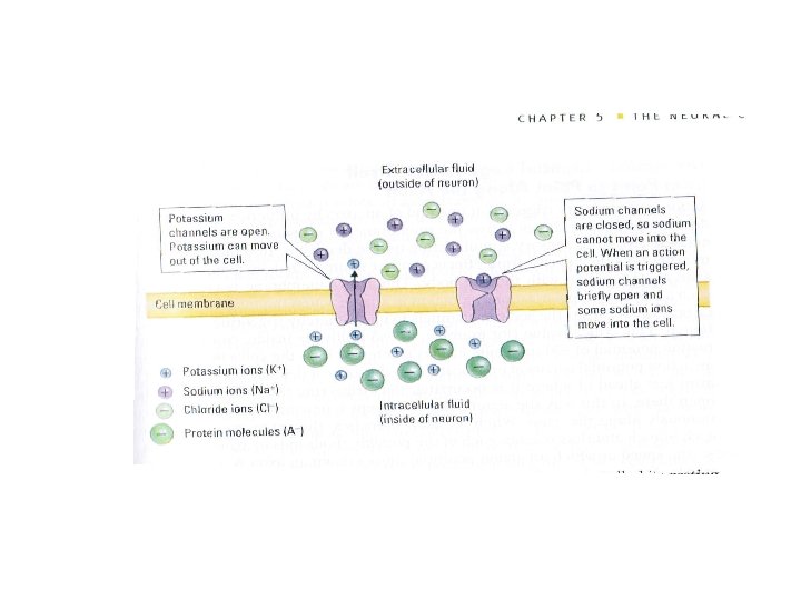

Resting membrane potential Animation

• Step 1: Resting State. Fig. 48. 9 Copyright © 2002 Pearson Education, Inc. , publishing as Benjamin Cummings

• Step 2: Threshold. Fig. 48. 9 Copyright © 2002 Pearson Education, Inc. , publishing as Benjamin Cummings

• Step 3: Depolarization phase of the action potential. Fig. 48. 9 Copyright © 2002 Pearson Education, Inc. , publishing as Benjamin Cummings

• Step 4: Repolarizing phase of the action potential. Fig. 48. 9 Copyright © 2002 Pearson Education, Inc. , publishing as Benjamin Cummings

• Step 5: Undershoot. Fig. 48. 9 Copyright © 2002 Pearson Education, Inc. , publishing as Benjamin Cummings

How Neurons send messages down their axons • Action potential

How Neurons send messages down their axons • The action potential is repeatedly regenerated along the length of the axon. – An action potential achieved at one region of the membrane is sufficient to depolarize a neighboring region above threshold. • Thus triggering a new action potential. • The refractory period assures that impulse conduction is unidirectional. Copyright © 2002 Pearson Education, Inc. , publishing as Benjamin Cummings

Copyright © 2005 Pearson Education, Inc. publishing as Benjamin Cummings

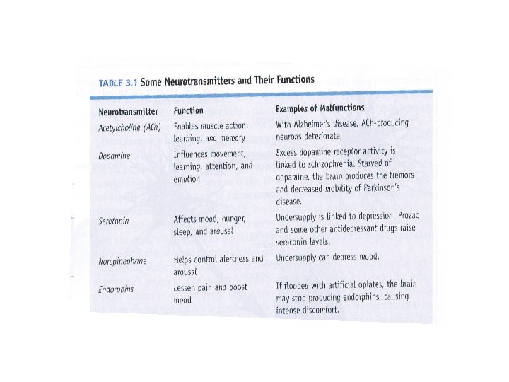
Transmission at the synapse • Synapse: Place where an axon of one neuron can send signals to the membrane(on a dendrite or cell body) of another neuron. • Neurotransmitter: A chemical that carries a signal from the terminal button on one neuron to the dendrite or cell body of another neuron.

Steps: • Action potential reaches the terminal button. • Vesicles release neurotransmitters into synaptic cleft. • Neurotransmitters diffuse across cleft and bind to specific receptors on receiving neuron. • Sodium channels open. • Sodium ions enter receiving neuron which can lead to an action potential. • Excess neurotransmitters reabsorbed back into the vesicles of the sending neuron (reuptake)

Transmission across the synapse Copyright © 2005 Pearson Education, Inc. publishing as Benjamin Cummings

Synaptic transmission • Nervous system animation by Mc. Graw-Hill – Chemical synapse – Synaptic transmission. Synaptic Transmission You. Tube

Mirror neurons • Video clip 2 • Video clip

Copyright © 2005 Pearson Education, Inc. publishing as Benjamin Cummings
 Central and peripheral nervous systems. (b) Spinal nerves, cranial nerves,")
Fig. 2. 6 (a) Central and peripheral nervous systems. (b) Spinal nerves, cranial nerves, and the autonomic nervous system. Copyright © 2010 by Worth Publishers

Fig. 2. 8 Sympathetic and parasympathetic branches of the autonomic nervous system.

Copyright © 2010 by Worth Publishers

Figure 2. 25 FIGURE 2. 25 This simplified drawing shows the main structures of the human brain and describes some of their most important features. (You can use the color code in the foreground to identify which areas are part of the forebrain, midbrain, and hindbrain. ) Copyright © 2010 by Worth Publishers

Hindbrain – Medulla: Connects brain with the spinal cord and controls vital life functions such as heart rate and breathing – Pons (Bridge): Acts as a bridge between medulla and other structures • Influences sleep and arousal – Cerebellum: Located at base of brain • Regulates posture, muscle tone, and muscular coordination of skilled movements Copyright © 2010 by Worth Publishers

Copyright © 2010 by Worth Publishers
 • Reticular Formation: Inside medulla and brainstem – Associated with")
Subcortex: Reticular Formation (RF) • Reticular Formation: Inside medulla and brainstem – Associated with alertness, attention, and some reflexes (breathing, coughing, sneezing, vomiting) – Reticular Activating System (RAS): Part of RF that keeps it active and alert • RAS acts like the brain’s alarm clock • Activates and arouses cerebral cortex Copyright © 2005 Pearson Education, Inc. publishing as Benjamin Cummings
")
• The Reticular System, Arousal, and Sleep. – The reticular activating system (RAS) of the reticular formation. • Regulates sleep and arousal. • Acts as a sensory filter. Fig. 48. 21 Copyright © 2002 Pearson Education, Inc. , publishing as Benjamin Cummings

Limbic System – Amygdala: Associated with fear responses – Hippocampus: Associated with storing permanent memories; helps us navigate through space

Hypothalamus • Not technically a part of the limbic system • Principle task is to regulate the internal environment of the body • Hypothalamus: Regulates emotional behaviors and motives (e. g. , sex, hunger, rage, hormone release)

Figure 2. 26 FIGURE 2. 26 Parts of the limbic system. Although only one side is shown here, the hippocampus and the amygdala extend out into the temporal lobes at each side of the brain. The limbic system is a sort of “primitive core” of the brain strongly associated Copyright ©with 2010 by emotion. Worth Publishers
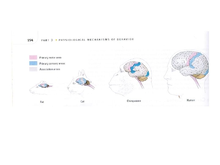

Cerebral cortex • Definition: Outer layer of the cerebrum; contains 70% of neurons in CNS • Cerebrum: Two large hemispheres that cover upper part of the brain • Corticalization: Increase in size and wrinkling of the cortex • Cerebral Hemispheres: Right and left halves of the cerebrum • Corpus Callosum: Bundle of fibers connecting cerebral hemispheres Copyright © 2005 Pearson Education, Inc. publishing as Benjamin Cummings

. Copyright © 2005 Pearson Education, Inc. publishing as Benjamin Cummings

The Frontal Lobes n Extend from front of brain to skull’s top center n Largest of brain lobes n Contain motor cortex, Broca’s area, & frontal association areas n Motor cortex – Rear of frontal lobes – Controls voluntary body movement – Participates in learning and cognitive events n Broca’s area – Control’s production of speech sounds n Broca’s asphasia – Physical inability to create speech or speech sounds – Caused by damage to Broca’s area n Frontal association areas – Thinking, motivation, planning, impulse control, personality (Phineas Gage) Copyright © 2011 Pearson Education, Inc. All Copyrights © 2010 reserved. by Worth Publishers

Fig. 48. 25 Copyright © 2002 Pearson Education, Inc. , publishing as Benjamin Cummings

The Temporal Lobes Involved in the reception and interpretation of auditory information n The primary auditory cortex – The part of each temporal lobe where hearing registers in the cerebral cortex – Wernicke’s area--Language area of the left temporal cortex – Comprehends spoken word, formulates coherent speech, and written language n Wernicke’s aphasia – Asphasia caused by damage to Wernicke’s area. – Speech is fluent and clearly articulated but doesn’t make sense to listeners Copyright © 2011 Pearson Education, Inc. All rights reserved.

The Occipital Lobes n The lobes that are involved in the reception and interpretation of visual information; they contain the primary visual cortex n Primary visual cortex – The area at the rear of the occipital lobes where vision registers in the cerebral cortex Copyright © 2011 Pearson Education, Inc. All rights reserved.

The Cerebral Hemispheres n Lateralization – Functional specialization of one of the cerebral hemispheres n Left hemisphere – Controls: n Right side of body n Most functions of speech and written language – Coordinates complex movements n Right hemisphere – Controls left side of body – Specialized for: n Visual-spatial perception n Interpreting nonverbal behavior – Right hemisphere damage can cause: n Attentional deficits n Inability to view objects in the left visual field Copyright © 2011 Pearson Education, Inc. All rights reserved. Copyright © 2010 by Worth Publishers

Right Brain/Left Brain • Left hemisphere better at math, judging time and rhythm, and coordinating order of complex movements – Processes information sequentially and is involved with analysis • Right hemisphere good at perceptual skills, and at expressing and detecting other’s emotions – Processes information simultaneously and holistically Copyright © 2010 by Worth Publishers

Copyright © 2005 Pearson Education, Inc. publishing as Benjamin Cummings

Fig. 2. 21 The left and right brain have different information processing styles. The right brain gets the big pattern; the left focuses on small details. Copyright © 2010 by Worth Publishers

Split Brain Research • Severed corpus callosum Copyright © 2010 by Worth Publishers
: Computer-enhanced X-ray image of the brain")
Researching the Brain • Computed Tomographic Scanning (CT): Computer-enhanced X-ray image of the brain or body • Magnetic Resonance Imaging (MRI): Uses a strong magnetic field, not an X-ray, to produce an image • Functional MRI (f. MRI): MRI that also records brain activity • Positron Emission Tomography (PET): Computer-generated color image of brain activity, based on glucose consumption in the brain

Plasticity of the brain • Anatomic structure and functional organization of the brain is more flexible or “plastic” than widely assumes. • Research has shown that: – Experience can cause structural changes in the brain. – Damage to brain tissue can lead to neural reorganization. – Plasticity diminishes with age Copyright © 2010 by Worth Publishers

Endocrine System n A system of ductless glands that manufacture hormones and secrete them into the blood stream to affect other parts of the body. n Hormone – Chemical made and secreted in one part and affects another part of the body. n Pituitary gland – “the master gland” – Controls growth hormone and activates other endocrine glands n Adrenal glands – Hormones that prepare body for emergencies and stress also releases corticoids and some sex hormones Copyright © 2011 Pearson Education, Inc. All Copyrights © 2010 reserved. by Worth Publishers

Figure 2. 27 Copyright © 2010 by Worth Publishers
- Slides: 52