Biology 323 Human Anatomy for Biology Majors Lecture

Biology 323 Human Anatomy for Biology Majors Lecture 13 Dr. Stuart S. Sumida Gut Tube: Development, Structure, Function
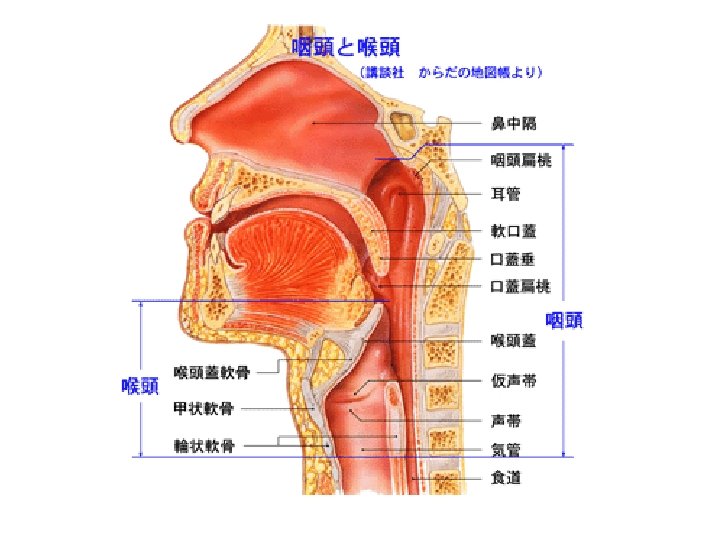

1. Implications of Gut Development Foregut Development Midgut Development Hindgut Development 2. Circulation – Part I

Dorsal vs. Ventral Mesentery Components
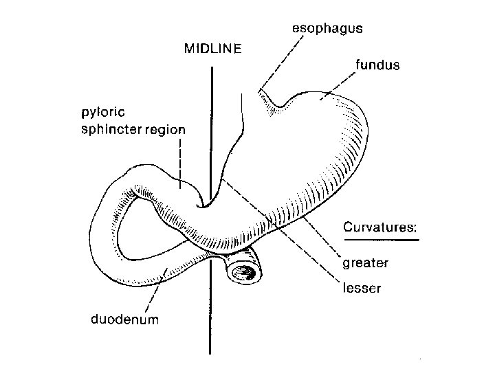

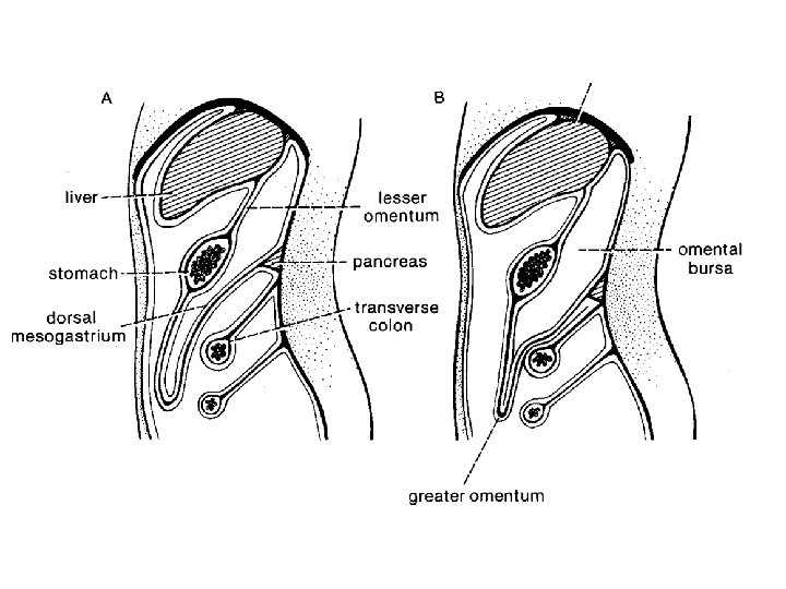
Early Development of the Stomach

Continued Development of the Stomach

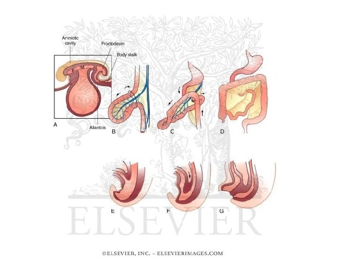
Rotation of the Foregut

Development of diverticula of the foregut

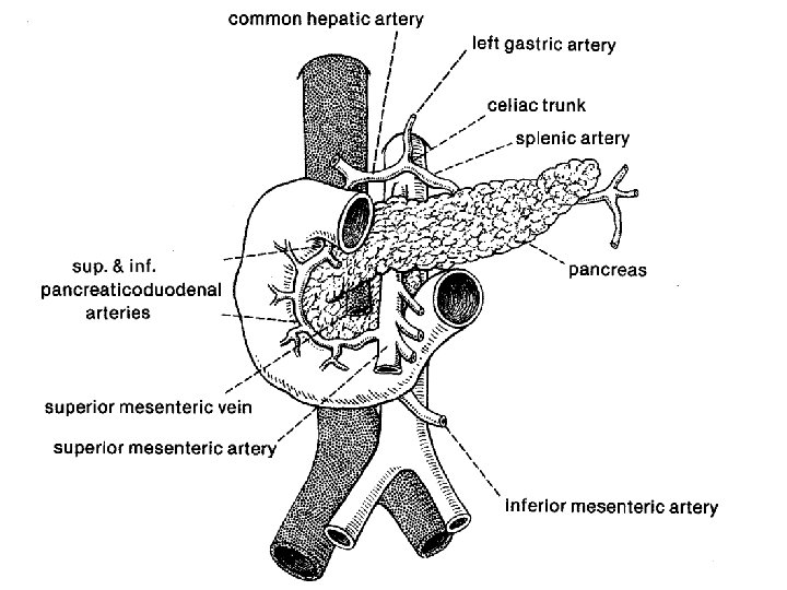
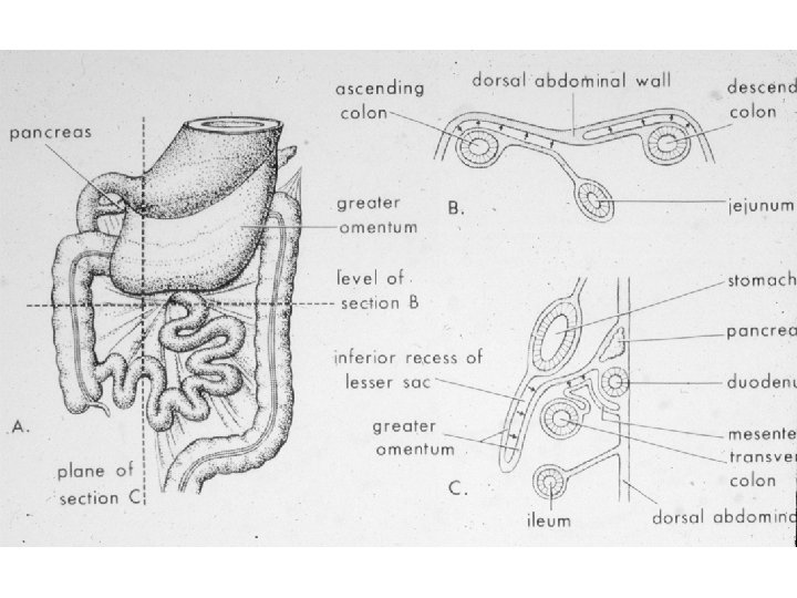
Pancreas Development

Development of Diverticula of Foregut • Hepatic Diverticulum • Pancreas originally two separate lobes (ventral part of hepatic diverticulum, dorsal independent. • Rotation brings dorsal (tail) and ventral components of pancreas together.

Development of Diverticula of Foregut • Hepatic Diverticulum • Pancreas originally two separate lobes (ventral part of hepatic diverticulum, dorsal independent. • Rotation brings dorsal (tail) and ventral components of pancreas together.

Development of Diverticula of Foregut • Hepatic Diverticulum • Pancreas originally two separate lobes (ventral part of hepatic diverticulum, dorsal independent. • Rotation brings dorsal (tail) and ventral components of pancreas together.

Development of Diverticula of Foregut • Hepatic Diverticulum • Pancreas originally two separate lobes (ventral part of hepatic diverticulum, dorsal independent. • Rotation brings dorsal (tail) and ventral components of pancreas together.


Hepatic Portal Vein Bile Duct

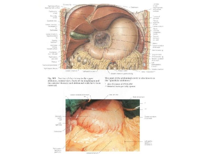
Liver Stomach
")
Liver: Dorsal View Ventral View Ligamentum teres (= old ductus venosus)

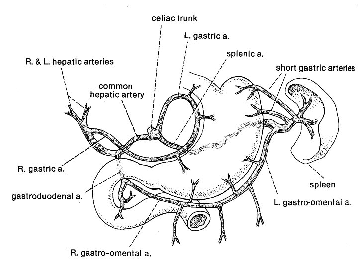
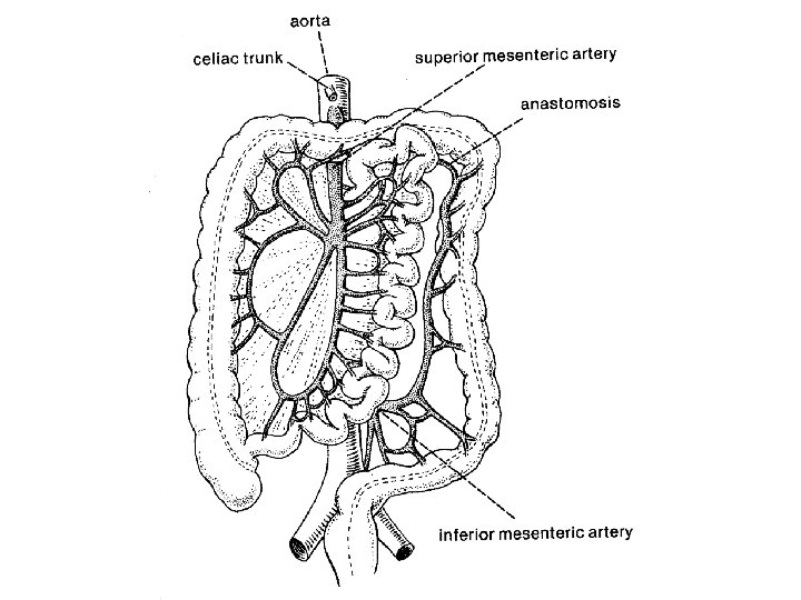
Foregut - Neurovascular Service: Unpaired Branch of Abdominal Aorta: Celiac Artery Unpaired Tributary of Hepatic Portal Vein: Splenic Vein Sympathetic Nerve: Greater Splanchnic Nerve Sympathetic Nerve Segmental Levels: T 5 -9 Sympathetic Ganglion: Celiac Ganglion Parasympathetic Nerve: Vagus Nerve (CN – X)


1. Implications of Gut Development Foregut Development Midgut Development Hindgut Development 2. Circulation – Part I

Dorsal vs. Ventral Mesentery Components

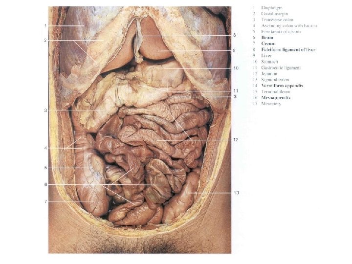
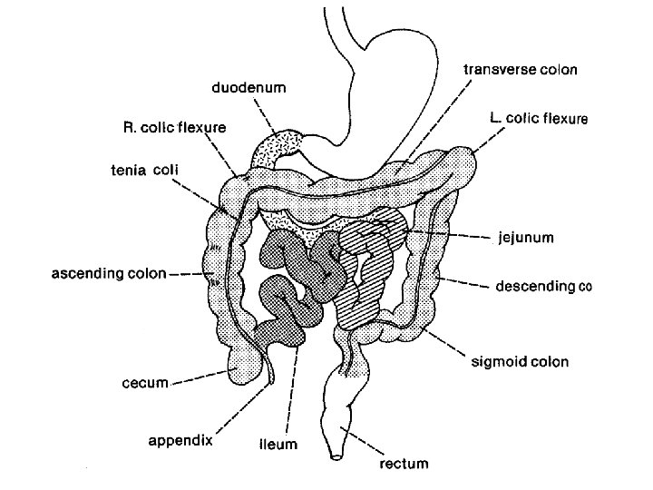
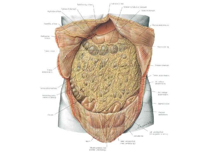
“Repackaging” of the Midgut
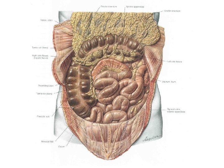

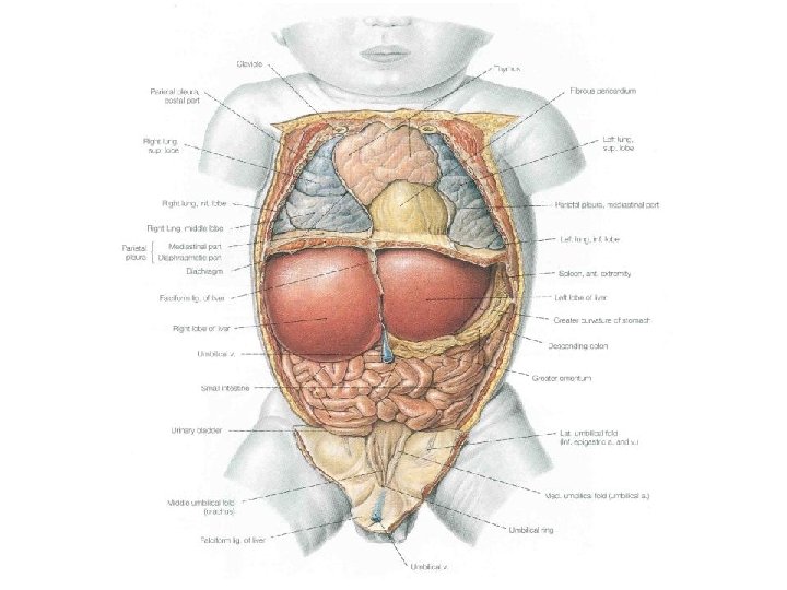
Retroperitoneal components of abdominal cavity

Midgut - Neurovascular Service: Unpaired Branch of Abdominal Aorta: Superior Mesenteric Artery Unpaired Tributary of Hepatic Portal Vein: Superior Mesenteric Vein Sympathetic Nerve: Lesser Splanchnic Nerve Sympathetic Nerve Segmental Levels: T 10 -11 Sympathetic Ganglion: Superior Mesenteric Ganglion Parasympathetic Nerve: Vagus Nerve (CN – X)

1. Implications of Gut Development Foregut Development Midgut Development Hindgut Development 2. Circulation – Part I


Review of Retroperitoneal components of abdominal cavity

Innervation of Bladder Sympathetic: predominantly L 1 -2 via hypogastric plexus Parasympathetic: S 2 -4 (as you would expect of a hindgut derivative).

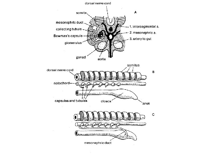
Developing

Hepatic Portal Vein and Its Tributaries

Sympathetic Supply of the Abdominal Gut
")
Parasympathetic Supply to the Hindgut: S 2 -4)

Hindgut - Neurovascular Service: Unpaired Branch of Abdominal Aorta: Inferior Mesenteric Artery Unpaired Tributary of Hepatic Portal Vein: Inferior Mesenteric Vein Sympathetic Nerve: Least Splanchnic Nerve + Lumbar splanchnics Sympathetic Nerve Segmental Levels: T 12, L 1(2) Sympathetic Ganglion: Superior Mesenteric Ganglion Parasympathetic Nerve: Pelvic Outflow, S 2 -4

Foregut, Midgut, & Hindgut Summary Boundaries Sympathetic nerve Levels Foregut Through first ½ of duodenum Greater Splanchnic T 5 -9 Midgut 2 nd ½ of duodenum through left colic flexure Lesser Splanchnic T 10 -11 Hindgut Left colic flexure to anus Least Splanchnic + Lumbar splanchnics T 12, L 1(2) Embryonic Gut

Summary Continued…. . Embryonic Preaortic gut ganglia Arteries of Abdominal aorta Hepatic portal vein Parasymp. innervation Foregut Celiac ganglion Celiac artery Splenic vein Vagus Nerve (X) Midgut Superior mesenteric ganglion Superior mesenteric artery Superior mesenteric vein Vagus Nerve (X) Hindgut Inferior mesenteric ganglion Inferior mesenteric artery Inferior mesenteric vein Sacral Outflow (S 1 -S 4)
- Slides: 52