Biology 30 Cell Division Where is DNA found

Biology 30 Cell Division

Where is DNA found? What do you think? • Put these terms in order from biggest to smallest – Gene -DNA – Organism -Cell – Chromosome -Nucleus

Where is DNA found?

Terminology Sister chromatids • Chromosome =___________ Telomere • Chromatin = DNA that is unwound (not compressed into a Centromere chromosome); found - where chromatids when cell is not dividingjoin together • ________= a chromosome duplicates to make 2 identical copies (DNA replication)

Chromosomes, genes and DNA • Genes are DNA sequences located on ________ Strand 1: A T G C C G A A Strand 2: T A C G G C T T

Chromosomes, Genes and DNA • Chromosomes are just tightly wound chromatin • Only visible during mitosis • Otherwise, exist as a stringy mass called _____ • __________________

Label diagram on pg. 3 DNA + Histones = Chromatin

Human DNA Fun Facts: - Human DNA is 3 billion base pairs and about 2 m in length. -50% of the genes found in humans are also found in bananas -We have 65% genetic similarity to fruit flies

Why do cells divide? • Cells grow because proteins and organelles accumulate in the cytoplasm • Cells divide because the ______ of the cell membrane becomes inadequate (volume vs. surface ratio) • Cells die and need to be replaced • In order to reproduce the cell must pass on it’s genetic information

What happens next? Our cells must be able to divide. There are 2 types of cell division: 1. Asexual cell division – occurs in somatic (body) cell division = Mitosis 2. Sexual cell division – occurs in sex cells (germ cells) = Meiosis

Asexual Reproduction • The cells in our bodies started from a single cell called a ____ (think back to reproduction) • We constantly replace old/dying cells and are able to heal wounds, thanks to cell division

The Cell Cycle has 4 major stages _____________________: - Cell spends 90% of time in Interphase - prepares cell for division; cell grows and makes organelles - DNA is in the form of chromatin - DNA replication occurs
 –growth phase")
Cell Cycle - Interphase has 3 parts 1. G 1 (Gap 1) –growth phase where cells grow and new organelles are made, _____________ 2. S (Synthesis) – genetic material in the cell doubles (DNA replication); therefore _______________________ 3. G 2 (Gap 2) – growth and preparation for division

The Cell Cycle: Summary • • • G 1, S and G 2 make up ______ This is the process of cell activity between cell division Cells grow and prepare for division in this stage ~ 90% of the cell cycle is interphase _____ (M) is the process by which the cell divides ~ 10% of the cell cycle is mitosis

Asexual Reproduction aka MITOSIS! • In asexual reproduction, cell division results in 2 identical “_____” cells being produced from a “parent” parent cell • Each human cell has ___ chromosomes in its nucleus

Cell Cycle - Mitosis • 10% of cell life cycle • The cell undergoes cell division In humans, millions of cells divide every second to maintain a total of ~60 trillion cells - some divide once a day (skin & hair), others less often (stomach lining) and some not at all (nerve & muscle cells)

Stages of Mitosis is a continual process, but we divide it into 4 phases Mitosis phases: 1. Prophase 2. Metaphase 3. Anaphase 4. Telophase

MITOSIS

Mitosis 1. _______ - chromatin condenses into distinct duplicated chromosomes - Nuclear membrane begins to disintegrate - In animal cells organelles called the _____ move to opposite sides of the cell (“poles”) - Astral rays (microtubles) form around centrioles

Be sure to label: Draw a prophase diagram 1. Sister chromatids/ chromosomes 2. Centrioles 3. Astral rays Centrioles 4. Nuclear memebrane Astral Rays Made of 2 sister chromatids, attached by a centromere Nuclear membrane disintegrates

Early Prophase

Late Prophase

Mitosis Step 2: Metaphase - Chromosomes line up at _________ and centromere attaches to spindle fibers that formed from elongated astral rays - At the end of this phase the centromere splits separating the sister chromatids - Nuclear membrane disappears

Draw a diagram Be sure to label: 1. Centromere 2. Equatorial plate Centromere 3. Spindle fibers 4. Centriole Spindle Fibres Centrioles Equatorial Plate

METAPHASE

Metaphase: Spindle Fibers
")
Mitosis Step 3: ______ - The spindle fibers contract, pulling the chromosomes (sister chromatids) to the opposite poles of the cell - Centromeres divide

Draw a diagram Be sure to include: 1. 2. 3. 4. Sister chromatids Spindle fibres Centromere Centrioles Centromere is split

ANAPHASE

Mitosis Step 4: _______ - Chromatids reach opposite poles; spindle and astral rays disappear Chromosomes unwind back into chromatin Nuclear membrane begins to reform Cell membrane pinches in the middle to divide the cell = _________

Cytokinesis • Cytoplasm begins to divide by forming a cleavage furrow at the equator and pinches off • Forming 2 daughter cells, with genetic information identical to each other • These cells will become the new parent cells • Cytokinesis in an animal cell Cleavage furrow

Draw a diagram • Be sure to liable: Part A: • Cleavage furrow Daughter cells • Nuclear membrane Part B: • Daughter cells • Nuclear membrane Nuclear • Chromatin membrane Chromatin

TELOPHASE

INTERPHASE PROPHASE

METAPHASE ANAPHASE Metaphase plate Spindle Daughter chromosomes TELOPHASE & CYTOKINESIS Cleavage furrow Nuclear envelope forming Nucleolus forming

Interphase Metaphase Prophase Anaphase Metaphase Telophase

Animals Versus Plants • There are 2 main differences in plant cell division 1. Plants do not contain centrioles - ________________________(_____), they just don’t have the centrioles 2. Plants do not undergo cytokinesis - __________________________

Cytokinesis • Animal Cytokinesis Cleavage furrow Cell Plate Plant

Mitosis

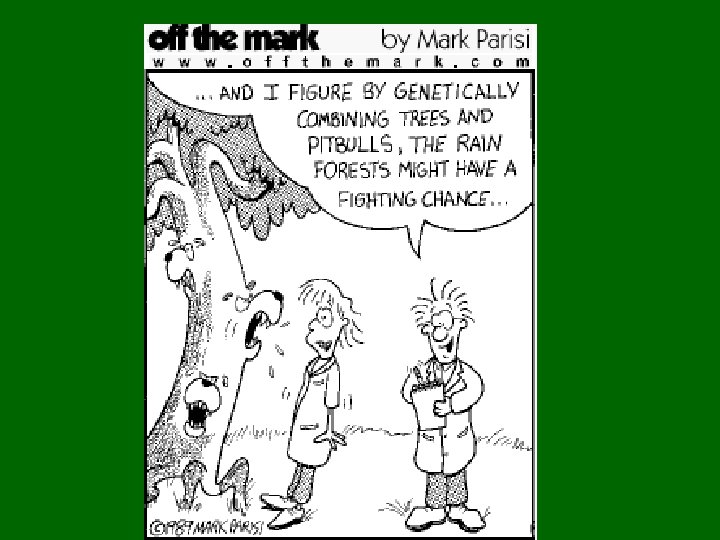
Biotechnology Genetic Engineering

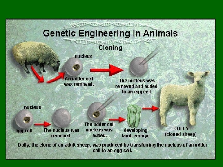
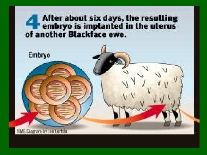
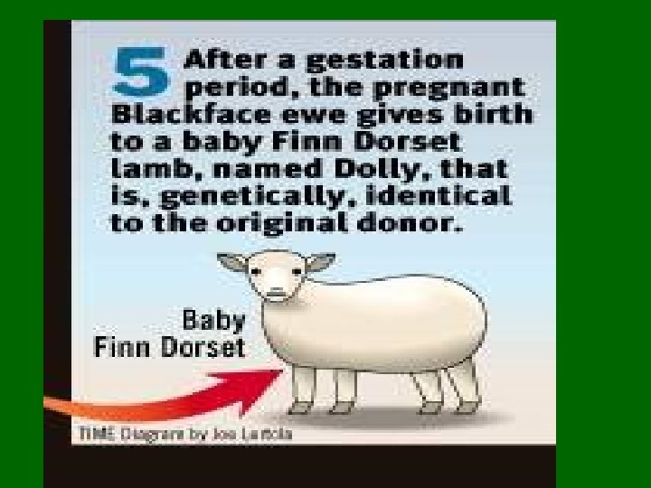
Cloning • Cloning is the process in which identical offspring are formed from a single cell or tissue • Many plants have the ability to naturally clone themselves; this is called ______ (i. e. strawberries, aspen trees) • Most animal cells are not totipotent (except salmanders) but technology has advanced to allow us to clone (i. e. Dolly the sheep)

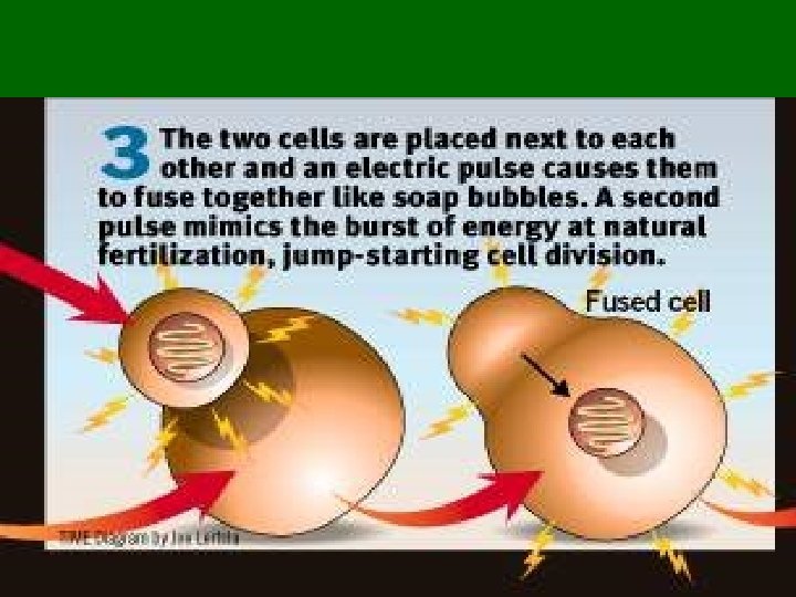
Cloning • There a few ways that cloning can occur 1. Nuclear transfer from blastula 2. Nuclear transfer with electrical or chemical shock 3. Identical twins

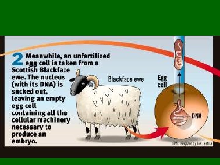
Nuclear transfer from blastula The nucleus of a cell from a blastula is removed and transplanted into an enucleated unfertilized egg Enucleated = cells that does not contain a nucleus - This egg begins to divide by Removing the nucleus from a mitosis, eventually forming an cell organism

Nuclear transfer from blastula Mitosis Remove Nucleus Fertilized Blastula Egg Stage Unfertilize Remove d Egg Nucleus Enucleate d Cell with transplanted nucleus begins to divide by mitosis

Why clone from a blastocyst? • Tried at gastrula stage but when they transplanted the nucleus the cell did not divide = no longer totipotent • Nuclear material must now be fixed and cells would have already started to differentiate

Nuclear transfer with electrical shock

Dolly and Telomeres • If cells divided without telomeres, they would lose the ends of their chromosomes, and the necessary information they contain. • The telomeres are disposable buffers blocking the ends of the chromosomes, are consumed during cell division, and are replenished by an enzyme, telomerase reverse transcriptase.

Identical Twins • After an egg has been fertilized, the newly formed zygote will divide and redivide through successive mitotic cycles to form the multicellular embryo • If a cell should break free from the forming embryo early in this process (prior to differentiation), it will develop into a second embryo having identical genetic information Are they clones?

• If both embryo successfully implant into the endometrium, they will both develop into fetuses, and eventually identical twins

Abnormal Cell Division: Cancer • The growth, differentiation and replication of cells is highly controlled • Uncontrolled cell division occurs when cells divide and they do not have a mechanism to stop • This results in a _____
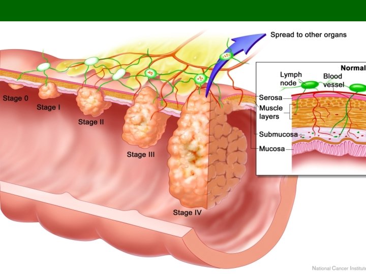
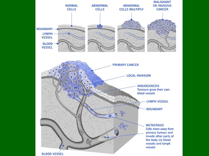

Cancer: Tumors • A benign tumor is a tumor that stays in one place, and often eventually stops dividing • A malignant tumor is a tumor that can metastasize (spread) • http: //www. pbs. org/wgbh/ nova/cancer/program. ht ml
 can")
Carcinogens • Carcinogenic or cancer-causing agents (x-rays, UV light, chemicals and some viruses) can cause mutations in the chromosomes that affect the replication process • Mutations can also occur on their own
 somatic cells without telomerase gradually lose telomeric sequences")
From Wikipedia… • Human (and other) somatic cells without telomerase gradually lose telomeric sequences as a result of incomplete replication (Counter et al. , 1992). • As human telomeres shorten, eventually cells reach their replicative limit and progress into senescence or old age. • Senescence involves p 53 and p. Rb pathways and leads to the halting of cell proliferation (Campisi, 2005). • Senescence may play an important role in suppression of cancer emergence, although inheriting shorter telomeres probably does not protect against cancer. [13] With critically shortened telomeres, further cell proliferation can be achieved by inactivation of p 53 and p. Rb pathways.

Progeria

Meiosis Sexual Cell Reproduction

Sexual Cell Reproduction • All living cells arise from pre-existing cells, tracing the lineage of every living things back to some primordial ancestor. If mitosis were the only process involved the production of new cells, then all cells would be exactly the same. • Instead, there exists an incredible variety among organisms

Gametes and the Life Cycle of a Sexual Organism Egg cell • The life cycle of a multicellular organism is the sequence of stages leading from the adults of one generation to the adults of the next Sperm cell Meiosis Fertilization Multicellular diploid adults (2 n = 46) Diploid zygote (2 n = 46) Mitosis and development Figure 8. 13

DNA Sequence • It is the sequence of the bases in a gene that holds the genetic code • And therefore holds the code for all cellular processes and the variety of inherited traits we see in organisms

Meiosis The cellular bases for sexual reproduction -the mixing of genetic traits produces offspring different from each parent - Fertilization requires 2 _______ (egg and sperm) -These gametes contain ½ the genetic information - Each sperm or egg produced carries 1 of over 8 million possible combinations of parental 2 n 46 chrom 2 n 2 n (46) n n 23 23

Meiosis versus Mitosis 3 Important Differences: 1. Takes place in 2 stages involving 2 successive divisions 2. The chromosomes arrange themselves in ________ (pair up with another chromosome of the same size and shape) 3. Therefore, the four daughter cells are not necessarily identical and have only ___________ present in the original parent cell.

Meiosis • The production of gametes with ______ the number of chromosomes as the original “parent” cell • During this process specialized cells in the gonads (ovary & testes), produce sex cells that contain only one set of chromosomes. • A human ______ with 46 chromosomes will undergo meiosis and produce gametes that have 23 chromosomes

Meiosis • The 46 chromosomes number is referred to as _____ and is written as ____ • The 23 chromosomes number is referred to as _____ and is written as ____ • Fertilization occurs when 2 gametes (_____) fuse, forming a _______ zygote (_____ chromosomes) Sperm Egg 23 Chromosomes (haploid) 23 chromosomes (haploid) Zygote 46 chromosomes (diploid)

Each chromosome has a partner • Keep in mind that the 23 chromosomes are not just any 23, but one member from each pair. • Each of the 23 chromosomes that you receive from your father is matched by 23 chromosomes from your mother • Example: Your father gives you a chromosome with genes that code for eye colour and so does your mother

Homologous Chromosomes • The paired chromosomes are called _______________ • Homologus Pairs = a pair of chromosomes that have similar lengths, shapes and carry genes controlling the same traits • However each chromosome does not necessarily carry the same genetic information For example: both chromosomes in a homologous pair may carry a gene for eye colour but one may code for blue and the other for brown eyes • Each cell produced during meiosis contains one member from each pair of homologous After DNA replication chromosomes

Phases of Meiosis • • • Meiosis I Interphase Prophase I Metaphase I Anaphase I Telophase I • • Meiosis II Prophase II Metaphase II Anaphase II Telophase II

Meiosis • Meiosis involves 2 cell divisions that produces 4 haploid cells • To keep things simple, in our example we will use cells that contain 2 n=4 chromosomes. • Therefore, the cell will contain two pairs of homologous chromosomes.
, must occur before")
Interphase • As in mitosis, interphase (cell growth and DNA replication), must occur before cell can replicate • Very important = ________________________________ Chromatin Centriole s

Interphase: DNA replication of homologous pairs Replication

1. Chromatin coils tightly to form chromosomes - DNA has already replicated Prophase 1 2. Homologous chromosomes pair up side by side in a process called _______. - When 2 homologous chromosomes are paired, the structure is called a ______ ( = ________) Paternal Homolog Maternal homolog
 pair up to form")
3. Chromosomes shorten & thicken - the homologous chromosomes (bivalents) pair up to form tetrads. • ___________________ Tetrad Prophase 1

Crossing Over • As the homologous chromosomes come close together, they often intertwine • Sometimes chromatids break & exchange segments. This process is called ____________ The area(s) where the chromatids overlap is called a ______ Here homologous chromosomes exchange genetic material

Crossing Over This leads to enormous genetic variation even between siblings • There are over 8 million possible combinations of parental chromosomes 5. The chiasma begins to disappear The nuclear membrane disintegrates **In females this is where meiosis stops until puberty (Prophase I)

Prophase I : 2 n = 6 or n=3 • Draw a germ cell in prophase I Homologous • Label: pair 1. Homologous chromosomes & tetrad 2. Nuclear membrane 3. Centrioles, astral rays Tetrad

Metaphase I • The tetrads line up on the _________ Metaphase plate • The homologous pairs line up randomly • There is a 50 -50 chance for the daughter cells to get either pair from each chromosome. You could get this one or that one Centriole

• Spindle fibres attach to the centromeres of the chromosomes • Each pair of sister chromatids from each homologous chromosome is ready to move to opposite poles of the cell Draw a diagram

Anaphase I • Homologous chromosomes separate and move to opposite poles • This process is known as _______ – the separation of paired genes • At this point, reduction division has occurred! • Each chromosome remains double stranded

Draw a diagram: 2 n = 6, n=3

Telophase I • Cytoplasm divides = Cytokinesis • A nuclear membrane begins to form • Chromosomes unwind into chromatin • The two new daughter cells contain 2 chromosomes each. Two haploid cells (cells with half the chromosome number) start to form. Chromosomes are half the number, but still double stranded.

Draw a diagram • Label the following: 1. Cleavage furrow 2. Daughter cells 3. Nuclear membrane 4. Chromatin

Meiosis I versus Mitosis • The first division is different from mitosis because the daughter cells are not identical • Each daughter cell contains 1 member of the chromosome pair • Remember homologous pairs are similar but they are not identical

Meiosis II ****The short phase between Meiosis I and II is referred to as interkinesis There may or may not be an interphase II depending on species • The next set of cell • Begin with the 2 daughter cells divisions will separate the from meiosis I chromatids Prophase II • DNA replication does ___ occur • Nuclear membrane dissolves, spindle fibres form

Draw a diagram

Metaphase II • The chromosomes , each with two sister chromatids, align on the equatorial plate • The centromeres attach to spindle fibres

Anaphase II • The centromeres separate and chromatids move towards opposite poles

Telophase II • 4 new cells will be formed Each of the new cells will contain only one member from each homologous pair = haploid (n) • The parent cell had 6 chromosomes; the daughter cells have 3 chromosomes each

Meiosis Summary • The haploid cells complete the meiotic cycle and differentiate to produce gametes (egg & sperm) Important to remember: -Meiosis I is referred to as _________ because the chromosome number is reduced by half - Meiosis II is called __________and is similar to mitosis as centromeres on sister chromatids separate and chromosome number remains unchanged
 Nuclear envelope")
Meiosis I MEIOSIS I: Homologous chromosomes separate INTERPHASE Centrosomes (with centriole pairs) Nuclear envelope PROPHASE I Sites of crossing over Spindle Chromatin Sister chromatids Tetrad METAPHASE I Metaphase plate ANAPHASE I Sister chromatids remain attached Homologous chromosomes separate

Meiosis II MEIOSIS II: Sister chromatids separate TELOPHASE I & CYTOKINESIS PROPHASE II METAPHASE II ANAPHASE II Sister chromatids separate TELOPHASE II & CYTOKINESIS Haploid daughter cells forming

Mitosis versus Meiosis Mitosis - the division of 1 cell into _ daughter cells to produce ___ (egg or sperm) ________ containing ½ the daughter cells genetic material - diploid (2 n) = contains 2 copies of - haploid (n) = contains 1 copy of the genome 2 n 2 n genome 46 chrom 2 n 2 n (46) n n (23) n n 23 23

Development of male and female gametes The Formation of sex cells during meiosis is referred to as _________ Sperm and egg production are different: • In males 4 viable sperm are produced Spermatogenesis • In females 3 of the cells produce are known as polar bodies and do not survive. Only one egg is formed Oogenesis

Differences • The cyctoplasm of the female oocyte does not divide equally; one of the daughter cells called an ootid, receives most of the cyctoplasm while the other cells called _____(“nurse cells”) die and are reabsorbed to provide nutrients • Sperms show equal division of cytoplasm; all 4 daughter cells become viable sperm

Spermatogenesis • Occurs in the __________ where ____ spermatogonia, ____ that are the precursors of sperm. • Spermatogonia divide by _______ to produce more spermatogonia ___ differentiate into spermatocytes

Spermatogenesis • _____ of each spermatocyte produces 4 _______ spermatids. This process takes over three weeks to complete • Then the spermatids differentiate into sperm, losing most of their cytoplasm in the process.

Sketch Spermatogeneis showing meiotic divisions 46 1. Spermatogonium 2. 1° Spermatocyte 3. 2° Spermatocyte 4. Spermatid 5. Sperm 1 st Meiotic division 46 23 23 2 nd Meiotic division 23 23

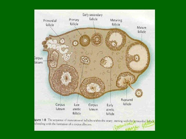
Oogenesis • Egg formation takes place in the _______ • the initial steps in egg production occur prior to birth. • Diploid stem cells called oogonia divide by mitosis to produce more oogonia and primary oocytes
 2° Oocyte")
Sketch Oogeneis showing meiotic divisions 46 1. Oogonium 2. Oocyte 3. a) 2° Oocyte b) 1 st polar body 4. a) Ootid b) polar bodies 5. Ovum 46 1 st Meiotic division 2 nd Meiotic division 23 23

• By the time the fetus is 20 weeks old, the process reaches its peak and all the oocytes that she will ever possess (~4 million of them) have been formed. • By the time she is born, 1– 2 million of these remain. Each has begun the first steps of the first meiotic division (______) and then stopped.

• No further development occurs until years later when the female becomes sexually mature. • Then the primary oocytes recommence their development, usually one at a time and once a month. Unfertilized oocyte

• The primary oocyte grows much larger and completes the _____, forming a large secondary oocyte and a small polar body that receives little more than one set of chromosomes. Which chromosomes end up in the egg and which in the polar body is entirely a matter of chance.

Meiosis: Male and Female Differences

• In humans, 22 of the 23 are homologous; these are the ______ • Thomas Hunt Morgan discovered that having 2 rod shaped (X chromosome) indicated female and 1 rod with 1 hooked shaped chromosome (Y chromosome) identified males • The X and Y are not homologous, they are the ________

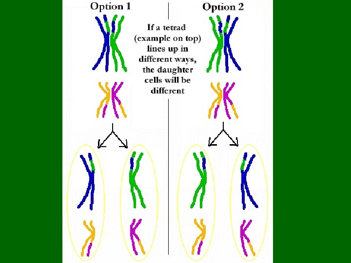
Meiosis and Variation • Unlike mitosis, meiosis does ____ produce identical cells • The cells produced only have half the number but the chromosomes therefore only ½ the genetic information • What chromosomes end up in what cell all depend upon how the chromosomes line up in ________

Meiosis and Variation • If the two blue chromosomes line up on the same side and the two red chromosomes line up on the same side • Then the daughter cells will have either the genetic information from the red ___ blue chromosome

Meiosis and Variation • If 1 red and 1 blue line up on the same side • The daughter cells will have genetic information from both red and blue chromosomes
 MEIOSIS Site of crossing over MEIOSIS I PROPHASE")
MITOSIS PARENT CELL (before chromosome replication) MEIOSIS Site of crossing over MEIOSIS I PROPHASE I Tetrad formed by synapsis of homologous chromosomes PROPHASE Chromosome replication Duplicated chromosome (two sister chromatids) 2 n = 4 Chromosomes align at the metaphase plate METAPHASE ANAPHASE TELOPHASE Sister chromatids separate during anaphase 2 n Chromosome replication 2 n Daughter cells of mitosis Tetrads align at the metaphase plate Homologous chromosomes separate during anaphase I; sister chromatids remain together No further chromosomal replication; sister chromatids separate during anaphase II METAPHASE I ANAPHASE I TELOPHASE I Haploid n=2 Daughter cells of meiosis I n n MEIOSIS II n n Daughter cells of meiosis II

Mistakes during Meiosis The movement of the chromosomes in a dividing cell is so precise that only 1 in every 100, 000 divisions will contain an error ___________ = ____________________________________
 •")
Non-disjunction • One daughter cell will be missing one of the chromosomes (___) • Other daughter cell will contain an extra chromosome (___) • In humans, nondisjunction produces gametes with _____ chromosomes

Non-disjunctions • When gamete with 24 chromosomes joins a normal gamete with ___ chromosomes, the zygote will contain ___ (instead of 46). This zygote will have 3 chromosomes in place of the normal pair = _____ • When the gamete with 22 chromosomes joins a normal gamete with ___ chromosomes, the zygote has ___; this zygote will have 1 chromosome in place of the normal pair = _______

KARYOTYPES • The best way to study non-disjunctions is by looking at karyotypes • Karyotypes are an inventory of an individuals chromosomes – A karyotype usually shows 22 pairs of autosomes and one pair of sex chromosomes

Preparation of a karyotype Blood culture Packed red And white blood cells Hypotonic solution Stain White Blood cells Centrifuge 3 2 1 Fixative Fluid Centromere Sister chromatids Pair of homologous chromosomes 4 5 Figure 8. 19

Human female chromosomes Hillaby - Biology 30 - 2009 124

Human female karyotype Hillaby - Biology 30 - 2009 125

Human male chromosomes Hillaby - Biology 30 - 2009 126

Human male karyotype Hillaby - Biology 30 - 2009 127

Hillaby - Biology 30 - 2009 128

129

Hillaby - Biology 30 - 2009 130

Non-disjunctions A number of disorders are caused by nondisjunctions 1. Down’s Syndrome 2. Edward’s Syndrome 3. Patau’s Syndrome 4. Turner’s Syndrome 5. Kleinfelter’s Syndrome 6. Other severe abnormalities

Non-disjunctions The risk of chromosomal abnormalities increases with maternal age because the egg cells are older Women over the age of 35, who have children increase their chance exponentially

Karyotype for Down’s Syndrome Trisomy 21

Down’s Syndrome Trisomy 21 Cause: - Non-disjunction of chromosome #21 - Individual has ___ chromosomes Karyotype: - has 3 chromosome #21 Symptoms: - mentally & physically delayed, large forehead, large space between eyes

What does this karyotype tell us?
 Cause: - non-disjunction of chromosome # 13 Karyotype: - has 3")
Patau’s Syndrome (trisomy) Cause: - non-disjunction of chromosome # 13 Karyotype: - has 3 chromosome #13 Symptoms: - child with multiple and severe abnormalities, and severe mental retardation - head very small, eyes absent or very small - hairlip, cleft palate, usually malformations of internal organs - most cases child dies soon after birth

What does this karyotype tell us?

Edward’s Syndrome: Trisomy 18 Cause: - Non-disjunction of chromosome #18 Karyotype: -has 3 of chromosome #18 Symptoms: -very small and weak, head flattened, hands short with very little development - severe mental handicap with life expectancy of less than one year

What does this Karyotype tell us?
 Cause: - Non-disjunction sex chromosomes Karyotype: - 2 X chromosomes")
Klinefelter’s Syndrome (Trisomy 23) Cause: - Non-disjunction sex chromosomes Karyotype: - 2 X chromosomes and a Y chromosome (XXY) Symptoms: - Appears male at birth but upon puberty releases high levels of female hormones - Sterile males, breast development, slightly feminized physique - Affects 1/1000 male births

What does this karyotype tell us?
 Cause: - Non-disjunction of X chromosome in females Karyotype: - females")
Turner Syndrome (Monosomy) Cause: - Non-disjunction of X chromosome in females Karyotype: - females have a single __ chromosome (XO) Symptoms: - sterile females, short stature, underdeveloped gonadal structures - Affects 1/5000 female births

Non-disjunction in Males XX XY O XY XXY Klinefelter X X XO Turner

Non-disjunction in females XX XY X Y XXY Klinefelter O XX XO Turner

Other possible abnormalites caused by non-disjunction -these infants do not live very long – a few days at the most; many are premature and stillborns -These photos may disturb you, you are not required to watch
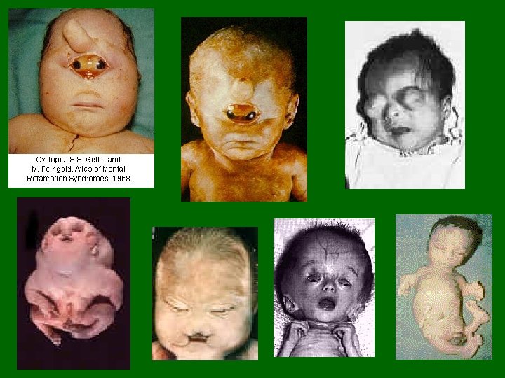

• Teratogenic compounds • Teratogen means, in Greek, "monster forming. " Teratogens are chemicals that cause abnormalities in embryos. • Includes: • Drugs • Infectious agents (viruses) • Radiation • The most well-known is thalidomide, a drug originally designed to combat morning sickness in pregnant women. It caused the long bones in the arms or legs of fetuses to not develop properly, resulting in babies with severely stunted arms or legs 147

AMNIOCENTESIS • Ultrasound - locate position of developing fetus within mother's womb • Amniocentesis - use of a syringe to Amniocentesis draw fluid from sac surrounding fetus • Analysis of fluid can identify certain types of disorders • Amniotic fluid contains cells from fetus • Cells are treated with special stains • Chromosomes are visible for microscopic examination • Amniocentesis – used at about the 14 th to 16 th week of pregnancy • Results available after several weeks 148

Hillaby - Biology 30 - 2009 149

• Can detect: • Down syndrome • Gender of fetus • Spina bifida - opening on baby’s spine which exposes spinal cord and causes paralysis (damage to nerve cells) • Hemophilia – blood clotting disorder • Amniocentesis process: • Syringe • Centrifugation of fetal cells • Culture • Karyotype • A camera mounted to microscope takes a picture of chromosomes Hillaby - Biology 30 - 2009 150
 - draws cells from outer membrane (undergoing mitosis)")
• Chorionic villus sampling (CVS) - draws cells from outer membrane (undergoing mitosis) surrounding embryo • Can be performed as early as 8 weeks into pregnancy • Results available after 24 hours • Many moral and social issues arise Hillaby - Biology 30 - 2009 151

Hillaby - Biology 30 - 2009 152
- Slides: 152