Biology 20 Chapter 8 Nutrients and the Digestive

Biology 20: Chapter 8 Nutrients and the Digestive System Nelson Pages 240 - 279
, vitamins, minerals, and nucleic acids")
8. 1 Essential Nutrients v. Proteins, carbohydrates, lipids (fats), vitamins, minerals, and nucleic acids v 3 major nutrient categories: 1. Carbohydrates 2. Lipids 3. Proteins Nutrition with Tim and Moby http: //www. brainpop. com/health /nutrition/
 Water Proteins Vitamins Macronutrients:")
Organic and Inorganic Nutrients Organic Inorganic Carbohydrates Minerals Lipids (fats) Water Proteins Vitamins Macronutrients: needed in large quantities by the body

1. Carbohydrates 1. • Energy nutrients • Plants synthesize carbohydrates • Photosynthesis lig 6 H 2 O + 6 CO 2 -----> C 6 H 12 O 6+ 6 O 2 ht

Carbohydrate Chemistry The most important energy source for the body; produced by plants. Exist as single sugars or chains of many sugar units. Classified by # of sugars they contain. Single sugars contain 1 carbon: 2 hydrogen: 1 oxygen. How many carbons does a triose sugar have? A hexose sugar? Sugars are often identified by the “-ose” suffix
 Monosaccharides v. Are simple sugars (single sugar units). v. Contain 3 -6")
a. ) Monosaccharides v. Are simple sugars (single sugar units). v. Contain 3 -6 carbons. o. Glucose, galactose, fructose In

Glucose Song! https: //www. youtube. com/watch? v=j. Jv. AL-ii. Ln. Q

b. Disaccharides v. Two monosaccharides form a disaccharide o Maltose= Maltose 2 glucose units o Sucrose = 1 glucose and 1 fructose. o Lactose(milk sugar) -1 glucose and 1 galactose.
 – water molecule is extracted from 2")
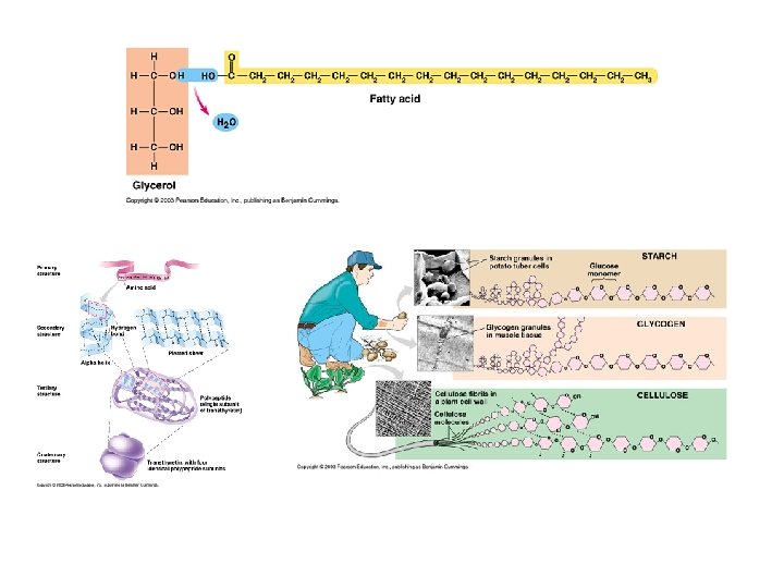
Dehydrolysis Synthesis v. Dehydrolysis synthesis (dehydration synthesis) – water molecule is extracted from 2 monosaccharide sugar molecules, bonds 2 sugars together. Requires energy. (opposite process is hydrolysis). In seeds of germinating plants
 Polysaccharides v. Are carbohydrates formed by more than 2 monosaccharides. v. Formed")
c. ) Polysaccharides v. Are carbohydrates formed by more than 2 monosaccharides. v. Formed by Dehydrolysis synthesis. o Starch p. Many glucose subunits p. A flat structure How plants store energy; made in leaves and stored in roots.
 o. Stored")
Glycogen v. Animals store carbohydrates in the form of glycogen (a polysaccharide) o. Stored in liver and muscles When glucose concentration in blood , glycogen is converted back into monosaccharide glucose units q Ø For energy

Types of Carbohydrates Monosaccharides Disaccharides Polysaccharides 14

Cellulose v. Contains many glucose; not a coiled structure. Exists in flat sheets. v. Cellulose o Cannot be digested by humans “fiber/roughage” o Holds water in large intestine, thus, helps eliminate wastes Structure of cellulose as it occurs in a plant cell wall.
 cannot digest plant cell walls 1 st")
Cows Ruminant animals (e. g. , cows) cannot digest plant cell walls 1 st stomach houses bacteria to help digest cellulose Cow then digests food, chews it again, and then diverts it to 2 nd second stomach

Importance of Carbohydrates food energy for cells, used in cellular respiration disaccharides and polysaccharides must be broken down into monosaccharides before they can be used in cellular respiration excess carbohydrates are stored as glycogen or fat 1 -2% of cell mass is carbohydrate

Tasks to be completed: Complete the practice problems 1 -10 in section 8. 1 Complete the chapter 8. 1 Review Questions 1 -6 on page 253 20

2. Lipids q Vary in chemical composition q Includes fats, oils, waxes, steroids, phospholipids… q Fats and oils are triglycerides; at room temperature, fats are solid(s), and oils are liquid (l). q Formed by dehydration synthesis.
 with 3")
a. Composition of Triglycerides Formed by combining 1 glycerol (3 C alcohol) with 3 fatty acids. Unsaturated: double bonds, more easily broken down (oils), from plant sources, reduce plaque build-up but high polyunsaturated products may cause cancer (breast and colon). Saturated: no double bonds, not easily broken down (fats), from animal sources, solids at room temperature, fats accumulate on arteries.
")
Saturated and Unsaturated fats 1 double bond = monounsaturated (Olive oil and canola oil) multiple double bonds = polyunsaturated

Saturated Fats Monounsaturated Fats Polyunsaturated Fats Beef Milk Olive oil Soybean Corn Pork Butter Canola oil Safflower Sesame Lamb Cheese Almond oil Sunflower Peanut Poultry Yogurt Cottonseed Others Coconut oil Linoleic Acid (Omega-6) Linolenic Acid (Omega-3) Soybean Flaxseed Safflower Soybean Sunflower Rapeseed (canola) Corn Pumpkin Wheat germ Walnut

b. Characteristics of lipids Insoluble in water and are hydrophobic. Lipids have twice the Energy as carbohydrates or proteins Polar end (negative end; replaces a fatty acid) is soluble in water Non-polar end is insoluble; Suited for cell membranes.

c. Waxes v. Long, stable molecules are insoluble in water vwaterproof coating for plant leaves and animal fur/feathers
 Functions of lipids Subcutaneous fat (under the skin) used for body temperature control.")
e) Functions of lipids Subcutaneous fat (under the skin) used for body temperature control. Energy storage. Protective coatings for organisms. Cell-surface recognition. Products include soaps, detergents, hormones, pheromones. Cholesterol: LDL (low density lipoproteins- “bad” ) and HDL (high Density lipoproteins- “good”). HDL break down LDL and carry it to the liver. Absorption of vitamins (A, D, E, K).

Fats and Diet: Saturated Fats are stable, thus stay in the body longer Saturated Fats increase the risk of various types cancer: breast, colon, prostate Saturated Fats contribute to obesity, which is linked to high blood pressure, and adult diabetes

Just for Fun 31

Ahhh more Jokes 32

So What Should I Eat? The current scientific thinking on fat consumption goes something like this: Limit fat intake to about 30 consume. percent of the total calories you Do not try to cut fat intake altogether, because you do need the essential fatty acids. A gram of fat has nine calories, meaning that if you consume 2, 000 calories in a day your total fat intake should hover around (2000 * 30 percent / 9 calories/gram) 67 grams of fat. When consuming fat, try to focus on mono-unsaturated fats like olive oil and canola oil, or on essential fatty acids. When consuming essential fatty acids, try to balance your intake of omega-6 and omega-3 fatty acids. Do that by consuming tuna/salmon/trout or omega-3 oils like flax seed oil.

Assignments to be completed: Read Section 8. 1 in Text - pages 242 -253 Complete the practice problems 1 -10 in section 8. 1 Complete the chapter 8. 1 Review Questions 1 -6 on page 253
 made up of hundreds of amino acids chemically bonded")
3. Proteins vlarge macromolecules (polymers) made up of hundreds of amino acids chemically bonded together v. Sequencing on amino acids is regulated by genes located on your chromosomes. There are 20 different amino acids found in proteins; 8 are essential and must be supplied by your diet. Basic Chemical Structure of amino acids Amino acids differ by the atoms attached at the “R” site

Proteins Essential Amino Acids occur primarily in animal sources. Proteins are essential for building, maintaining and repairing body tissues. Too much protein = Kidney Failure (clogs kidney with wastes). Too little = kwashiorkor (bellies swell because of water retention).

a. Formation of Proteins When proteins are made a water molecule is released, the covalent bond between the acid and amino group is a peptide bond. This occurs in the ribosome. Amino acids are joined using peptide bonds; the order and type of amino acid determines the type of protein. Dehydration synthesis of proteins

b. Classification of Proteins Dipeptide = two amino acids Polypeptide = a chain of 3 or more amino acids Protein = larger amino acid chain

Protein Organization There are 4 levels of organization: Primary Secondary Tertiary Quaternary
 organized in linear arrangement. Determined by DNA in")
1. Primary Proteins amino acids (AA’s) organized in linear arrangement. Determined by DNA in the nucleus of the cell. A single cell in the order changes the function of the protein (e. g. sickle cell anemia). 2. Secondary Proteins AA’s are arranged in coils. Hydrogen bonds between negative and positive end pull together into spiral.

3. Tertiary Proteins occur because of Rgroup interactions. 4. Quaternary Proteins interactions between more than 1 protein. Hemoglobin molecules = 4 globin molecules bonded together.

c. Functions of Proteins There are 7 functions of proteins in the body. 1. Enzymes: pepsin (in stomach) 2. Storage of amino acids: albumins ( in blood). 3. Transport: hemoglobin. 4. Movement: muscle fiber proteins. 5. Structural: collagen 6. Hormones: insulin 7. Protective: antibodies.

Denaturation and coagulation v. Exposing proteins to excess heat, radiation, or a change in p. H can alter bonds and shape of protein v. Denaturation otemporary change in shape v. Coagulation o. Permanent change in protein shape o Bonds holding a protein molecule are disrupted Example: frying an egg.

Nucleic Acids found in DNA and RNA. Contain Nitrogen and are processed by liver into uric acid (urea).

4. Vitamins Organic molecules needed in small quantities. needed to create coenzymes for biochemical reactions. Important vitamins: Vitamin A: “beauty” vitamin (skin/hair/nails) and visual pigment (at night). Vitamin B: energy metabolism. Vitamin C: bones/teeth, immune system, Connective tissue. Vitamin D: calcium absorption (bones/teeth).

5. Minerals Inorganic elements needed in small amounts. Common minerals: Calcium: growth of bones/teeth (rickets). Iron: blood hemoglobin (Anemia) Iodine: produce thyroxin (goiter). Potassium/sodium: nerve impulse (nerve disorders).

Assignments to be completed: Read Section 8. 1 in Text - pages 242 -253 Complete the practice problems 1 -10 in section 8. 1 Complete the chapter 8. 1 Review Questions 1 -6 on page 253

BIOLOGY 20 8. 2 Enzymes – Textbook Reference - Pages 254 258 Enzymes, Energy of Activation, Lock and Key Model vs. Induced Fit model: http: //www. sumanasinc. com/webcontent/animations/content/enzymes. html 49

Enzymes and Chemical Reactions Living systems depend on chemical reactions All chemical reactions in body called metabolism The rate of these reactions needs to be controlled almost every reaction requires an enzyme ENZYMES: functional 3 -D proteins that act as biological catalysts enzyme names often end with -ase and are named for their substrates 50

Catalysts: a substance that increases the rate of a chemical reaction without. . . becoming part of the product being changed itself each enzyme usually controls just one reaction; that is they are reaction- specific ie. enzymes that break down cornstarch cannot breakdown beef protein Example of enzyme function: digestion of egg whites (protein) outside body: 20 hours with strong acid at 100 o. C inside body: 2 hours with enzymes at body temp (37. 5 o. C) 51

1. Energy of Activation • a reaction will usually not proceed unless some energy is put into it the energy that must be supplied to cause a rxn is called the energy of activation enzymes lower the necessary energy of activation 52
 53")
Energy of Activation ( Page 254) 53
 describes how")
2. The Lock and Key Model (See Figure 2 – Page 254) describes how enzymes act as keys to “lock” or “unlock” substrates SUBSTRATE(S): reactant(s) in an enzymatic rxn ACTIVE SITE: region on enzyme where substrate(s) attach 54

Figure 2 – Page 254

ENZYME ACTIVITY – A LOCK AND KEY DIAGRAM 56

3. Induced- Fit Model replaces lock and key model of enzymes the enzyme changes shape to improve fit between active site and substrate which in turn increase rates of chemical reaction (analogy: handshake) check out: http: //programs. northlandcollege. edu/biology/Biology 1111/animations/enzym e. swf 57

4. FACTORS AFFECTING ENZYME ACTIVITY normally reactions are rapid Eg. H 2 O 2 ---- H 2 O and O 2 600, 000 times a sec!! rate of enzyme action is affected by various factors: a) Temperature increase temp; increase activity, to a point activity declines rapidly after a certain temperature enzyme is denatured at high temps active site can no longer bind substrate molecules 58
 p. H ( see figure 3 – P 255) enzyme has an optimum")
b) p. H ( see figure 3 – P 255) enzyme has an optimum p. H (works best at this p. H) change in acidity or basicity can alter enzyme shape active site can no longer bind substrate molecules http: //www. kscience. co. uk/animations/model. swf 59
 Substrate Concentration (Figure 4 - P 255) the greater the number of substrate")
c) Substrate Concentration (Figure 4 - P 255) the greater the number of substrate molecules, the greater the rate of reaction, up to a point D)END PRODUCT CONCENTRATION as enzymes work, they produce an end product some are poisonous to enzymes in high concentrations enzyme activity will then decrease to avoid this build up (feedback inhibition) – see figure 7 – page 257 http: //programs. northlandcollege. edu/biology/Biology 11 11/animations/enzyme. swf 60

Textbook figure 7 – p 257 61
 COMPETITIVE AND NON–COMPETITIVE INHIBITORS Competitive Inhibitors (figure 6 – Page 256) molecule so")
E) COMPETITIVE AND NON–COMPETITIVE INHIBITORS Competitive Inhibitors (figure 6 – Page 256) molecule so close to enzyme’s substrate that it competes for the active site inhibitor binds to enzyme and will not allow product to be produced Non- Competitive Inhibitors a chemical binds to a regulatory site causing the active site to change shape. http: //programs. northlandcollege. edu/biology/Biolog y 1111/animations/enzyme. swf An enzyme that changes shape due to a chemical binding to the regulatory site is known as allosteric activity 62

Figure 6 – Page 256 Competitive Inhibition

64
 Coenzymes molecule that assists an enzyme to complete a reaction (organic) eg: vitamins")
f) Coenzymes molecule that assists an enzyme to complete a reaction (organic) eg: vitamins g) Cofactors inorganic enzyme helpers; eg: minerals: Mg 2+, K+ 65
 Enzymes participate in")
6. Regulation of Enzyme Activity Negative Feedback/ Feedback Inhibition (P 257) Enzymes participate in a metabolic pathway where the substrate is modified by a number of enzymes before producing a final product As the final product accumulates within the cell, it binds to the regulatory site of an enzyme in the pathway, changing its shape, and thus preventing the substrate from binding The final product is no longer produced until concentrations are reduced 66
 Accumulation of substrate molecules causes these molecules to attach")
b. Precursor Activity (P 257) Accumulation of substrate molecules causes these molecules to attach to the regulatory site of one of the enzymes in a pathway, which improves fit between enzyme and substrate --- increases reaction rate A B C 67

FIGURE 8 – PAGE 257 – SUMMARY OF FEEDBACK INHIBITION AND PRECURSOR ACTIVITY 68

Tasks to be completed: Read Section 8. 2 in your textbook pages 254 -258 Complete section 8. 2 Questions Number’s 1 -13 – page 258 Workbook Questions 69

BIOLOGY 20 Chapter 8. 3 – 8. 4 Notes Pages 259 - 270

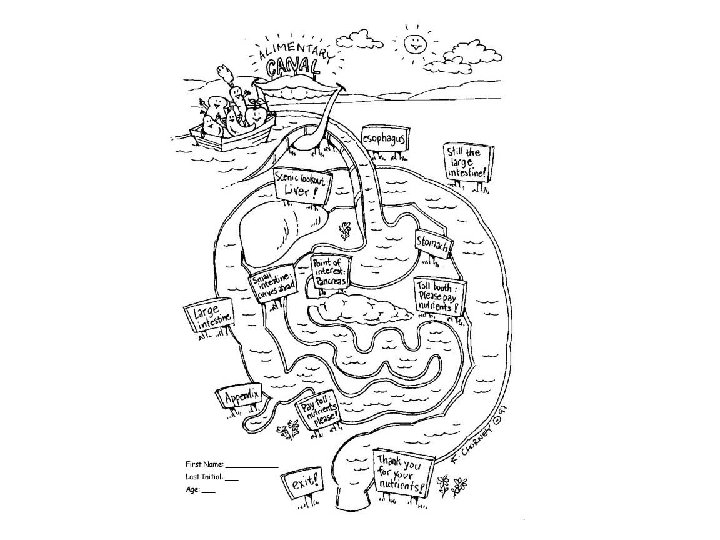
Can you determine the major organs of the digestive system? 71

How did you do? 72

DIGESTION What happens: Polymers monomers absorbed mitochondria ATP Cells require the following materials: Monosaccharides Amino acids Fatty acids and glycerol Other types of nutrients are too large to pass through the digestive tract into the bloodstream 73
 absorption of nutrients")
This process takes place in 4 steps ingestion chemical breakdown (digestion) absorption of nutrients egestion (elimination of wastes) There are two types of digestion: Physical digestion – breaking of food into smaller pieces, increasing its surface area Chemical digestion – breaking chemical bonds in food, using enzymes OVERVIEW OF DIGESTION ANIMATION: http: //www. biocourse. com/mhhe/bcc/resources/concept. xsp? id=0 00012109&type=MOVIE 74
 digestive system has two parts: Alimentary canal – a continuous, coiled,")
A mammalian (human) digestive system has two parts: Alimentary canal – a continuous, coiled, and hollow muscular tube that food passes through measures from 6. 5 to 9 m in length Click Image for Interactive Website the movement of materials is as follows: (IN) MOUTH PHARYNX ESOPHAGUS STOMACH SMALL INTESTINE LARGE INTESTINE (COLON) RECTUM ANUS (OUT) 75

Accessory Organs: Makes the chemicals needed for digestion and send them into the alimentary canal: Salivary glands, liver, gallbladder, pancreas 77
 The Mouth Physical digestion")
DIGESTION: A CLOSER LOOK Starting where it all begins…. 1) The Mouth Physical digestion teeth chewing action Important for physical digestion Each tooth is covered with enamel Hardest substance in body 8 sharp, dagger – shaped incisors Front of mouth Cut food 78

Premolars Broad, flattened Grind food Molars Broader and flatter than premolars Have cusps Crush food Wisdom teeth Do not usually emerge until 16 to 20 years of age tongue manipulation and taste 79
 into maltose (disac) – only starch")
Chemical digestion Salivary amylase breaks down starch (polysac) into maltose (disac) – only starch digestion takes place in the mouth Salivary glands in the mouth secrete saliva Components of saliva: Component Function water Dissolves ions in food for taste mucus Eases the passage of food by making it slippery Salivary amylase Chemical digestion Starch maltose 81
")
Mixture of saliva and food = bolus Swallowing moves materials to the pharynx (throat) This is an intersection that leads to the trachea and the esophagus The epiglottis prevents food from entering the trachea Food for thought – How are we able to swallow food upside down? 82

Gunther von Hagens 83
 Assists passage of bolus by")
The esophagus Straight, muscular tube, runs behind trachea (windpipe) Assists passage of bolus by creating waves of muscular contractions called peristalsis (involuntary) http: //www. westga. edu/~lkral/peristalsis/ http: //health. howstuffworks. com/adam-200088. htm 84
 The Stomach The cardiac sphincter muscle controls the movement of material from the")
3) The Stomach The cardiac sphincter muscle controls the movement of material from the esophagus to the stomach (sphincters insure one movement of food) J-shaped organ that can store up to 1. 5 L of food The walls of the stomach secrete gastric juice 86
 Rennin Pepsinogin (inactive) Pepsin (active) Function")
Gastric Juice Secretion: Component Mucus Hydrochloric acid (HCl) Rennin Pepsinogin (inactive) Pepsin (active) Function Protects the walls of the stomach Kills bacteria in food Helps with physical breakdown of food Starts to break down milk proteins (causes coagulation) Slows movement of milk proteins Lots in children, decreases in adulthood Pepsinogin is activated at low 87 p. H’s Breaks down proteins into

Physical digestion – churning of the stomach breaks up food Chemical digestion – proteins polypeptides – milk proteins coagulation The stomach is mainly a storage tank, very little chemical digestion takes place Alcohol and some drugs (aspirin) are absorbed into the blood The image above shows rugae on the surface of a dog's stomach. It takes 2 -6 hours for the stomach to empty Food leaves the stomach through the pyloric sphincter Material leaving the stomach is called chyme 88
: What are Ulcers? Helicobacter pylori seen under an electronic microscope")
Peptic Ulcers (Page 262): What are Ulcers? Helicobacter pylori seen under an electronic microscope Video animation on the stomach and ulcers What is the cause of Ulcers? How do you treat ulcers? 89

ASSIGNMENTS TO BE COMPLETED: Read pages 259 - 270 In Textbook Complete Section 8. 3 Questions: 2 -12 – Page 263 Label diagrams of digestive system in workbook – color diagrams according to instructions Begin completing the summary of digestive enzymes charts 90

DO NOW Label the diagram 13. 1. 12. 2. 3. 11. 10. 9. 4. 5. 6. 7. 8.
 The Small Intestine Major digestive organ Site of final preparation of food to")
4) The Small Intestine Major digestive organ Site of final preparation of food to be absorbed into the blood transport system Average length is 7 m 3 subdivisions of small intestine duodenum (first 25 cm) – chyme enters here jejunum (middle 2. 5 m) ileum (last 3. 6 m) Villi- fingerlike projections that extend into the small intestine and increase surface area 92

Main functions: break down food chemically absorb digested foods into blood and lymph make certain hormones small amounts of chyme enter periodically, soaked in pepsin and HCl secretions then work to break down the chyme into absorbable units the pancreas, liver and the small intestine itself release secretions into the duodenum 93
:")
The small intestine itself secretes enzymes Secretion is called intestinal juice: Peptidase (an erepsin): breaks down polypeptides into amino acids (completes protein digestion) maltase: maltose glucose + glucose sucrase: sucrose glucose + fructose lactase: lactose glucose + galactose in addition, mucus protects the intestine and slows food movement 94

thus chyme entering from the stomach is mixed with 3 secretions in the duodenum: 1. Pancreatic secretions 2. Bile from the gall bladder (liver) 3. Intestinal secretions The small subunits will be absorbed into the blood: Monosac’s, a. a’s, water soluble vitamins, minerals, and water Lymph: fatty acids and glycerol At the end of the ileum, all that remains is some water and indigestible food matter, and bacteria 95

Accessory Organ: Pancreas enzyme-rich pancreatic juice is secreted contains enzymes that breakdown all 3 major nutrient types pancreatic amylase – completes digestion of starch into maltose trypsinogen converted into trypsin (active form) in the small intestine by the enzyme enterokinase long chain polypeptides shorter chains lipase triglycerides fatty acids and glycerol 96

pancreatic juice also contains bicarbonate ions, which neutralizes the HCl of the chyme: raising the p. H to about 8 or 9 the pancreas is stimulated by: acids enter duodenum and cause prosecretin to be activated to secretin stimulates pancreas to release pancreatic juices including bicarbonate ions 97 Canine Pancreas

Accessory Organs: The Liver and Gallbladder the liver produces bile, bile which emulsifies fats (emulsification is a physical process that breaks up fat globules) bile (not an enzyme) is composed of: bile salts (emulsify fats) bile pigments (colours feces) – products of old RBC’s processed by liver cholesterol 98

bile is sent to the gallbladder where it is stored Gallbladder stimulated by intestinal secreted hormone cholescystokinin (CCK) from the gallbladder, bile is sent to the duodenum through the bile duct if bile salts crystallize, a gallstone forms preventing bile from entering the duodenum leads to build up of pigments in tissues resulting in jaundice and decreased fat digestion

What are other functions of the liver? q Removes the toxic nitrogen group from amino acids q Converts glucose to glycogen and glycogen to glucose q Stores glycogen q Converts harmful compounds such as alcohol to less harmful ones 100

101
– Obstruction of bile duct causing a yellowish")
Disorders of the liver: Jaundice (P 268)– Obstruction of bile duct causing a yellowish discoloration of the skin Cirrhosis (P 268) – Chronic inflammation of the liver, caused by alcohol abuse, nutritional deprivation or infections. Replaces damaged tissue with fibrous tissue (that doesn’t do much) 102
 Functions: Resident bacteria metabolize some of the remaining nutrients,")
5. The large Intestine (Colon) Functions: Resident bacteria metabolize some of the remaining nutrients, releasing gases (methane, H 2 S) Resident bacteria also make some vitamins (K and some B) Absorbs most water from feces and has limited nutrient absorption; vitamins, some ions The solid dried out product (feces) is delivered to the rectum Mucus is produced by the walls along the way to ease the passage 103

Fibre increases the strength of colon contractions that move feces towards the rectum (adds bulk) Appendicitis – 104

Rectum storage site of feces when feces enter, the walls expand, and send a message to the brain The Anus voluntary sphincter that is external passage for wastes (defecation) 105

ASSIGNMENTS TO BE COMPLETED: Read pages 264 - 270 In Textbook Chapter review questions page 278 # 1 -13, 16, 17, 19 106
- Slides: 101