BIOLOGY 2 E Module 3 CELL STRUCTURE Power

BIOLOGY 2 E Module 3: CELL STRUCTURE Power. Point Image Slideshow This work is licensed under a Creative Commons Attribution. Non. Commercial-Share. Alike 4. 0 International License.

rior organelles and structures of a Eukaryotic animal cell plant cell

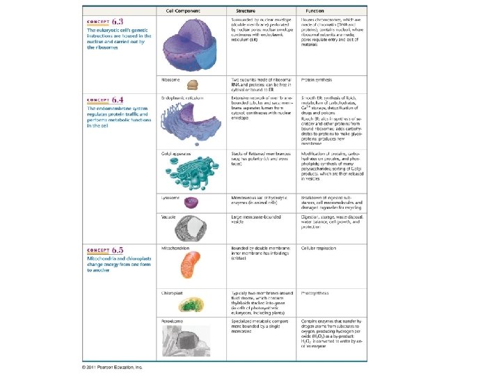
Nucleus • The Nuclear Envelope is a double membrane • Separates DNA containing nucleoplasm from cytoplasm • Separates transcription from translation • the outer membrane of the nuclear envelope is contagious with the endoplasmic reticulum. • Nuclear Pores perforate the nuclear membrane • Connect nucleoplasm to cytoplasm • Regulate flow of molecules back & forth

• Within the nucleus, DNA and associated proteins are organized into fibrous material, chromatin • When the cell prepares to divide, the chromatin fibers coil up to be seen as separate structures, chromosomes
")
Nucleolus • The nucleolus is a region inside nucleus where ribosomal RNA (r. RNA) is synthesized and ribosomes are assembled from r. RNA & proteins

Ribosomes • Made of a small and a large subunit • Slightly larger in eukaryotes • Made of special RNA (r. RNA) and proteins • During protein synthesis, ribosomes assemble amino acids into proteins

• Free ribosomes in the cytosol synthesize proteins that function within the cytosol • Bound ribosomes attached to the outside of the endoplasmic reticulum synthesize membrane or export proteins

Endomembrane system • The endomembrane system consists of internal membranes and organelles in eukaryotic cells that work together to modify, package, and transport lipids and proteins • nuclear envelope, • endoplasmic reticulum, • Golgi apparatus, • lysosomes, • vacuoles, • plasma membrane • Transfer of vesicles, sacs of membrane, among organelles of the endomembrane system
 • Interconnected membranous sacs and tubules. • Modifies proteins (rough ER)")
Endoplasmic reticulum (ER) • Interconnected membranous sacs and tubules. • Modifies proteins (rough ER) and synthesizes lipids (smooth ER). • The hollow portion of the ER tubules is called the lumen or cisternal space. • The membrane of the ER is continuous with the nuclear envelope.

Rough Endoplasmic Reticulum • The RER also makes proteins destined to for the membrane or secretion • Ribosomes attached to the cytoplasmic surface manufacture proteins • New proteins are modified (by folding or the acquisition of side chains) in the lumen of the RER • Modified proteins are either incorporated into cellular membranes or secreted from the cell (e. g. protein hormones, enzymes)

Protein destinations in the cell

Smooth Endoplasmic Reticulum The SER is continuous with the RER but has few or no ribosomes on its cytoplasmic sur • Synthesis of Phospholipids, Oils, Steroids • Detoxification of medications and poisons • Storage of Ca++

Translocation of phospholipids across the ER membrane • Phospholipids are synthesized on the cytosolic side of the ER membrane • Phospholipids are then translocated across the membrane by phospholipid flippases

Golgi Apparatus • Lipids or proteins within transport vesicles still need to be sorted, packaged, and tagged so that get to the right place • This occurs in the Golgi apparatus (also called the Golgi body) which consists of a series of flattened membranes. The receiving side of the Golgi apparatus is called the cis face; the opposite side is the trans face. • Transport vesicles from the ER fuse with the cis face and empty their contents into the lumen of the Golgi apparatus. • As the proteins and lipids travel through the Golgi, they are further modified so they can be sorted. • This often involves adding short chains of sugar molecules

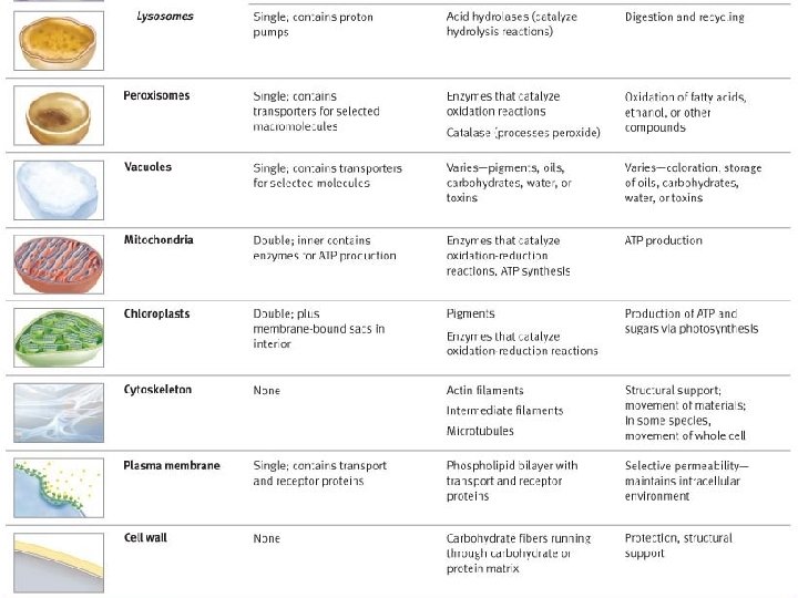
Lysosomes • Are primarily in animal cells • Contain digestive enzymes • The lysosomal enzymes (hydrolases) and membrane are synthesized by ER and then transferred to the Golgi • Interior is acidic • Breakdown large biomolecules in food vacuoles (pinocytosis or phagocytosis) or excess secretory vesicles. • Can fuse with and breakdown worn-out organelles, a process called autophagy, which renews the cell • Play a critical role in the programmed destruction of cells in multicellular organisms • Massive leakage from lysosomes can destroy a cell by autodigestion https: //www. youtube. com/watch? v=to. BTf. M 8 li-c

Peroxisomes • Peroxisomes are small rounded organelles enclosed by a lipid bilayer • Reactions that break down fatty acids and amino acids occur here • Peroxisomes may detoxify poisons (hydrogen peroxide) Fatty acid oxidation in peroxisomes

Mitochondrion • Site for cellular respiration and production of large quantities of ATP • Double membrane • Inner membrane is folded • Folds are called cristae with many copies of the electron transport chain and ATP synthase (proteins) • Area enclosed by the membrane is the mitochondrial matrix • Contains ribosomes and DNA ATP synthase

Mitochondrial diseases
 • Chloroplasts are double-membrane organelles; have their own ribosomes and")
Chloroplasts (plants and algae) • Chloroplasts are double-membrane organelles; have their own ribosomes and DNA like mitochondria • The inner membrane encloses an aqueous fluid (stroma) that contains a set of interconnected and stacked fluid-filled membrane sacs called thylakoids • Each stack of thylakoids is a granum (plural = grana). 1. outer membrane 2. intermembrane space 3. inner membrane (1+2+3: envelope) 4. stroma (aqueous fluid) 5. thylakoid lumen (inside of thylakoid) 6. thylakoid membrane 7. granum (stack of thylakoids) 8. thylakoid 9. starch 10. ribosome 11. plastidial DNA 12. plastoglobule (drop of lipids)

The Central Vacuole • Plant cells have a large vacuole that occupies most of the area of the cell • This central vacuole helps regulate water concentration under changing environmental conditions, and contributes to cell expansion. Organisms whose cells have cell walls (plants, fungi, bacteria and some protists) prefer hypotonic extracellular solutions. plasmolysis – plasma membrane detaches from the cell wall The pressure exerted the cell wall (turgor pressure) is critical to organismal growth & functions.
 • Centrosome functions as the microtubule organizing center (MTOC) •")
Microtubule organizing center (MTOC) • Centrosome functions as the microtubule organizing center (MTOC) • In animal cells, the MTOC is called the centrosome and often contains a • In plant cells the MTOC lacks centrioles NOTE: This slide has been updated. The slide in the lecture in

Plant Cell Walls • The cell wall is a rigid protective structure external to the plasma membrane • Plant cell walls differ from prokaryotes because they are made up of cellulose rather than peptidoglycan. Cellulose molecule

Contrasting Animal and Plant Cells • Both plants and animal cells have microtubule organizing centers (MTOCs), called centrosomes (in animal cells), but animal cells have centrioles associated with the MTOC/centrosomes • Animal cells have lysosomes, plant cells do not • Plant cells have vacuoles, animal cells do not • Plant cells have a cell wall, chloroplasts and other specialized plastids and a large central vacuole - animal cells do not NOTE: This slide has been updated. The slide in the lecture in

Endosymbiotic theory • It is hypothesized that mitochondria and chloroplasts originated as independent prokaryotic organisms via endosymbiosis. • These became endosymbionts of the ancestors of the eukaryotes. • Evidence: • Mitochondria and chloroplasts have their own DNA and ribosomes. At the DNA level, mitochondria’s closest living relatives are the α-proteobacteria. Chloroplast’s closest living relative are the Cyanobacteria. • The size of these organelles is similar to that of free-living prokaryotes. • Mitochondria and chloroplasts have double membrane (one coming from the endosymbiont and one from the host plasma membrane).

• The Endosymbiont theory http: //glencoe. mcgraw-hill. com/sites/9834092339/student_view 0/chapter 4/animation_-

The Cytoskeleton The cytoskeleton is a network of protein fibers with several functions • It helps maintain the shape of the cell • Hold some organelles in specific positions • Allows movement of cytoplasm and vesicles within the cell • Enables cells within multicellular organisms to move
 • Intermediate filaments are found throughout the")
Three Components of Cytoskeleton Microfilament (actin filament) • Intermediate filaments are found throughout the cell and hold organelles in place. • Microfilaments thicken the periphery of the cell; like rubber bands, they resist tension. • Microtubules are found in the interior of the cell where they maintain cell shape.
 internal skeleton for s")
Microtubules • Form rigid yet dynamic (easily assembled and disassembled) internal skeleton for s • Provide framework for motor proteins to move structures within cell • Made of tubulin dimers: 13 chains of dimers surround central cavity of microtubule Vesicle ATP Receptor for motor protein Motor protein (ATP powered) Microtubule of cytoskeleton (a) Motor proteins that attach to receptors on organelles can “walk” the organelles along microtubules or, in some cases, microfilaments. Microtubule Vesicles

Microtubules form Cilia & Flagella • Ultrastructure • Same 9+2 array of microtubules • 9 doublets on outside • 2 unfused in center • Spokes connect doublets to middle • Cilia shorter and more numerous • Beating patterns differ
 • Involved in movement • Whole cell")
Microfilaments • dynamic (easily assembled and disassembled) • Involved in movement • Whole cell or internal parts • Determine & stabilize shape • Made from actin monomers

Intermediate filaments • Several strands of fibrous proteins twisted together. • Most diverse group of cytoskeletal elements, e. g. keratin, the fibrous protein in ha • Their function is structural, e. g. maintaining the shape of the cell, and anchor the n • They don’t participate in cell movement • Stable (i. e. , not dynamic).

Extracellular Structures Collagen Animals • Extracellular matrix binds cells together and helps their adhesion, migration, proliferation, and differentiation • Collagens • Proteoglycans EXTRACELLULAR MATRIX Fibronectin Polysaccharide molecule Proteoglycan complex Integrins Plasma membrane Plants • Cell wall • Support • Barrier to infection • Plasmodesmata connect cells • Middle lamella with sticky polysaccharides that holds cells together Proteoglycan complex Microfilaments CYTOPLASM
 cells")
Intercellular junctions Animal tissues • • • At tight junctions (fluid blocking junctions) cells are stitched/pressed together with proteins, preventing passage of fluid between cells. Found in epithelial cells of internal organs and cavities. Desmosomes (anchoring junctions) fasten cells together into strong sheets that resist mechanical stress. Join adjacent cells in tissues that stretch (e. g. heart, lungs, muscles) Gap junctions (communicating junctions) Made of transmembrane proteins that provide cytoplasmic channels between adjacent cells. Tight junctions prevent fluid from moving across a layer of cells Tight junction Intermediate filaments Desmosome Gap junction Ions or small molecules Space between cells Plasma membranes of adjacent cells Plant tissues • plasmodesmata (communicating junctions) plasma membrane connections between cells that provide cytoplasmic channels between adjacent cells Extracellular matrix

What you need to know • How are prokaryotic cells similar and different? Know the structures and their functions! • Why does cell size matter (surface-to-volume ratio)? • The three domains of cellular life. • Describe the information flow of the central dogma. • How do plant and animal cells differ? • Know the structure and function of all of the organelles in a eukaryotic cell • Describe endosymbiotic theory (mechanisms and evidence). • Describe the endomembrane system. • Describe how a protein that will be excreted is translated, processed, and delivered in a eukaryote. A protein that will be embedded in the plasma membrane? A cytosolic protein? • Describe the structure of the extracellular matrix. What is its function? • Describe the type of cell junctions and give examples of where you would find them.
- Slides: 36