Biologically Important Molecules II Biologically Important Molecules I

Biologically Important Molecules – II !

Biologically Important Molecules I. Water II. Carbohydrates

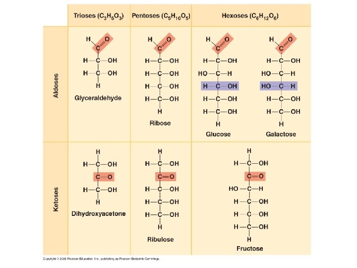
II. Carbohydrates A. Structure 1. monomer = monosaccharide typically 3 -6 carbons, and Cn. H 2 n. On formula

II. Carbohydrates A. Structure 1. monomer = monosaccharide typically 3 -6 carbons, and Cn. H 2 n. On formula have carbonyl and hydroxyl groups

II. Carbohydrates A. Structure 1. monomer = monosaccharide typically 3 -6 carbons, and Cn. H 2 n. On formula have carbonyl and hydroxyl groups carbonyl is either ketone or aldehyde


II. Carbohydrates A. Structure 1. monomer = monosaccharide typically 3 -6 carbons, and Cn. H 2 n. On formula have carbonyl and hydroxyl groups carbonyl is either ketone or aldehyde in aqueous solutions, they form rings

II. Carbohydrates A. Structure 1. monomer = monosaccharide typically 3 -6 carbons, and Cn. H 2 n. On formula have carbonyl and hydroxyl groups carbonyl is either ketone or aldehyde in aqueous solutions, they form rings examples:

II. Carbohydrates A. Structure 1. monomer = monosaccharide 2. polymerization: dehydration synthesis reaction

II. Carbohydrates A. Structure 1. monomer = monosaccharide 2. polymerization 3. Polymers = polysaccharides

Disaccharides

Polysaccharides

Polysaccharides

Polysaccharides The ‘cross-linkages’ in cellulose are not digestible by starch-digesting enzymes, so animals cannot eat wood unless they have bacterial endosymbionts. Decomposing fungi and bacteria also have these enzymes, and can access the huge amount of energy in cellulose.

Polysaccharides H-bonds link cellulose molecules together

Polysaccharides glucosamine
 - structural")
II. Carbohydrates A. Structure B. Function - energy storage (short and long) - structural (cellulose and chitin) CO 2 Glucose, Cellulose, Starch H 2 O

Biologically Important Molecules I. Water II. Carbohydrates III. Lipids

III. Lipids - not true polymers or macromolecules; an assortment of hydrophobic, hydrocarbon molecules classes as fats, phospholipids, waxes, or steroids.

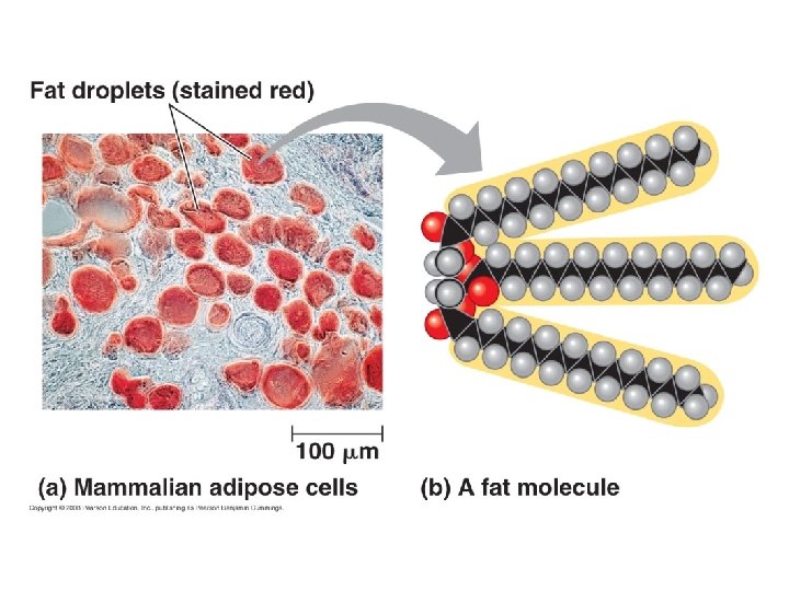
III. Lipids A. Fats - structure
 with three fatty acids")
III. Lipids A. Fats - structure glycerol (alcohol) with three fatty acids
")
(or triglyceride)
 Straight chains pack")
III. Lipids A. Fats - structure -saturated fats (no double bonds) Straight chains pack tightly; solid at room temperature like butter and lard. Implicated in plaque buildup in blood vessels (atherosclertosis) Animal fats (not fish oils)
 Plant and fish")
III. Lipids A. Fats - structure -unsaturated fats (no double bonds) Plant and fish oils Kinked; don’t pack – liquid at room temperature. “Hydrogenation” can make them saturated and solid, but the process also produces trans-fats (trans conformation around double bond) which may contribute MORE to atherosclerosis than saturated fats)
")
III. Lipids A. Fats - structure - functions - long term energy storage (dense) not vital in immobile organisms (mature plants), so it is metabolically easier to store energy as starch. But in seeds and animals (mobile), there is selective value to packing energy efficiently. In animals, fat is stored in adipose cells
")
III. Lipids A. Fats - structure - functions - long term energy storage (dense) - insulation (subcutaneous fat) - cushioning

III. Lipids A. Fats B. Phospholipids - structure Glycerol 2 fatty acids phosphate group (and choline) Hydrophilic and hydrophobic regions

III. Lipids A. Fats B. Phospholipids - function selective membranes In water, they spontaneously assemble into micelles or bilayered liposomes.

III. Lipids A. Fats B. Phospholipids C. Waxes - structure An alcohol and fatty acid Wax Alcohol Fatty Acid Carnuba CH 3(CH 2)28 CH 2 -OH CH 3(CH 2)24 COOH Beeswax CH 3(CH 2)28 CH 2 -OH CH 3(CH 2)14 COOH Spermacetic CH 3(CH 2)14 CH 2 -OH CH 3(CH 2)14 COOH

III. Lipids A. Fats B. Phospholipids C. Waxes - structure - function Retard the flow of water (plant waxes) Structural (beeswax) Signals – waxes on the exoskeleton can signal an insect’s sexual receptivity.

III. Lipids A. Fats B. Phospholipids C. Waxes D. Steroids - structure typically a four-ring structure with side groups cholesterol and its hormone derivatives

Cholesterol

Biologically Important Molecules I. III. IV. Water Carbohydrates Lipids Proteins

IV. Proteins A. structure - monomer: amino acids

IV. Proteins A. structure - monomer: amino acids Carboxyl group Amine group

IV. Proteins A. structure - monomer: amino acids 20 AA’s found in proteins, with different chemical properties. Of note is cysteine, which can form covalent bonds to other cysteines through a disulfide linkage.

IV. Proteins A. structure - monomer: amino acids - polymerization: dehydration synthesis The bond that is formed is called a peptide bond

IV. Proteins A. structure - monomer: amino acids - polymerization: dehydration synthesis - polymer: polypeptide

IV. Proteins A. structure - monomer: amino acids - polymerization: dehydration synthesis - polymer: polypeptide May be 1000’s of aa’s long Not necessarily functional (“proteins” are functional polypeptides) Sequence determines the function

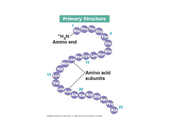
IV. Proteins A. structure - monomer: amino acids - polymerization: dehydration synthesis - polymer: polypeptide - protein has 4 levels of structure 1 o (primary) = AA sequence

IV. Proteins A. structure - monomer: amino acids - polymerization: dehydration synthesis - polymer: polypeptide - protein has 4 levels of structure 1 o (primary) = AA sequence 2 o (secondary) = pleated sheet or helix

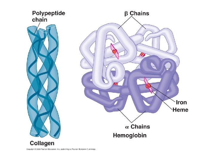
The result of H-bonds between neighboring AA’s… not involving the side chains. Some proteins are functional as helices - collagen

IV. Proteins A. structure - monomer: amino acids - polymerization: dehydration synthesis - polymer: polypeptide - protein has 4 levels of structure 1 o (primary) = AA sequence 2 o (secondary) = pleated sheet or helix 3 o (tertiary) = folded into a glob

The three dimensional structure of the protein is stabilized by all types of bonds between the side chains… ionic between charged AA’s, Hydrogen bonds between polar AA’s, van der Waals forces, and even covalent bonds between sulfurs.

IV. Proteins A. structure - monomer: amino acids - polymerization: dehydration synthesis - polymer: polypeptide - protein has 4 levels of structure 1 o (primary) = AA sequence 2 o (secondary) = pleated sheet or helix 3 o (tertiary) = folded into a glob 4 o (quaternary) = >1 polypeptide

Actin filament in muscle is a sequence of globular actin proteins…
 http: //3 dotstudio. com/prenhall/muscle. jpg")
50 myofibrils/fiber (cell) http: //3 dotstudio. com/prenhall/muscle. jpg
 - structural (actin/collagen/etc. ) -")
IV. Proteins A. structure B. functions! - catalysts (enzymes) - structural (actin/collagen/etc. ) - transport (hemoglobin, cell membrane) - immunity (antibodies) - cell signaling (surface antigens)

IV. Proteins A. structure B. functions! C. designer molecules If protein function is ultimately determined by AA sequence, why can’t we sequence a protein and then synthesize it?

IV. Proteins A. structure B. functions! C. designer molecules If protein function is ultimately determined by AA sequence, why can’t we sequence a protein and then synthesize it? Folding is critical to function, and this is difficult to predict because it is often catalyzed by other molecules called chaparones

IV. Proteins A. structure B. functions! C. designer molecules If protein function is ultimately determined by AA sequence, why can’t we sequence a protein and then synthesize it? Folding is critical to function, and this is difficult to predict because it is often catalyzed by other molecules called chaparones Perhaps by analyzing large numbers of protein sequences and structures, correlations between “functional motifs” and particular sequences will be resolved.
- Slides: 55