
Biological Image Processing Physiology and Function C 428615

Biological Image Processing Physiology and Function C 428/615 Martin Jagersand With illustrations from: Tutis Vilis Talia Konkle and others as cited
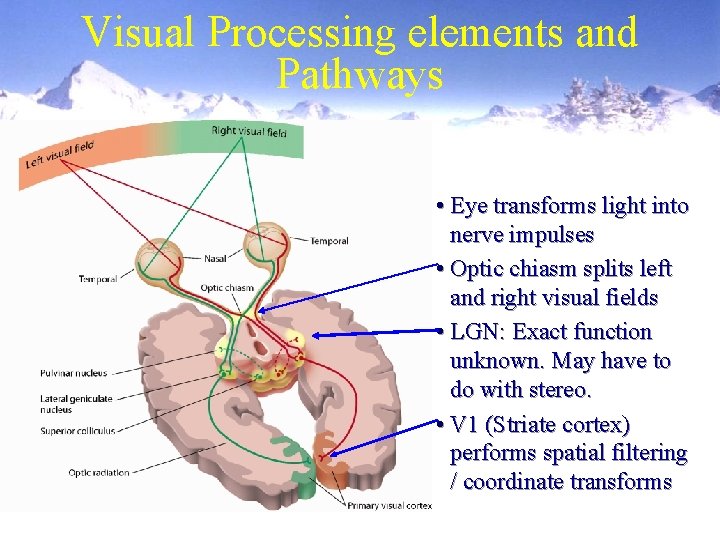
Visual Processing elements and Pathways • Eye transforms light into nerve impulses • Optic chiasm splits left and right visual fields • LGN: Exact function unknown. May have to do with stereo. • V 1 (Striate cortex) performs spatial filtering / coordinate transforms
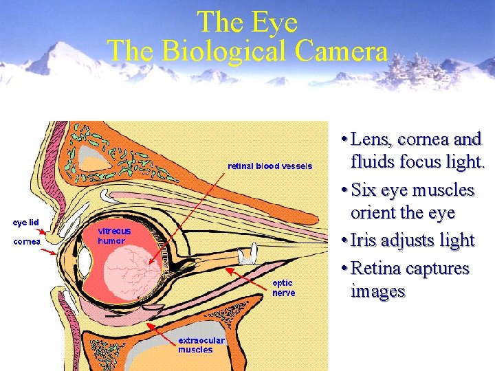
The Eye The Biological Camera • Lens, cornea and fluids focus light. • Six eye muscles orient the eye • Iris adjusts light • Retina captures images
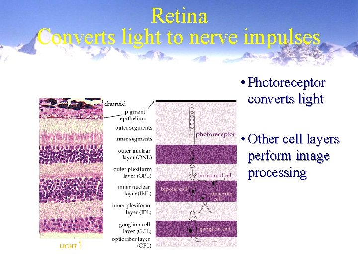
Retina Converts light to nerve impulses • Photoreceptor converts light • Other cell layers perform image processing
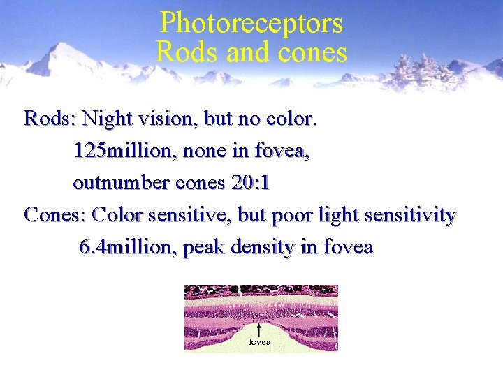
Photoreceptors Rods and cones Rods: Night vision, but no color. 125 million, none in fovea, outnumber cones 20: 1 Cones: Color sensitive, but poor light sensitivity 6. 4 million, peak density in fovea
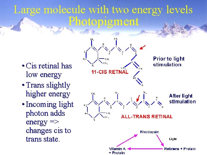
Large molecule with two energy levels Photopigment • Cis retinal has low energy • Trans slightly higher energy • Incoming light photon adds energy => changes cis to trans state.
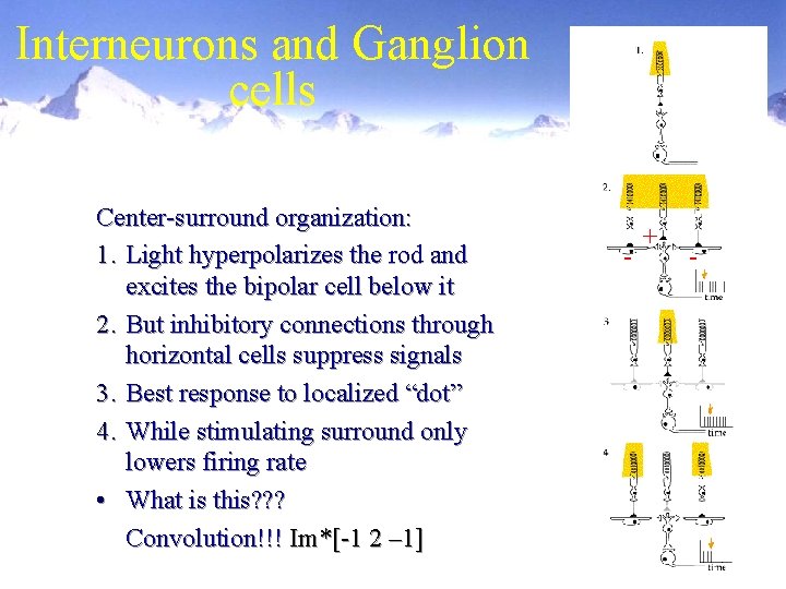
Interneurons and Ganglion cells Center-surround organization: 1. Light hyperpolarizes the rod and excites the bipolar cell below it 2. But inhibitory connections through horizontal cells suppress signals 3. Best response to localized “dot” 4. While stimulating surround only lowers firing rate • What is this? ? ? Convolution!!! Im*[-1 2 – 1] - + -
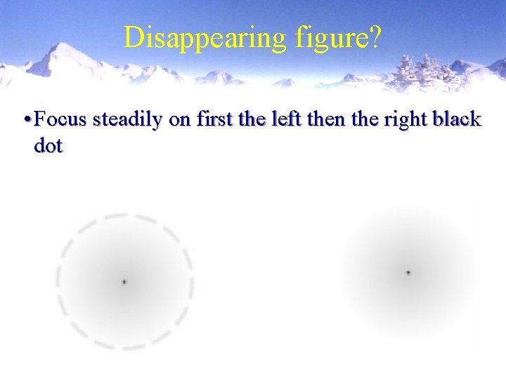
Disappearing figure? • Focus steadily on first the left then the right black dot
The visual system adjusts itself. • In time. • In space. • Why does it adjust? – – sensitivity and gain? neural fatigue? adjustment of priors? (effects in the opposite direction) Error correction?
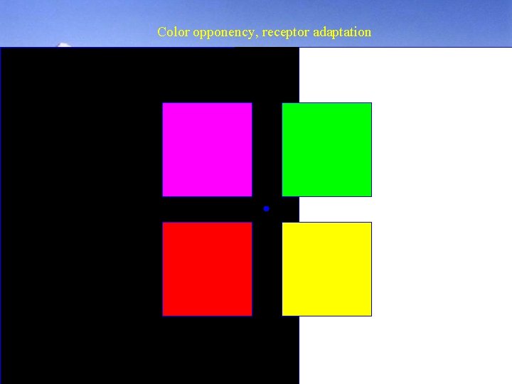
Color opponency, receptor adaptation
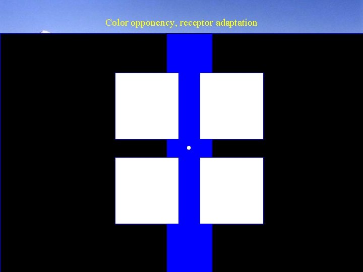
Color opponency, receptor adaptation
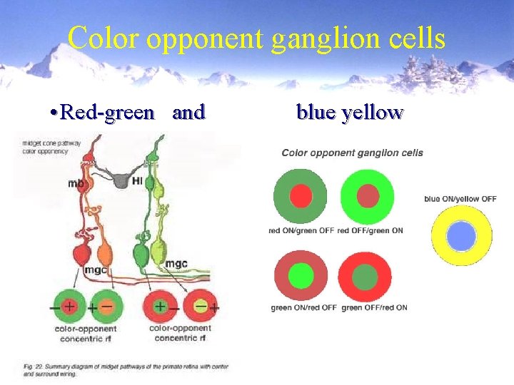
Color opponent ganglion cells • Red-green and opponency blue yellow
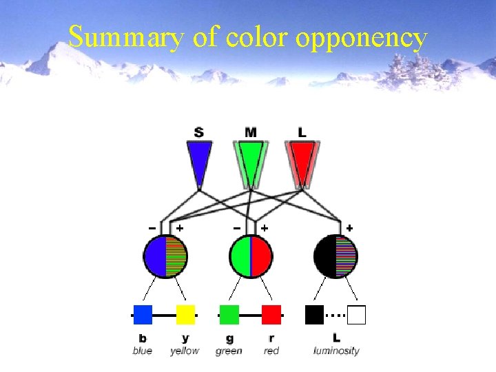
Summary of color opponency
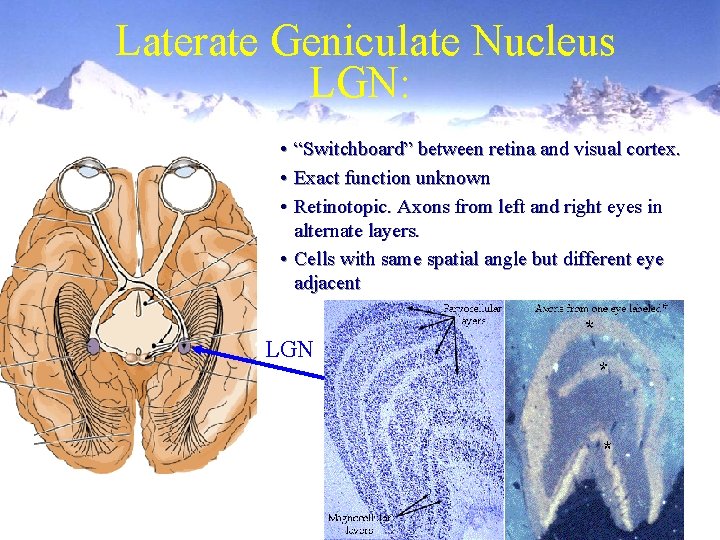
Laterate Geniculate Nucleus LGN: • “Switchboard” between retina and visual cortex. • Exact function unknown • Retinotopic. Axons from left and right eyes in alternate layers. • Cells with same spatial angle but different eye adjacent LGN

Visual Cortex: P and M cells
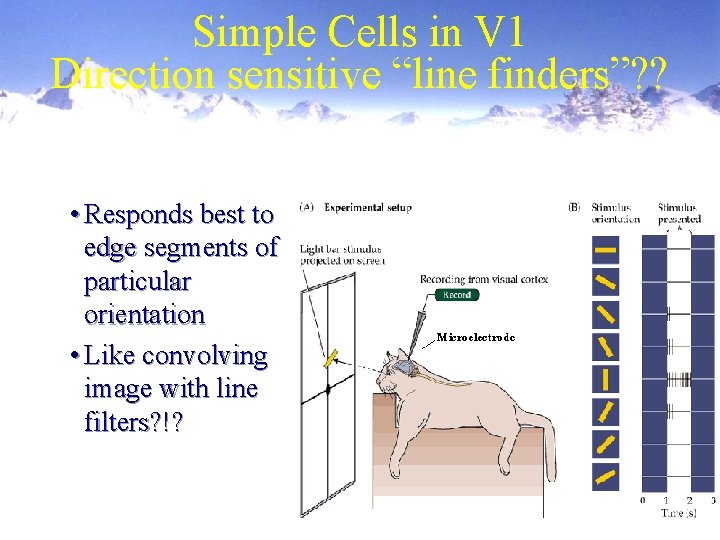
Simple Cells in V 1 Direction sensitive “line finders”? ? • Responds best to edge segments of particular orientation • Like convolving image with line filters? !?
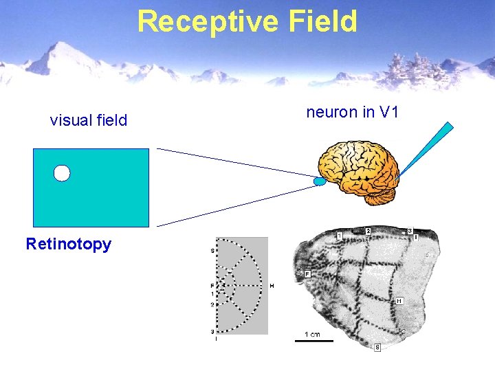
Receptive Field visual field Retinotopy neuron in V 1
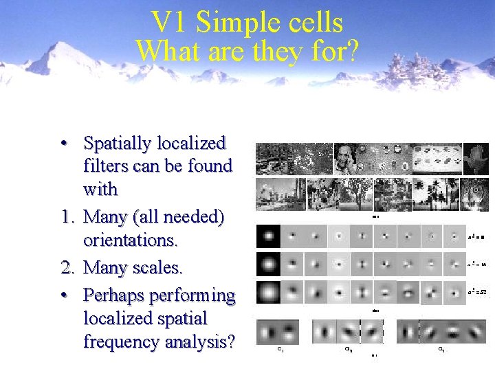
V 1 Simple cells What are they for? • Spatially localized filters can be found with 1. Many (all needed) orientations. 2. Many scales. • Perhaps performing localized spatial frequency analysis?
Complex and Hyper-complex cells More cells in V 1: • Complex cells: No specific retinotopic region. Responds to particular orientation or direction over a wide area. • Hypercomplex cells: End stop. Only responds to a terminating line.
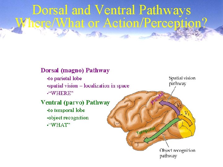
Dorsal and Ventral Pathways Where/What or Action/Perception?
Example responses Higher cells in the M pathway • Follower neurons: Responds to particular object moving regardless of where in the visual field. • Reach neurons: Responds when reaches to contact a specific 3 D location. • Attention neurons: Responds when gaze is directed to a specific object. • Gaze neurons: Responds when gaze is directed to a specific 3 D location.

Kaniza triangle
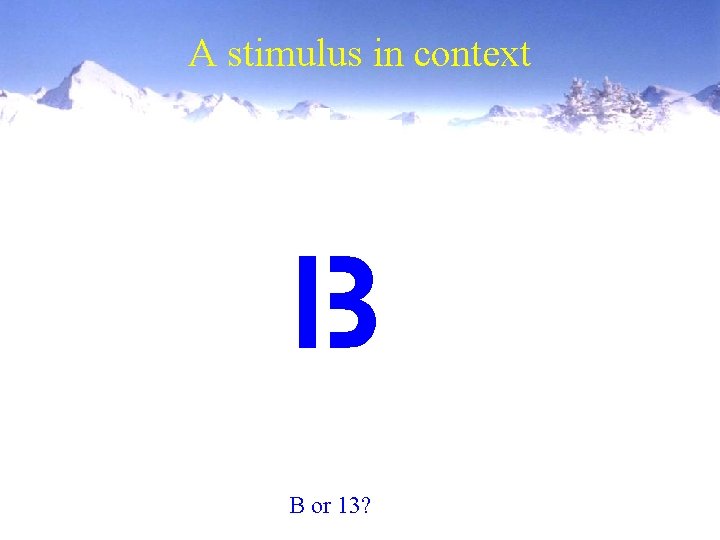
A stimulus in context 2 ABC 4 B or 13?
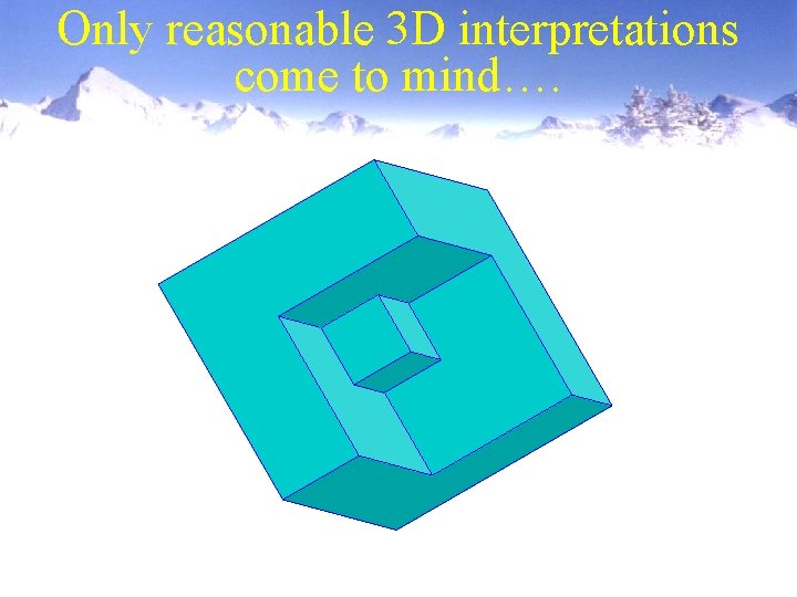
Only reasonable 3 D interpretations come to mind….
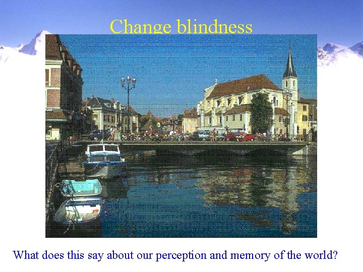
Change blindness What does this say about our perception and memory of the world?

Get Ready… I’m going to show you a picture and I want you to remember it! (you’ll have 3 seconds)
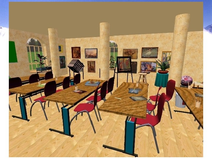
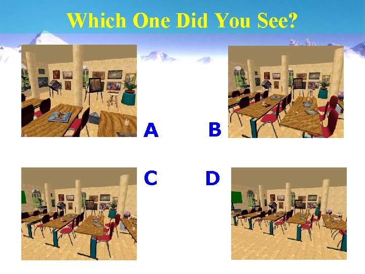
Which One Did You See? A B C D
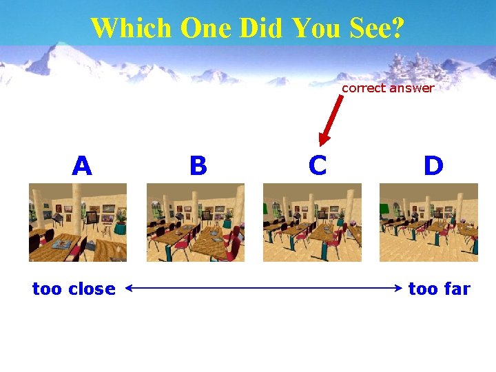
Which One Did You See? correct answer A too close B C D too far
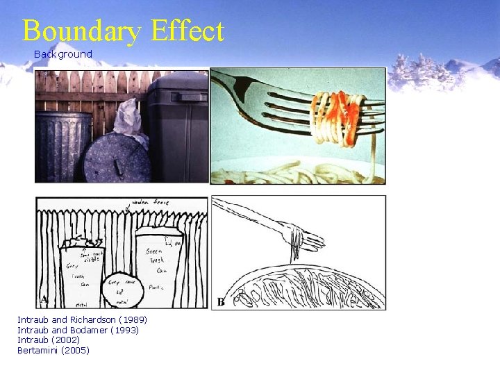
Boundary Effect Background Intraub and Richardson (1989) Intraub and Bodamer (1993) Intraub (2002) Bertamini (2005)

Attention
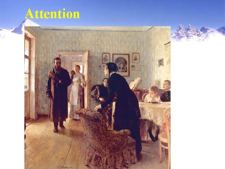
Attention
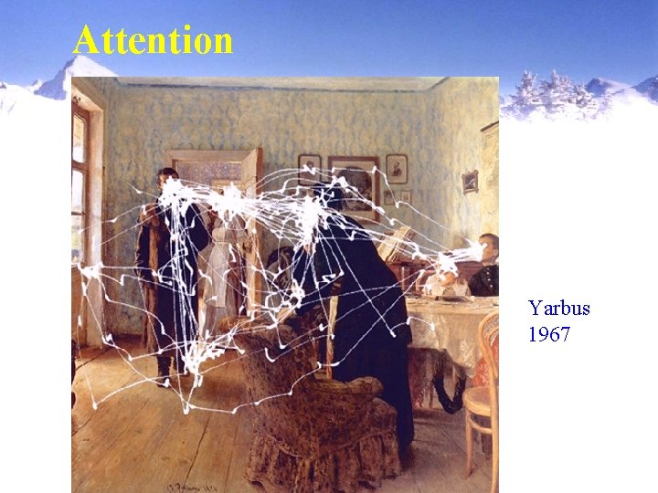
Attention Yarbus 1967
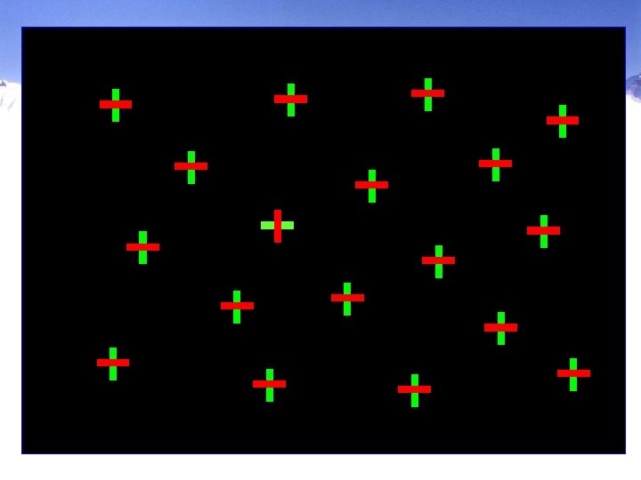
Visual Search for the red vertical
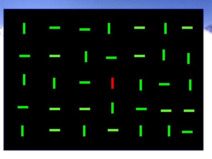
Pretty easy. Search for the red vertical again
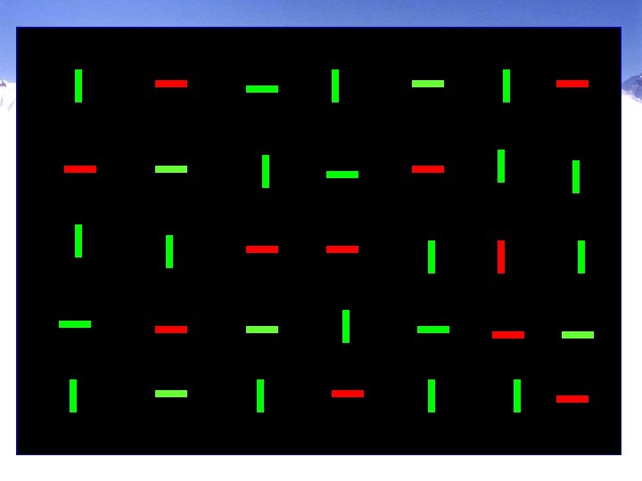
Harder? Guiding attention by color… pretty easy Guiding attention by color AND orientation, more difficult one more time, find the red vertical
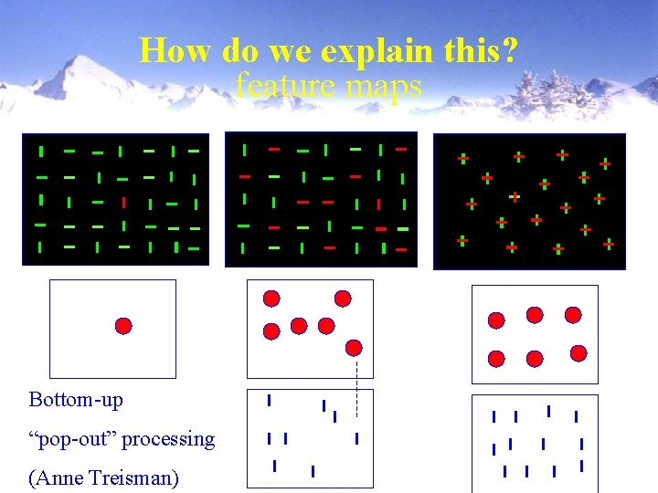
How do we explain this? feature maps Bottom-up “pop-out” processing (Anne Treisman)
Bio motion (Gestalt, or expectations? ) • http: //www. biomotionlab. ca/Demos/BMLwalker. html
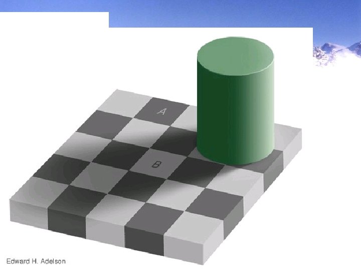
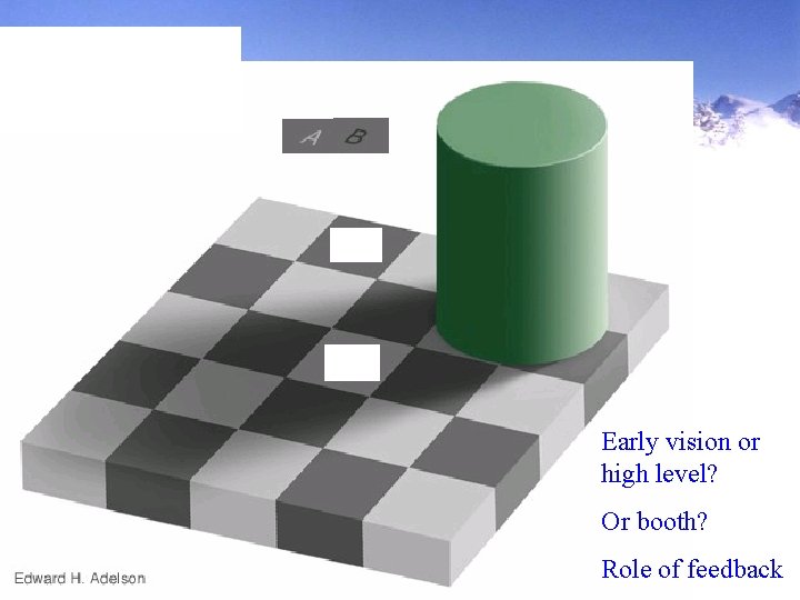
Early vision or high level? Or booth? Role of feedback
Conclusions: Biological vision • The visual system provides researchers with a window into the brain. • A fair amount is known about the eye, retina and early retinotopic areas like striate cortex (V 1). • Less is known about the function of higher areas.
Questions
- Slides: 45