BIOLOGICAL CLASSIFICATION BIOLOGY CLASS XI CHAPTER02 Carolus Linnaeus

BIOLOGICAL CLASSIFICATION BIOLOGY , CLASS –XI, CHAPTER-02
 ØIn 1731 he first published Systema Naturae. ØIn 1753 he")
Carolus Linnaeus- (1707 -1778) ØIn 1731 he first published Systema Naturae. ØIn 1753 he published Species Plantarum. ØDeveloped binomial naming system in 1753 ØFather of taxonomy.


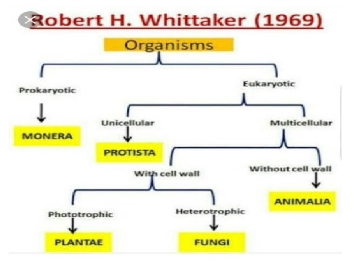
LINNAEUS 1735 HAECKEL 1866 CHATTON 1925 COPELAND 1938 TWO KINGDOMS THREE KINGDOMS TWO EMPIRES FOUR KINGDOMS PROKARYOTA (NOT TREATED) MONERA RH WOESE ET AL. WHITTAKER 1977 1990 1969 FIVE KINGDOMS SIX KINGDOMS THREE DOMAINS EUBACTERIA ARCHAEBACTERIA ARCHAEA MONERA PROTISTA EUKARYOTA PLANTAE ANIMALIA EUCARYA PLANTAE FUNGI ANIMALIA PLANTAE ANIMALIA
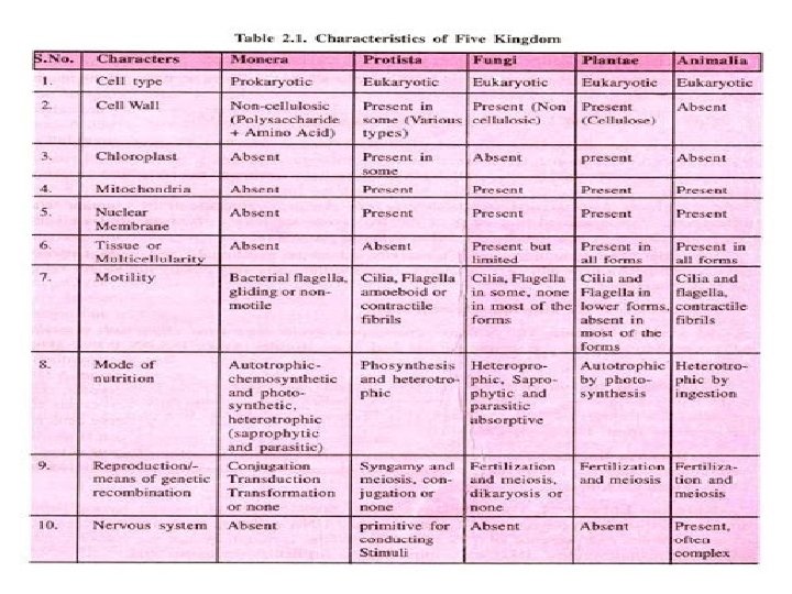

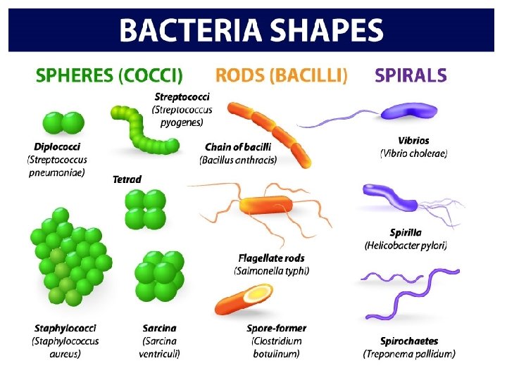
1. MONERA Bacteria are the sole members of the Kingdom Monera. They are the most abundant micro-organisms. Bacteria occur almost everywhere. Bacteria are grouped under four categories based on their shape: The spherical Coccus (pl. : cocci), The rod-shaped Bacillus (pl. : bacilli), The comma-shaped Vibrium (pl. : vibrio) and The spiral Spirillum (pl. : spirilla).

Prokaryotic Shapes Most prokaryotes have one of 3 basic shapes -Bacillus = Rod-shaped -Coccus = Spherical -Spirillum = Helical-shaped 23

• DNA is circular. DNA combined with non histone protein is known as nucleiod. It is equivalent to one chromosome. • Besides nuclear DNA, in some bacteria extra-chromosomal DNA are present which is knwon as plasmid. • In prokaryotes ribosomes are of 70 s type. There are three types of plasmid: • • F-factor or fertility factor: It is responsible for transfer of genetic material. • R-factor or resistance factor: It provides resistance against drugs. • Colicinogenic factor: It produces colicines which kill other bacteria.

Conjugation E. coli 25
Archaebacteria")
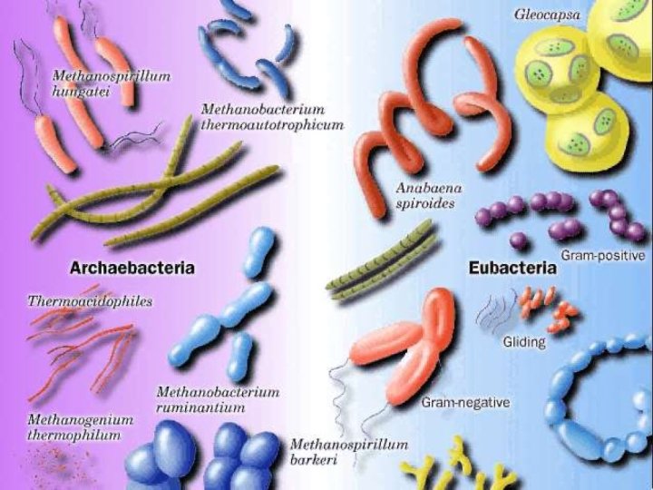
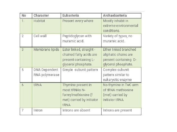
1. MONERA • Kingdom Monera is NOT monophyletic • Two main branches – (A)Archaebacteria = extreme environments – (B) Eubacteria or True Bacteria
 ARCHAEBACTERIA These bacteria are special since they live in some of the most")
(A) ARCHAEBACTERIA These bacteria are special since they live in some of the most harsh habitats such as extreme Salty areas (halophiles), Hot springs (thermoacidophiles)and Marshy areas (methanogens).

Methanogens Methane producing bacteria These bacteria convert CO 2 of swampy areas into methane. These bacteria convert the organic substance present in cow dung into methane by fermentation. (Gobar gas fermenter) Example: Methanococcus. They are also present in the rumen of cattle, where it digests the cellulose by fermentation and convert it into methane. Example: Rumenococcus

HALOPHILES Halophiles means Salt loving. They are found in extreme salty areas such as the Great Salt Lake and the Dead Sea. Halophiles are surrounded by purple membrane in which a pigment, bacteriorhodopsin is found due to this reason membrane absorbs the bright light and directly forms ATP i. e. they cannot prepare food like eubacteria. Instead of it they directly form ATP. Therefore They are non photosynthetic

Thermoacidophiles • Thermoacidophiles means heat and acid loving. • These archaebacteria are found at those places where temperature is approx 80 o. C – 100 o. C and medium is acidic. • These are aerobic bacteria, which have the capacity to oxidise sulphur to H 2 SO 4 at high temperature and high acidity. • Some of these bacteria are able to reduce sulphur to H 2 S under anerobic conditions. • They are found in hot sulphur springs such as Yellowstone National park.

“Heat-loving” prokaryotes

Extreme halophiles
 Eubacteria pneumonia cyanobacteria anthrax")
(B) Eubacteria pneumonia cyanobacteria anthrax
 EUBACTERIA There are thousands of different eubacteria or ‘true bacteria’. They are characterised")
(B) EUBACTERIA There are thousands of different eubacteria or ‘true bacteria’. They are characterised by the presence of a rigid cell wall, and if motile, a flagellum. Eubacteria may be of following types: 1. Cyanobacteria 2. Chemosynthetic 3. Heterotrophic and 4. Mycoplasma
 and are photosynthetic")
Types of Eubacteria • Cyanobacteria have chlorophyll (similar to green plants) and are photosynthetic autotrophs. • Cyanobacteria were the first organisms that produced O 2 on our earth and also known as BGA. • The cyanobacteria are unicellular (Spirullina), colonial (Anabaena) or filamentous (Oscillatoria). • They may be freshwater or marine • Some of these organisms can fix atmospheric nitrogen in specialised cell called heterocysts. Example: Nostoc and Anabaena.

Chemosynthetic Autotrophic • These are nonphotosynthetic autotrophs. They use chemical energy instead of light energy for food synthesis. Chemical energy is obtained from oxidation of chemical compounds. Photolysis of water does not take place here, so hydrogen is received from other sources like inorganic sulphur compounds (H 2 S) or organic compound (Amino acids, Fatty acid etc. ) • Example: Sulphur producing bacteria. Heterotrophic Bacteria • These are the most abundant in nature. Many of them have a significant impact on human affairs. They are helpful in making curd from milk, production of antibiotics, fixing nitrogen in legume roots, etc. • Some are pathogens causing damage to human being, crops, farm animals and pets. Cholera diseases caused by different bacteria. Mycoplasma • Mycoplasma are organisms that completely lack a cell wall. • These are the smallest living cells known and can survive without oxygen. • Many mycoplasma are pathogenic in animals and plants.

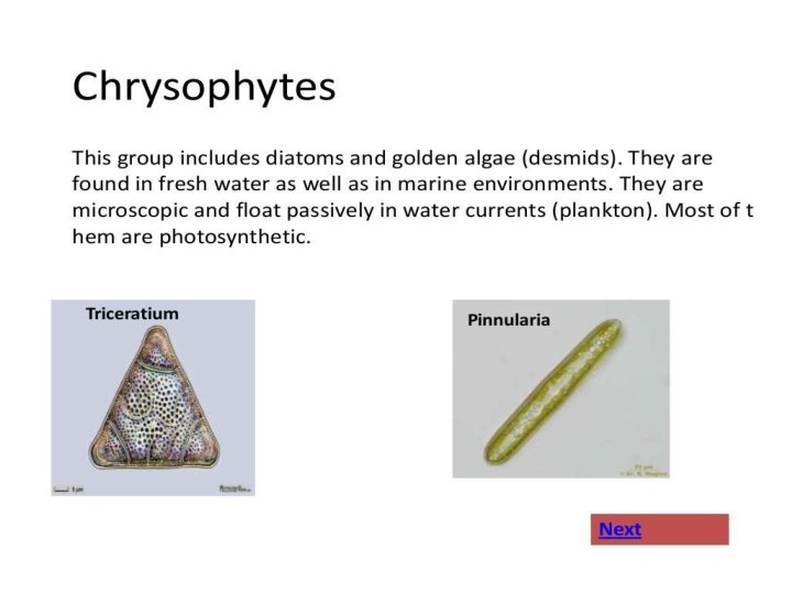
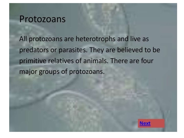
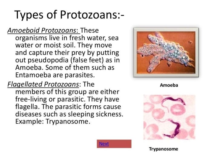
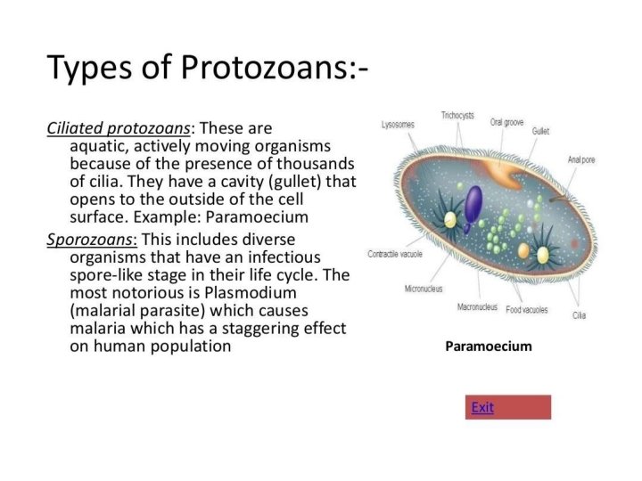
2. PROTISTA All single-celled eukaryotes are placed under Protista. Members of Protista are primarily aquatic. Being eukaryotes, the protistan cell body contains a well defined nucleus and other membrane-bound organelles. Some have flagella or cilia. Protists reproduce asexually and sexually by a process involving cell fusion and zygote formation.
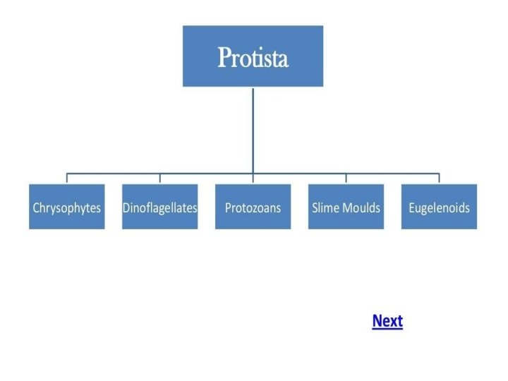

Animal-like Protists Plant-like Protists Amoeba Cilliates Flagellates Diatoms Dinoflagellates
 African Sleeping Sickness Malaria Vector")
Disease Protist Ameobic dysentery Entameoba histolytica Giardaisis (beaver fever) African Sleeping Sickness Malaria Vector (carrier) Symptoms Details Water Diarrhea Can get from tap water in some places Giardia Water Diarrhea, vomiting Don't drink water from streams Trypanosoma Tse tse fly Uncontrolled sleepiness, confusion Plasmodium Anopheles mosquito Fever, chills, death Only found in isolated areas lives in blood Can be treated with quinine lives in blood results in millions deaths per year
")
Unicellular (yeast)
 Fusion of protoplasms")
FUNGI-SEXUAL CYCLE The sexual cycle involves the following three steps: (i) Fusion of protoplasms between two motile or non-motile gametes called plasmogamy. (ii) Fusion of two nuclei called karyogamy and (iii) Meiosis in zygote resulting in haploid spores.

Sexual cycle-Fungi Two haploid hyphae of compatible mating types come together and fuse. In some fungi the fusion of two haploid cells immediately results in diploid cells (2 n). In other fungi ascomycetes and basidiomycetes), an intervening dikaryotic stage (n + n, i. e. , two nuclei per cell) occurs; such a condition is called a dikaryon and the phase is called dikaryophase of fungus. Later, the parental nuclei fuse and the cells become diploid. The fungi form fruiting bodies in which reduction division occurs, leading to formation of haploid spores.

Generalized life cycle of fungi

Life cycle

Multicellular fungi consist of long, slender filaments called hyphae. Some hyphae are continuous-Others are divided by septa. A mass of connected hyphae is called a mycelium-It grows through and digests its substrate

Mycorrhiza e • Mutualism between fungi and the roots of 90% of all vascular plants • Increases absorption of phosphorous, zinc & other nutrients

Fungal Parasites and Pathogens Largest Organism? Armillaria –a pathogenic fungus – 8 hectares 46

Penicillin • Fungus infection in fish Ringworm

4. PLANTAE Kingdom Plantae includes all eukaryotic chlorophyll-containing organisms commonly called plants. A few members are partially heterotrophic such as the insectivorous plants or parasites. Bladderwort and Venus fly trap are examples of insectivorous plants and Cuscuta is a parasite. The plant cells have an eukaryotic structure with prominent chloroplasts and cell wall mainly made of cellulose.

Cont. Life cycle of plants has two distinct phases – the diploid sporophytic and the haploid gametophytic – that alternate with each other. The lengths of the haploid and diploid phases, and whether these phases are free– living or dependent on others, vary among different groups in plants. This phenomenon is called alternation of generation.

5. ANIMALIA This kingdom is characterised by heterotrophic eukaryotic organisms that are multicellular and their cells lack cell walls. They directly or indirectly depend on plants for food. They digest their food in an internal cavity and store food reserves as glycogen or fat. Their mode of nutrition is holozoic – by ingestion of food. They follow a definite growth pattern and grow into adults that have a definite shape and size.

Cont. Higher forms show elaborate sensory and neuromotor mechanism. Most of them are capable of locomotion. The sexual reproduction is by copulation of male and female followed by embryological development.

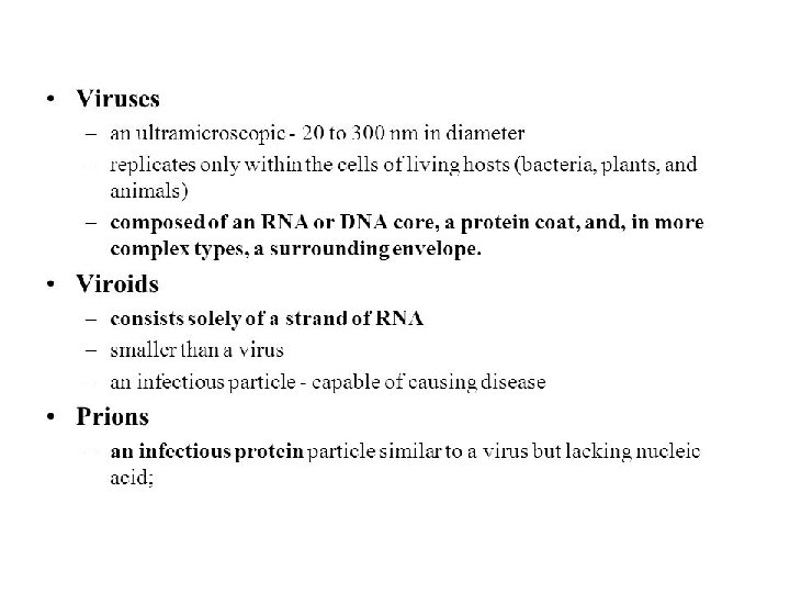
VIRUSES Viruses did not find a place in classification since they are not truly ‘living’, if we understand living as those organisms that have a cell structure. The viruses are non-cellular organisms that are characterised by having an inert crystalline structure outside the living cell. Once they infect a cell they take over the machinery of the host cell to replicate themselves, killing the host. It is known as connecting link between living and non living.

VIRUSES The name virus that means venom or poisonous fluid was given by Pasteur. D. J. Ivanowsky (1892) recognised certain microbes as causal organism of the mosaic disease of tobacco. These were found to be smaller than bacteria because they passed through bacteria-proof filters. M. W. Beijerinek (1898) demonstrated that the extract of the infected plants of tobacco could cause infection in healthy plants and called the fluid as Contagium vivum fluidum (infectious living fluid). W. M. Stanley (1935) showed that viruses could be crystallised and crystals consist largely of proteins. They are inert outside their specific host cell. Viruses are obligate parasites.
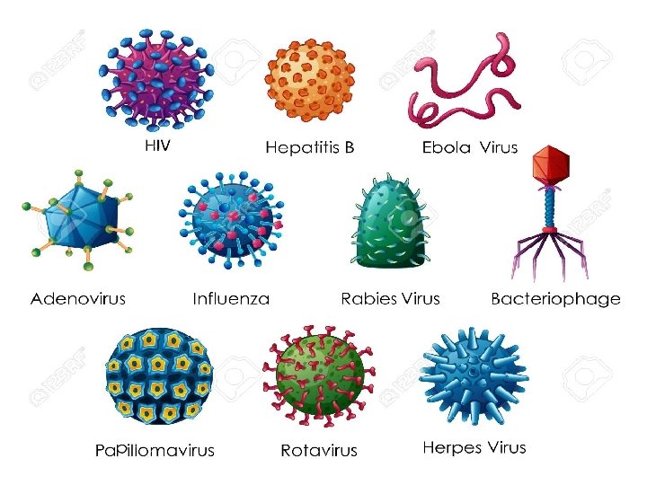

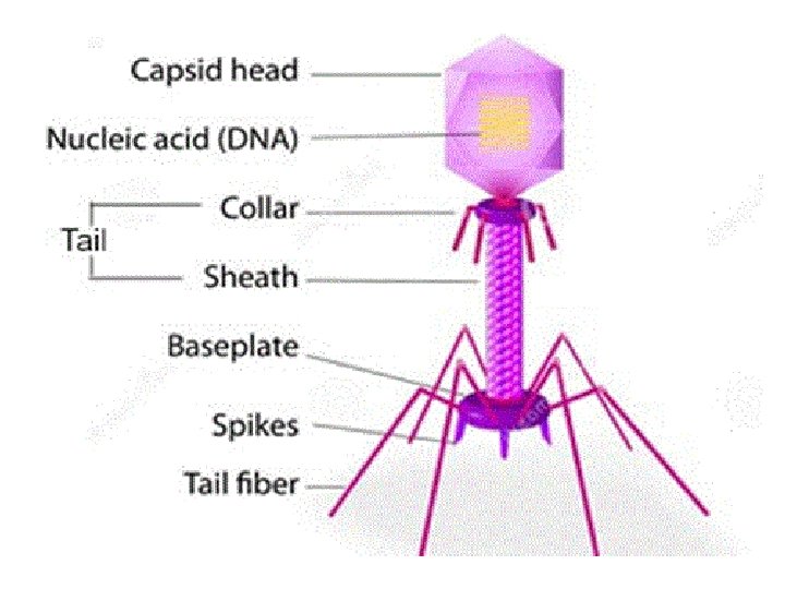
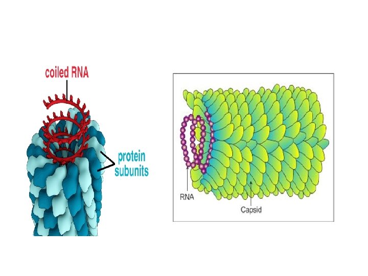
STRUCTURE OF VIRUSES It has proteins, genetic material either RNA or DNA. No virus contains both RNA and DNA. A virus is a nucleoprotein and the genetic material is infectious. Viruses that infect plants have single stranded RNA and viruses that infect animals have either single or double stranded RNA or double stranded DNA. Bacterial viruses or bacteriophages (viruses that infect the bacteria) are usually double stranded DNA viruses. The protein coat called capsid made of small subunits called capsomeres, protects the nucleic acid. These capsomeres are arranged in helical or polyhedral geometric forms.
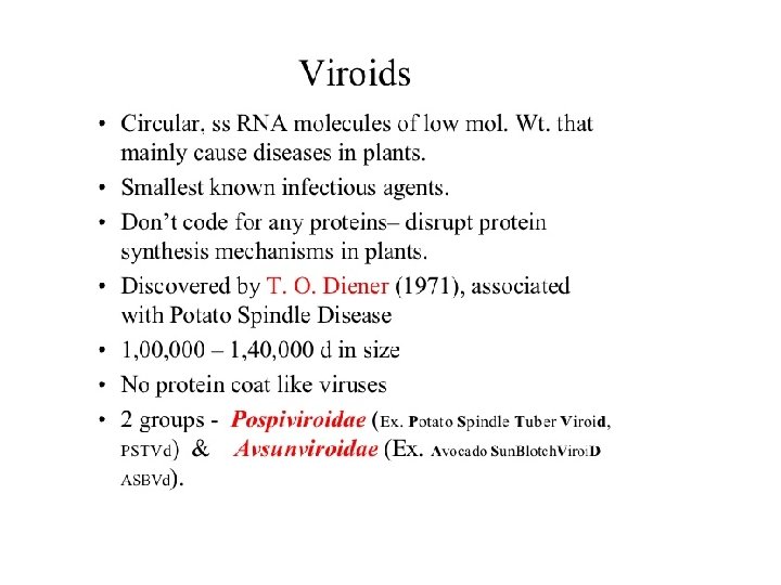

VIROIDS It is a free RNA. It lacked the protein coat that is found in viruses, hence the name viroid. The RNA of the viroid was of low molecular weight.

END OF CHAPTER THANK YOU !
- Slides: 66